LBH589對腎癌細胞株OS-RC-2增殖的影響及機制探討
2016-01-20 13:55:10馬俊健陳婷劉彬徐米清廣州醫科大學附屬第二醫院廣州54002廣州市番禺區婦幼保健院廣州心血管疾病研究所
山東醫藥 2015年31期
馬俊健,陳婷,劉彬,徐米清( 廣州醫科大學附屬第二醫院,廣州5400;2廣州市番禺區婦幼保健院; 廣州心血管疾病研究所)
LBH589對腎癌細胞株OS-RC-2增殖的影響及機制探討
馬俊健1,2,陳婷1,劉彬3,徐米清1
( 1廣州醫科大學附屬第二醫院,廣州511400;2廣州市番禺區婦幼保健院; 3廣州心血管疾病研究所)
摘要:目的觀察LBH589對腎癌細胞株OS-RC-2細胞增殖的影響,并探討其機制。方法將人腎癌細胞株OS-RC-2細胞分為DMSO組、N-乙酰半胱氨酸( NAC)組、LBH589組和NAC + LBH589組,NAC組加入5 mmol/L NAC刺激2 h,LBH489組加入50 nmol/L LBH589刺激48 h,NAC + LBH589組先加入5 mmol/L NAC預處理2 h,洗脫后再以50 nmol/L LBH589刺激48 h。EdU法測算細胞增殖情況,流式細胞術檢測細胞凋亡、活性氧簇( ROS),Western blotting法檢測細胞p-STAT3、Cyclin D1蛋白。結果DMSO組、NAC組、LBH589組、NAC + LBH589組EdU染色陽性細胞比例分別為53%±4%、52%±5%、7%±3%、29%±6%,細胞凋亡率分別為0.25%±0.13%、0.18%±0.15%、11.35%±2.40%、1.92%±0.63%,ROS分別為0.60%±0.26%、0.33%±0.25%、12.2%± 2.65%、5.33%±1.05%,DMSO組、NAC + LBH589組與LBH589組比較,P均<0.05。DMSO組、NAC組、LBH589組、NAC + LBH589組p-STAT3相對表達量分別為1.00±0.07、1.02±0.07、0.49±0.04、0.52±0.05,Cyclin D1相對表達量分別為1.00±0.08、1.03±0.08、0.45±0.04、0.49±0.03,DMSO組與LBH589組比較,P均<0.05。結論LBH589對OS-RC-2腎癌細胞生長有抑制作用,其可能與誘導細胞凋亡、抑制細胞增殖、升高ROS水平有關。LBH589能抑制STAT3信號通路,進而使Cyclin D1表達下降,而LBH589誘導ROS升高不是通過抑制STAT3信號通路從而使腎癌細胞生長抑制。
關鍵詞:腎腫瘤;腎癌;細胞增殖;細胞凋亡;組蛋白去乙酰化酶抑制劑
Effect of LBH589 on cell proliferation of renal carcinoma cell line OS-RC-2 and its mechanism
MA Jun-jian1,CHEN Ting,LIU Bin,LI Ai-qun,XU Mi-qing
( 1 The Second Affiliated Hospital of Guangzhou Medical University,Guangzhou 511400,China)
Abstract:Objective To observe the influences of LBH589 on cell proliferation of renal cancer cell line OS-RC-2 and to explore its mechanism.Methods OS-RC-2 cells were divided into the DMSO group,N-acetyl cysteine ( NAC) group,LBH589 group and NAC +LBH589 group.NAC group was stimulated with 5 mmol/L of NAC for 2 h,LBH489 group was stimulated with 50 nmol/L of LBH589 for 48 h,and the NAC +LBH589 group was pre-treated with 5 mmol/L of NAC for 2 h and then after elution,the group was stimulated with 50 nmol/L of LBH589 for 48 h.EdU assay was used to measure cell proliferation,flow cytometry was used to detect the apoptosis and reactive oxygen species ( ROS),while Western blotting was used to detect p-STAT3 and Cyclin D1 proteins.Results The proportions of positive Edu cells in the DMSO group,NAC group,LBH589 group and NAC +LBH589 group were respectively 53%±4%,52%±5%,7%±3% and 29%±6%.The apoptosis rates of the corresponding groups were 0.25%±0.13%,0.18%±0.15%,11.35%±2.40% and 1.92%± 0.63%.ROS of the corresponding groups were 0.60%±0.26%,0.33%±0.25%,12.2%±2.65% and 5.33%± 1.05%.Significant difference was found between the DMSO,NAC +LBH589 groups and the LBH589 group ( all P<0.05).The relative expression of p-STAT3 was 1.00±0.07,1.02±0.07,0.49±0.04 and 0.52±0.05 in the DMSO group,NAC group,LBH589 group and NAC +LBH589 group.The relative expression of Cyclin D1 was 1.00±0.08,1.03±0.08,0.45 ±0.04 and 0.49±0.03 in the corresponding groups.Significant difference was found between the DMSO group and the LBH589 group ( all P<0.05).Conclusions LBH589 inhibits the growth of renal cancer cell line OS-RC-2,which may be related to induction of apoptosis,inhibition of cell proliferation and the increased ROS level.LBH589 can inhibit STAT3 sig-
naling pathway,thus decrease the expression of Cyclin D1.However,LBH589-induced ROS increase is not achieved through the inhibition of STAT3 signaling pathway to inhibit the growth of renal cancer cells.
Key words:kidney neoplasms; renal cell carcinoma; cell proliferation; apoptosis; histone deacetylase inhibitor
腎細胞癌( RCC)的治療以根治性手術為主,放療及化療效果不理想,手術后復發率大于20%[1]。因此,尋找和研發新的抗腎癌藥物十分必要。組蛋白去乙酰化酶抑制劑( HDACIs)可通過細胞周期阻滯、誘導分化和凋亡等機制[2,3]對血液系統腫瘤和一些實體腫瘤有明顯抑制作用[4,5]。但HDACIs對腎癌細胞抑制作用的研究尚未見報道。HDACIs能提高細胞內活性氧簇( ROS)水平,降低抗氧化系統作用,抑制腫瘤細胞增殖,誘導細胞凋亡。LBH589為一種新的HDACIs類藥物。本研究觀察了LBH589誘導的氧化應激對腎癌細胞株OS-RC-2增殖的影響,并探討其可能的機制。
1 材料與方法
1.1細胞及試劑LBH589、N-乙酰半胱氨酸( NAC)購自Sigma公司;腎癌細胞株OS-RC-2(中國科學院細胞庫) ;胎牛血清、RPMI1640完全培養基、胰蛋白酶購自Gibco公司; MTT細胞增殖及細胞毒性檢測試劑盒、細胞凋亡檢測試劑盒、EdU試劑盒購自廣州銳博公司;總ROS檢測試劑盒購自Enzo Life Sciences公司;兔抗人p-STAT3、Cyclin D1、GAPDH單抗,羊抗兔二抗購自美國Santa Cruzs公司。
1.2細胞培養將人腎癌細胞株OS-RC-2細胞培養于含100 mL/L血清、100 kU/L青霉素、100 mg/L鏈霉素的RPMI1640培養液中,待細胞貼壁生長達到80%以上時進行傳代培養。細胞進行藥物刺激時,各實驗組培養基的DMSO終濃度為0.1%。
1.3 LBH589藥物濃度篩選采用MTT法檢測細胞生長情況。取對數生長期OS-RC-2細胞接種于96孔板,每孔0.2 mL。24 h后棄孔內液體,每孔分別加入100 μL濃度為0、10、50、100、500、1 000 nmol/L LBH589的培養基,藥物處理24、48、72 h,每組5個復孔,每孔加50 μL 1×MTT,培養4 h,棄上清,每孔加150 μL DMSO,測D550值,實驗重復5次,取平均值。MTT實驗結果顯示,LBH589呈時間和劑量依賴性抑制OS-RC-2細胞增殖,LBH589作用OS-RC-2細胞24、48、72 h,IC50分別為220、50、38 nmol/L,因此選擇50 nmol/L LBH589處理OS-RC-2細胞。
1.4細胞分組及LBH589干預將生長狀態良好的細胞接種于培養板,分為DMSO組、NAC組、LBH589組和NAC + LBH589組。各設3個復孔,NAC組加入5 mmol/L NAC刺激2 h,LBH489組加入50 nmol/L LBH589刺激48 h,NAC + LBH589組先加入5 mmol/L NAC預處理2 h,洗脫后再以50 nmol/L LBH589刺激48 h。
1.5細胞增殖檢測采用EdU法。取各組細胞行EdU標記、細胞固化、EdU染色、Hoechst33342染色,激光共聚焦顯微鏡觀察并計算每個視野中的EdU染色陽性細胞百分比,共計算10個視野。激光共聚焦顯微鏡圖像分析顯示新增殖細胞經EdU染色后表現為紅色熒光,所有活細胞經Hoechst33342染色后表現為藍色熒光,Overlay為紅色熒光和藍色熒光的重疊圖片。
1.6細胞凋亡檢測采用流式細胞術。取各組對數生長期OS-RC-2細胞接種于培養皿,藥物刺激后PBS 洗2次,胰蛋白酶消化細胞,收集至相應的流式管內,行AnnexinV、PI染色,流式細胞儀檢測,分析凋亡細胞百分率,實驗重復5次,取平均值。
1.7細胞ROS檢測采用流式細胞技術。取各組對數生長期OS-RC-2細胞接種于培養皿。藥物刺激后PBS洗2次,胰蛋白酶消化細胞,收集至相應的流式管內,離心,PBS洗2次,每個流式管各加500 μL ROS Detection Solution,37℃避光30 min,流式儀檢測分析細胞內ROS表達水平,實驗重復5次,取平均值。
1.8細胞p-STAT3、Cyclin D1表達檢測采用Western blotting法。取各組對數生長期細胞藥物處理后棄培養液,PBS洗2次,加入細胞裂解液、蛋白酶抑制劑,收集裂解混合液,12 000 r/min離心15 min,BCA法測定濃度。取30 μg蛋白標本為上樣量,經SDS-PAGE電泳后,轉移至PVDF膜,最后加入GAPDH、p-STAT3、Cyclin D1一抗和二抗,之后曝光顯影,重復5次,以Image J2x軟件進行灰度分析,取平均值。
1.9統計學方法采用SPSS16.0統計軟件。計量資料以珋x±s表示,組間比較用單因素方差分析。P <0.05為差異有統計學意義。
2 結果
2.1各組細胞增殖情況比較DMSO組、NAC組、LBH589組、NAC + LBH589組EdU染色陽性細胞比例分別為53%±4%、52%±5%、7%±3%、29%± 6%,DMSO組、NAC + LBH589組與LBH589組比較,P均<0.05。
2.2各組細胞凋亡率比較DMSO組、NAC組、LBH589組、NAC + LBH589組細胞凋亡率分別為0.25%±0.13%、0.18%±0.15%、11.35%± 2.40%、1.92%±0.63%,DMSO組、NAC + LBH589組與LBH589組比較,P均<0.05。
2.3各組細胞ROS比較DMSO組、NAC組、LBH589組、NAC + LBH589組細胞ROS分別為0.60%±0.26%、0.33%±0.25%、12.2%± 2.65%、5.33%±1.05%,DMSO組、NAC + LBH589組與LBH589組比較,P均<0.05。
2.4各組細胞p-STAT3和Cyclin D1蛋白表達比較結果見表1。
表1 各組OS-RC-2細胞p-STAT3和Cyclin D1蛋白表達比較(±s)
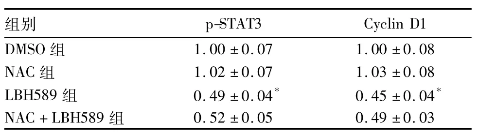
表1 各組OS-RC-2細胞p-STAT3和Cyclin D1蛋白表達比較(±s)
注:與DMSO組比較,*P<0.05。
組別p-STAT3 Cyclin D1 DMSO組1.00±0.07 1.00±0.08 NAC組 1.02±0.07 1.03±0.08 LBH589組 0.49±0.04* 0.45±0.04*NAC + LBH589組0.52±0.05 0.49±0.03
3 討論
本實驗結果表明,LBH589對OS-RC-2腎癌細胞增殖有明顯的抑制作用,其抑制率具有濃度依賴性和時間依賴性。EdU增殖試驗結果顯示OS-RC-2細胞經50 nmol/L LBH589處理48 h后,細胞增殖數量明顯減少。流式細胞術結果同樣顯示,50 nmol/L LBH589刺激OS-RC-2細胞48 h,其細胞凋亡比例比DMSO組明顯增加。ROS的大量產生可能是細胞凋亡的關鍵事件之一[6,7]。LBH589能顯著誘導OS-RC-2細胞ROS生成,NAC能減少LBH589作用細胞后ROS產生,從而減弱LBH589抑制細胞增殖及促細胞凋亡作用。這可能與LBH589處理細胞后細胞的氧化還原狀態如ROS的堆積密切相關,而NAC作為一種強大的抗氧化劑及自由基清除劑能夠保護腎癌細胞,對抗LBH589對OS-RC-2細胞的生長抑制作用。
STAT3持續激活可導致細胞異常增殖和惡性轉化,目前已被公認為癌基因[8,9]。它能調控目標基因的表達,如通過上調Bcl-xL、McL-1、Cyclin D1和c-myc基因的作用參與腫瘤的形成[10,11]。Cyclin D1是參與細胞周期的重要調節因子,過度表達的Cyclin D1可以調節細胞周期前進,加快腫瘤細胞的增殖速度。LBH589作用OS-RC-2腎癌細胞后p-STAT3、Cyclin D1的表達明顯下降,LBH589抑制p-STAT3及下游靶基因蛋白Cyclin D1的表達使細胞周期進程受阻從而使細胞增殖受到抑制。NAC預處理后再以LBH589刺激48 h,p-STAT3、Cyclin D1蛋白表達與LBH589組相比沒有統計學差異。這可能因為LBH589誘導細胞產生的大量ROS并不是通過抑制STAT3信號通路使腎癌細胞生長抑制,而是通過其他的方式和途徑引起細胞凋亡,這需要通過進一步的實驗來證實。
可見,LBH589對OS-RC-2腎癌細胞生長有抑制作用,其抗腫瘤機制可能與誘導細胞凋亡、抑制細胞增殖、升高ROS水平有關。LBH589能抑制STAT3信號通路,進而使Cyclin D1表達下降,而LBH589誘導ROS升高不是通過抑制STAT3信號通路從而使腎癌細胞生長抑制。
參考文獻:
[1]Jemal A,Siegel R,Ward E,et al.Cancer statistics,2008[J].CA Cancer J Clin,2008,58( 2) : 71-96.
[2]Ahmad M,Hamid A,Hussain A,et al.Understanding histone deacetylases in the cancer development and treatment: an epigenetic perspective ofcancer chemotherapy[J].DNA Cell Biol,2012,31 ( Suppl 1) : 62-71.
[3]XU M,Hong M,Xie H.Histone deacetylase inhibitors induce human renal cell carcinoma apoptosis through p-JNK activation[J].J South Med Univ,2013,33( 10) : 1409-1415.
[4]Dashwood RH,Myzak MC,Ho E.Dietary HDAC inhibitors: time to rethink weak ligands in cancer chemoprevention[J].Carcinogenesis,2006,27( 2) : 344-349.
[5]Vanhaecke T,Papeleu P,Elaut G,et al.Trichostatin A-like hydroxamate histone deacetylase inhibitors as therapeutic agents: toxicological point of view[J].Curr Med Chem,2004,11( 12) :1629-1643.
[6]Liu YB,Gao X,Deeb D,et al.Pristimerin Induces Apoptosis in Prostate Cancer Cells by Down-regulating Bcl-2 through ROS-dependentUbiquitin-proteasomal Degradation Pathway[J].J Carcinog Mutagen,2013,Suppl 6: 005.
[7]Kim TH,Kim JS,Kim ZH,et al.Khz-cp ( crude polysaccharide extract obtained from the fusion of Ganoderma lucidum and Polyporus umbellatusmycelia) induces apoptosis by increasing intracellular calcium levels and activating P38 and NADPH oxidase-dependent generation of reactive oxygen species in SNU-1 cells[J].BMC Complement Altern Med,2014,14: 236.
[8]Zhao J,Lin W,Cal Z,et al.Total alkaloids of Rubus aleaefolius Poir.inhibit the STAT3 signaling pathway leading to suppression of proliferation and cell cycle arrest in a mouse model of hepatocellular carcinoma[J].Oncol Rep,2013,30( 3) : 1309-1314.
[9]Lu L,Li C,Li D,et al.Cryptotanshinone inhibits human glioma cell proliferation by suppressing STAT3 signaling[J].Mol Cell Biochem,2013,381( 1-2) : 273-282.
[10]Grabenstatter HL,Del Angel YC,Carlsen J,et al.The effect of STAT3 inhibition on status epilepticus and subsequent spontaneous seizures in the pilocarpinemodel of acquired epilepsy[J].Neurobiol Dis,2014,62: 73-85.
[11]Guo S,Zhi Y,Yang H,et al.Bcl-2 expression is associated with poor prognosis of solitary plasmacytoma of bone[J].Ann Hematol,2014,93( 3) : 471-477.
收稿日期:( 2015-05-27)
通信作者簡介:徐米清( 1964-),男,主任醫師,主要研究方向為腎臟疾病的診斷與治療。E-mail: gyeyxu@163.com
作者簡介:第一馬俊健( 1988-),男,碩士研究生,主要研究方向為腎臟疾病的診斷與治療。E-mail: mjjtt121@163.com
基金項目:廣東省高等學校優秀青年教師培養計劃項目( Yq2013133)。
文章編號:1002-266X( 2015)31-0008-03
文獻標志碼:A
中圖分類號:R737.11
doi:10.3969/j.issn.1002-266X.2015.31.003
猜你喜歡
中國中藥雜志(2016年22期)2017-02-13 17:28:52
右江醫學(2016年4期)2017-01-05 16:26:48
科技視界(2016年15期)2016-06-30 12:27:37
中國實用醫藥(2016年11期)2016-05-04 21:56:32
課程教育研究·學法教法研究(2016年6期)2016-04-26 10:06:57
新課程·中學(2015年9期)2015-10-26 19:00:20
山東體育學院學報(2015年3期)2015-08-14 20:30:25
中國當代醫藥(2015年18期)2015-08-06 18:09:34
中國現代醫生(2015年13期)2015-06-17 10:19:05
