Notch信號相關受體/配體在皮膚惡性黑素瘤中的表達
2017-11-02 02:50:57劉白俞婉婷程偉邵雪寶姜祎群
中華皮膚科雜志 2017年4期
關鍵詞:信號
劉白 俞婉婷 程偉 邵雪寶 姜祎群
210042南京,中國醫學科學院 北京協和醫學院 皮膚病研究所病理科
Notch信號相關受體/配體在皮膚惡性黑素瘤中的表達
劉白 俞婉婷 程偉 邵雪寶 姜祎群
210042南京,中國醫學科學院 北京協和醫學院 皮膚病研究所病理科
目的檢測Notch信號通路中Notch1、Notch4、Jagged1以及Dll4在皮膚惡性黑素瘤組織中的表達,初步探討Notch信號通路在黑素瘤發病機制中的作用。方法免疫組化SP法檢測40例惡性黑素瘤及15例色素痣石蠟標本中Notch1、Notch4、Jagged1以及Dll4的表達模式和表達強度。采用SPSS21.0軟件進行卡方檢驗及Spearman秩相關分析。結果在40例黑素瘤組織(31例陽性表達)及15例色素痣組織(3例陽性表達)Notch1的表達率差異有統計學意義(χ2=15.281,P=0.000),原位(18例)及侵襲性黑素瘤(22例)間Notch1表達強度差異無統計學意義(χ2=0.631,P=0.427)。Notch4、Jagged1以及Dll4的表達率在惡性黑素瘤與色素痣之間差異均有統計學意義(均P<0.05),三者表達強度在原位與侵襲性黑素瘤之間差異亦均有統計學意義(P<0.05)。在黑素瘤組織中,Notch1與Jagged1的表達呈正相關(rs=0.350,P=0.027),與Dll4的表達亦呈正相關(rs=0.562,P=0.000),但是Jagged1與Dll4的表達呈負相關(rs=-0.734,P=0.000)。結論Notch信號通路異常可能是黑素瘤的發病機制之一,但具體作用機制有待進一步研究。
痣和黑素瘤;受體,Notch;免疫組織化學;Jagged 1;Dll4
Notch信號通路是一個進化保守的信號通路,在生物體發育成熟過程中影響細胞的增殖、分化、凋亡,調控細胞周期,起到決定細胞命運、組織模式及形態轉化的作用。研究發現,Notch信號通路在腫瘤的發生機制中發揮重要作用,許多腫瘤都與此通路的異常相關,目前已經成為研究的熱點之一。有報道在肝癌[1?2]、肺癌[3?4]、乳腺癌[5?6]、胃癌[7]、皮膚腫瘤(如基底細胞癌、鱗狀細胞癌)[8?9]等中均發現Notch信號通路異常表達。Massi等[10]首次報道,人類惡性黑素瘤組織標本中Notch蛋白及其配體的表達上調,提示Notch信號通路激活可能與黑素瘤發生相關。我們利用免疫組化的方法,研究Notch1、Notch4、Jagged1以及Dll4在黑素瘤中的表達情況及其相互之間的關系,希望能夠初步了解Notch信號通路在黑素瘤發生、發展中的作用。
材料和方法
一、樣本選取
選用中國醫學科學院皮膚病研究所病理科2011—2015年存檔的皮膚惡性黑素瘤標本40份及色素痣標本15份,均經4%甲醛固定,常規石蠟包埋。皮膚黑素瘤40例中,男21例,女19例,年齡24~86歲,平均59.78歲;病程0.5個月至20年;皮損位于肢端者32例,非肢端者8例。原位黑素瘤18例,侵襲性黑素瘤22例,Clark分級Ⅰ~Ⅱ級27例,Ⅲ~Ⅴ級13例。15例色素痣均為皮內痣,其中男7例,女8例;年齡8~59歲,平均29.733歲;皮損位于面部12例,胸部1例,四肢2例。
二、主要試劑
兔單克隆抗體抗Notch1抗體、抗Jagged1抗體、抗Dll4抗體,小鼠單克隆抗體抗Notch4抗體均來自英國Abcam公司;二抗為Alexa Flour?488標記山羊抗小鼠IgG,來自南京福麥斯生物技術有限公司;顯色劑二氨基聯苯胺(DAB)、S100、HMB?45、Melan?A、Ki67均來自丹麥Dako公司。
三、方法
1.免疫組化染色:石蠟包埋組織經4 μm厚連續切片,60℃干烤1 h,分別進行HE和免疫組化染色。免疫組化染色采用SP法,DAB顯色,用磷酸鹽緩沖液代替一抗作陰性對照。
2.甲苯胺藍雙重復染法[11]:將切片常規脫蠟至水,按免疫組化染色常規步驟進行,蘇木素復染,1%鹽酸乙醇分化、水洗;滴加甲苯胺藍工作液,覆蓋全部組織,室溫孵育30 min,水洗;用1∶500冰醋酸分化,鏡下觀察,黑素顆粒呈現墨綠色,與DAB染色陽性棕黃色顆粒區別明顯,水洗終止反應;常規脫水、封固。
3.免疫組化陽性判讀:HE染色、免疫組化染色切片由兩位高年資病理醫師采用雙盲法進行診斷及染色結果判讀。每張切片隨機選取5個高倍視野(×200),每個高倍視野計數50個腫瘤細胞,觀察陽性細胞染色強度,計數陽性細胞百分比。染色強度計分標準:不著色0分,弱著色(淺黃色)1分,中等著色(棕黃色)2分,強著色(深褐色)3分。陽性細胞百分比計分標準:按平均每視野所見陽性細胞計數,無細胞著色0分,<25%為1分,25%~75%為2分,>75%為3分。表達強度為染色強度計分與陽性細胞百分比計分相乘得出最終積分:計0分為陰性表達,>0分為陽性表達;0~4分為低表達,>4分為高表達。蛋白表達率=陽性表達例數/總例數×100%。
4.統計學方法:采用SPSS21.0軟件對實驗數據進行統計學分析,計數資料比較采用卡方檢驗,等級資料采用Spearman秩相關分析。以P<0.05為差異有統計學意義。
結果
一、Notch1表達情況
Notch1廣泛表達于細胞胞質,少量表達于細胞核,細胞核表達均出現在胞質陽性的細胞中。40例原發性黑素瘤中,31例陽性表達;15例色素痣中,3例陽性表達(圖1)。侵襲性黑素瘤與原位黑素瘤比較,Notch1表達強度差異無統計學意義(P= 0.427),但惡性黑素瘤與色素痣比較,Notch1表達率差異有統計學意義(P=0.000),見表1、2。Notch1在腫瘤周邊正常皮膚處呈陰性表達。不論在原發性黑素瘤還是侵襲性黑素瘤中,Notch1的表達在腫瘤的中心及邊緣位置均無差別。
二、Notch4表達情況
Notch4在細胞質/細胞膜中均表達。40例原發性黑素瘤患者中36例陽性表達;15例色素痣中6例為陽性表達(圖1)。侵襲性黑素瘤與原位黑素瘤之間Notch4表達強度差異有統計學意義(P=0.033),惡性黑素瘤與色素痣之間Notch4表達率差異亦有統計學意義(P=0.000)。見表1,2。Notch4在腫瘤周圍正常組織血管內皮細胞、毛囊及汗腺中可見陽性表達。在原發性黑素瘤和侵襲性黑素瘤中,Notch4的表達在腫瘤的中心及邊緣之間均無差別。
三、Jagged1表達情況
Jagged1表達于細胞膜中。40例原發性黑素瘤患者中34例陽性;15例色素痣中1例為陽性表達(圖1)。侵襲性黑素瘤與原位黑素瘤之間Jagged1表達強度差異有統計學意義(P=0.011),惡性黑素瘤與色素痣之間Jagged1表達率差異亦有統計學意義(P=0.000)。見表1、2。Jagged1在腫瘤周圍正常組織無表達。在原發性黑素瘤和侵襲性黑素瘤中,Jagged1的表達在腫瘤的中心及邊緣之間均無差別。

圖1 Notch1、Notch4、Jagged1、Dll4在原發性惡性黑素瘤及色素痣中的表達(免疫組化×100) Notch1、Notch4、Jagged1、Dll4在色素痣中不表達。Notch1在原位黑素瘤及侵襲性黑素瘤中廣泛表達于腫瘤細胞胞質,少量表達于細胞核;Notch4主要表達于原位黑素瘤及侵襲性黑素瘤細胞胞質/胞膜;Jagged1表達于原位黑素瘤及侵襲性黑素瘤細胞胞膜;Dll4表達于原位黑素瘤及侵襲性黑素瘤細胞胞膜/胞質
四、Dll4表達情況
Dll4表達于細胞膜/質中。40例原發性黑素瘤標本中38例陽性;15例色素痣標本均為陰性,見圖1。侵襲性黑素瘤與原位黑素瘤之間Dll4表達強度差異有統計學意義(P=0.002),惡性黑素瘤與色素痣之間Dll4表達率差異亦有統計學意義(P= 0.000)。見表1、2。Dll4在腫瘤周圍正常皮膚組織血管內皮細胞中可見陽性表達。在原發性黑素瘤和侵襲性黑素瘤中,Dll4的表達在腫瘤的中心及邊緣之間均無差別。
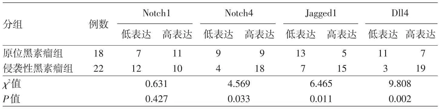
表1 Notch信號相關受體/配體在原位及侵襲性黑素瘤組織中的表達強度比較(例)
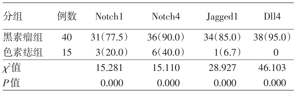
表2 Notch信號相關受體/配體在原發性黑素瘤及色素痣中的表達率比較[例(%)]
五、黑素瘤中Notch1、Jagged1、Dll4表達的相關性
Notch1表達與Jagged1表達呈正相關(rs= 0.350,P=0.027),與Dll4表達呈正相關(rs=0.562,P=0.000);Jagged1和Dll4的表達呈負相關(rs= -0.734,P=0.000)。
討論
人類惡性黑素瘤組織標本中Notch蛋白及其配體表達失調,提示Notch信號通路的激活可能與黑素瘤發生、發展相關,已有研究表明,Notch信號通路的上調是黑素瘤發病的早期事件[10,12]。我們利用免疫組化檢測40例原發性黑素瘤及15例色素痣組織標本中Notch1、Notch4、Jagged1以及Dll4的表達及相互聯系,希望能夠初步了解Notch信號通路在黑素瘤發生、發展中所發揮的作用。
多項研究表明,黑素瘤發生、發展依賴于Notch1信號的表達,Notch1信號通過自身加工和調節蛋白酶(如furin蛋白)進行自我激活[13?14]。我們的研究發現,Notch1在腫瘤中的表達明顯高于色素痣,但是在原位黑素瘤與侵襲性黑素瘤中表達強度無明顯差異,提示Notch1在黑素瘤的發生過程中可能發揮重要作用,同時其他機制對黑素瘤中Notch1表達的影響可能導致其對惡性黑素瘤侵襲能力的影響較小。
我們還發現,Notch4不但在黑素瘤中陽性表達,在腫瘤周圍正常皮膚處血管、毛囊及汗腺中也不同強度地表達。考慮腫瘤周圍內皮細胞出現Notch信號的陽性表達,提示其可能影響腫瘤的侵襲及轉移活性。Hardy等[15]研究揭示,Notch4和Nodal在侵襲性黑素細胞系中多重表達與腫瘤侵襲性相關,說明同時表達Notch4和Nodal的細胞亞群可能會保留特殊的性質,如細胞可塑性增強[15],并且對Notch4血管生成擬態的形成起到部分作用[16]。Notch4功能抑制會下調Nodal信號的表達,而且破壞、削弱侵襲性黑素細胞中的血管生成擬態網絡[17]。在我們所檢測的40例黑素瘤組織標本中,Notch4呈強弱不等的陽性表達,明顯高于色素痣,且在原位和侵襲性黑素瘤中的表達有統計學差異。表明Notch4表達上調可以促進黑素瘤發生、進展,上調的Notch4信號可能通過自身信號或者對其他相關信號通路的影響發揮促癌作用。
在原發性膠質母細胞瘤中,Jagged1和Dll4在微血管形成過程中起到相反的作用,但對疾病的預后具有一致性[18]。那么在黑素瘤中是否具有類似情況呢?有文獻報道,Dll4通過Notch1信號抑制內皮細胞活化,而Jagged1不但抑制Notch1/Dll4信號,同時保持血管生長與成熟的平衡[19],二者在腫瘤血管形成過程中具有相反作用[20?22]。在我們的研究中,Notch1的表達與Jagged1、Dll4的表達呈正相關,Jagged1的表達與Dll4的表達呈負相關,并且具有統計學意義,與文獻研究結果一致,提示Notch1和Jagged1、Dll4兩兩之間具有協同表達效應,并形成負反饋回路,調節腫瘤血管生成途徑,從而增加侵襲和轉移活性,但具體作用機制需要進一步探討。
總之,在原發性皮膚惡性黑素瘤中Notch信號通路表達失調,Notch1、Notch4、Jagged1以及Dll4均呈陽性表達,初步提示Notch信號通路異常可能是黑素瘤的發病機制之一,但具體作用機制有待進一步的研究探索。本文僅通過免疫組化的方法證明Notch信號通路參與黑素瘤的發生、發展,方法單一,具有一定的局限性。今后可通過運用q?PCR等方法進一步證實實驗結果,并可通過體外培養黑素細胞系檢測上述調控關系。
[1]Gao J,Song Z,Chen Y,et al.Deregulated expression of Notch receptors in human hepatocellular carcinoma[J].Dig Liver Dis, 2008,40(2):114?121.DOI:10.1016/j.dld.2007.08.001.
[2]Gramantieri L,Giovannini C,Lanzi A,et al.Aberrant Notch3 and Notch4 expression in human hepatocellular carcinoma[J].Liver Int,2007,27(7):997?1007.DOI:10.1111/j.1478?3231.2007. 01544.x.
[3]Konishi J,Kawaguchi KS,Vo H,et al.Gamma?secretase inhibitor prevents Notch3 activation and reduces proliferation in human lung cancers[J].Cancer Res,2007,67(17):8051?8057.DOI: 10.1158/0008?5472.CAN?07?1022.
[4]Donnem T,Andersen S,Al?Shibli K,et al.Prognostic impact of Notch ligands and receptors in nonsmall cell lung cancer: coexpression of Notch?1 and vascular endothelial growth factor?A predicts poor survival[J].Cancer,2010,116(24):5676?5685. DOI:10.1002/cncr.25551.
[5]Chen J,Imanaka N,Chen J,et al.Hypoxia potentiates Notch signaling in breast cancer leading to decreased E?cadherin expression and increased cell migration and invasion[J].Br J Cancer,2010,102(2):351?360.DOI:10.1038/sj.bjc.6605486.
[6]Peng GL,Tian Y,Lu C,et al.Effects of Notch?1 down?regulation on malignant behaviors of breast cancer stem cells[J].J Huazhong Univ Sci Technolog Med Sci,2014,34(2):195?200. DOI:10.1007/s11596?014?1258?4.
[7]Brzozowa M,Mielańczyk L,Michalski M,et al.Role of Notch signaling pathway in gastric cancer pathogenesis[J].Contemp Oncol(Pozn),2013,17(1):1?5.DOI:10.5114/wo.2013.33765.
[8]Nowell C,Radtke F.Cutaneous Notch signaling in health and disease[J].Cold Spring Harb Perspect Med,2013,3(12): a017772.DOI:10.1101/cshperspect.a017772.
[9]South AP,Purdie KJ,Watt SA,et al.NOTCH1 mutations occur early during cutaneous squamous cell carcinogenesis[J].J Invest Dermatol,2014,134(10):2630?2638.DOI:10.1038/jid.2014.154.
[10]Massi D,Tarantini F,Franchi A,et al.Evidence for differential expression of Notch receptors and their ligands in melanocytic nevi and cutaneous malignant melanoma[J].Mod Pathol,2006, 19(2):246?254.DOI:10.1038/modpathol.3800526.
[11]吳瓊,李阿梅,邵雪寶,等.甲苯胺藍染色在黑素瘤免疫組化復染中的應用[J].臨床與實驗病理學雜志,2015,(8):941?942. DOI:10.13315/j.cnki.cjcep.2015.08.029.
[12]Murtas D,Piras F,Minerba L,et al.Activated Notch1 expression is associated with angiogenesis in cutaneous melanoma[J].Clin Exp Med,2015,15(3):351?360.DOI:10.1007/s10238?014?0300?y.
[13]Qiu H,Tang X,Ma J,et al.Notch1 autoactivation via transcriptionalregulation offurin,which sustains Notch1 signaling by processing Notch1?activating proteases ADAM10 and membrane type 1 matrix metalloproteinase[J].Mol Cell Biol, 2015,35(21):3622?3632.DOI:10.1128/MCB.00116?15.
[14]Basak A,Chen A,Scamuffa N,et al.Blockade of furin activity and furin?induced tumor cells malignant phenotypes by the chemi?cally synthesized human furin prodomain[J].Curr Med Chem, 2010,17(21):2214?2221.DOI:10.2174/092986710791331040.
[15]Hardy KM,Kirschmann DA,Seftor EA,et al.Regulation of the embryonic morphogen Nodal by Notch4 facilitates manifestation of the aggressive melanoma phenotype[J].Cancer Res,2010,70(24):10340?10350.DOI:10.1158/0008?5472.CAN?10?0705.
[16]Bedogni B.Notch signaling in melanoma:interacting pathways and stromal influences that enhance Notch targeting[J].Pigment Cell Melanoma Res,2014,27(2):162?168.DOI:10.1111/pcmr. 12194.
[17]Kirschmann DA,Seftor EA,Hardy KM,et al.Molecular pathways: vasculogenic mimicry in tumor cells:diagnostic and therapeutic implications[J].Clin Cancer Res,2012,18(10):2726?2732. DOI:10.1158/1078?0432.CCR?11?3237.
[18]Qiu XX,Chen L,Wang CH,et al.The vascular notch ligands delta?like ligand 4(DLL4)and Jagged1(JAG1)have opposing correlations with microvascularization but a uniform prognostic effect in primary glioblastoma:a preliminary study[J].World Neurosurg,2016,88:447?458.DOI:10.1016/j.wneu.2015.10.058.
[19]Pedrosa AR,Trindade A,Fernandes AC,et al.Endothelial Jagged1 antagonizes Dll4 regulation of endothelial branching and promotes vascular maturation downstream of Dll4/Notch1[J]. Arterioscler Thromb Vasc Biol,2015,35(5):1134?1146.DOI: 10.1161/ATVBAHA.114.304741.
[20]Benedito R,Rocha SF,Woeste M,et al.Notch?dependent VEGFR3 upregulation allows angiogenesis without VEGF?VEGFR2 signal?ling[J].Nature,2012,484(7392):110?114.DOI:10.1038/ nature10908.
[21]Siekmann AF,Affolter M,Belting HG.The tip cell concept 10 years after:new players tune in for a common theme[J].Exp Cell Res,2013,319(9):1255?1263.DOI:10.1016/j.yexcr.2013.01. 019.
[22]Zhang J,Ye J,Ma D,et al.Cross?talk between leukemic and endothelial cells promotes angiogenesis by VEGF activation of the Notch/Dll4 pathway[J].Carcinogenesis,2013,34(3):667?677.DOI:10.1093/carcin/bgs386.
Expression of Notch pathway receptors and ligands in cutaneous malignant melanoma
Liu Bai,Yu Wanting,Cheng Wei,Shao Xuebao,Jiang Yiqun
Department of Pathology,Institute of Dermatology,Chinese Academy of Medical Sciences and Peking Union
Jiang Yiqun,Email:yiqunjiang@qq.com
ObjectiveTo determine the expression of Notch pathway receptors(Notch1 and Notch4)and ligands(Jagged1 and Dll4)in cutaneous malignant melanoma(CMM)tissues,and to preliminarily explore the role of the Notch signaling pathway in the pathogenesis of CMM.MethodsImmunohistochemical study was performed to determine the expression pattern and intensity of Notch1, Notch4,Jagged1 and Dll4 in 40 paraffin?embedded CMM specimens and 15 paraffin?embedded pigmented nevus specimens.Statistical analysis was carried out by chi?square test and Spearman rank correlation analysis with the SPSS 21.0 software.ResultsNotch1 was detected in 31(77.5%)of 40 CMM specimens, as well as in 3 of 15 pigmented nevus specimens,and the positive rates significantly differed between the two groups(χ2=15.281,P<0.001).However,no significant difference in the expression intensity of Notch1 was observed between 18in situmelanoma tissues and 22 invasive melanoma tissues(χ2=0.631,P=0.427).In addition,the positive rates of Notch4,Jagged1 and Dll4 were also significantly higher in the CMM group than those in the pigmented nevus group(allP<0.05),and the expression intensity of Notch4, Jagged1 and Dll4 significantly differed betweenin situand invasive melanoma tissues(allP<0.05).In CMM tissues,the expression of Notch1 was positively correlated with that of Jagged1(rs=0.350,P= 0.027)and Dll4(rs=0.562,P<0.001),while the expression of Jagged1 was negatively correlated with that of Dll4(rs=-0.734,P<0.001).ConclusionAbnormality of the Notch signaling pathway may be involved in the pathogenesis of melanoma,but further researches are still needed to elucidate the detailed mechanism.
Nevi and melanomas;Receptors,Notch;Immunohistochemistry;Jagged1;Dll4
姜祎群,Email:yiqunjiang@qq.com
10.3760/cma.j.issn.0412?4030.2017.04.005
Medical College,Nanjing 210042,China
2016?07?20)
(本文編輯:尚淑賢)
猜你喜歡
鴨綠江(2021年35期)2021-04-19 12:24:18
考試與評價·高一版(2020年6期)2020-11-02 02:45:24
媽媽寶寶(2019年10期)2019-10-26 02:45:34
中國生殖健康(2019年3期)2019-02-01 06:12:26
鐵道通信信號(2018年11期)2019-01-19 01:15:08
電子制作(2018年11期)2018-08-04 03:25:42
鐵道通信信號(2018年2期)2018-04-18 12:18:10
鐵道通信信號(2016年11期)2016-06-01 12:11:32
鑿巖機械氣動工具(2016年3期)2016-03-01 04:00:25
中國病理生理雜志(2015年8期)2015-12-21 12:38:06