甘露糖醛酸寡糖對H2O2誘導衰老心肌細胞的影響
2019-09-10 07:22:44牟婕馮文靜毛擁軍
青島大學學報(醫學版) 2019年4期
關鍵詞:自噬
牟婕 馮文靜 毛擁軍

[摘要] 目的 探討甘露糖醛酸寡糖(M3K)對過氧化氫(H2O2)誘導的心肌細胞衰老的作用及其可能機制。方法 用100 μmol/L H2O2構建H9c2心肌細胞衰老模型,應用不同濃度(10、50、100 mg/L)的M3K干預衰老心肌細胞,觀察M3K對心肌細胞衰老的影響。實驗分為對照組、H2O2組、H2O2+M3K低劑量組、H2O2+M3K中劑量組和H2O2+M3K高劑量組。用倒置顯微鏡觀察各組心肌細胞數量,Western blot檢測衰老標志蛋白P21和P53的表達,PT-RCR檢測自噬相關基因beclin1、Atg7和LC3-Ⅱ的mRNA表達。結果 倒置顯微鏡觀察顯示,與對照組相比,H2O2組心肌細胞數量明顯減少;與H2O2組相比,M3K干預各組心肌細胞數量呈劑量依賴性增多,差異有統計學意義(F=8.685,P<0.05)。Western blot檢測顯示,H2O2組心肌細胞P21和P53蛋白表達較對照組明顯增加,M3K干預各組心肌細胞P21和P53蛋白表達較H2O2組明顯降低,差異有顯著意義(F=31.507、13.111,P<0.05)。PT-RCR檢測顯示,H2O2組心肌細胞beclin1、Atg7和LC3-Ⅱ的mRNA表達較對照組顯著降低,M3K干預各組三者的mRNA表達較H2O2組顯著增加(F=10.134~61.041,P<0.05)。結論 M3K可以延緩H2O2誘導的心肌細胞衰老,其機制可能與自噬的激活有關。
[關鍵詞] 甘露糖;寡糖類;衰老;肌細胞,心臟;自噬;過氧化氫
[中圖分類號] R329.25 ?[文獻標志碼] A ?[文章編號] ?2096-5532(2019)04-0411-05
[ABSTRACT] Objective To investigate the effect of mannuronic acid oligosaccharides (M3K) on hydrogen peroxide (H2O2)-induced cardiomyocyte senescence and the possible mechanism. ?Methods H9c2 cardiomyocytes were treated with 100 μmol/L H2O2 to establish a model of senescence, and then the senescent H9c2 cardiomyocytes were treated with different concentrations of M3K (10, 50, and 100 mg/L) to observed the effect of M3K on cardiomyocyte senescence. The cardiomyocytes were divided into control group, H2O2 group, H2O2+low-dose M3K group, H2O2+medium-dose M3K group, and H2O2+high-dose M3K group. Inverted microscopy was used to measure the number of cardiomyocytes, Western blot was used to measure the expression of the senescence marker proteins P21 and P53, and RT-PCR was used to measure the mRNA expression of the auto-phagy-related genes beclin1, Atg7, and LC3-Ⅱ. ?Results Inverted microscopy showed that compared with the control group, the H2O2 group had a significant reduction in the number of cardiomyocytes, and compared with the H2O2 group, the H2O2+low-dose M3K group, the H2O2+medium-dose M3K group, and the H2O2+high-dose M3K group had a significant increase in the number of cardiomyocytes in a dose-dependent manner (F=8.685,P<0.05). Western blot showed that compared with the control group, the H2O2 group had significant increases in the protein expression of P21 and P53 in cardiomyocytes, and compared with the H2O2 group, the H2O2+low-dose M3K group, the H2O2+medium-dose M3K group, and the H2O2+high-dose M3K group had significant reductions in the protein expression of P21 and P53 (F=31.507,13.111;P<0.05). RT-PCR showed that compared with the control group, the H2O2 group had significant reductions in the mRNA expression of beclin1, Atg7, and LC3-Ⅱ, and compared with the H2O2 group, the H2O2+low-dose M3K group, the H2O2+medium-dose M3K group, and the H2O2+high-dose M3K group had significant increases in the mRNA expressions of beclin1, Atg7, and LC3-Ⅱ (F=10.134-61.041,P<0.05). ?Conclusion M3K can delay H2O2-induced cardiomyocyte senescence, possibly by activating autophagy.
[KEY WORDS] mannose; oligosaccharides; aging; myocytes, cardiac; autophagy; hydrogen peroxide
衰老是一個復雜的過程,隨著年齡的增加,機體發生退行性改變,進而引起各種衰老相關疾病。目前,世界人口老齡化加劇,隨著老齡人口的增加,衰老相關疾病逐漸增多,其中心血管疾病是導致老年人死亡的主要原因[1-2]。機體衰老后心臟會發生一系列病理性改變,如心肌纖維化、心肌肥厚、心功能不全等[3]。有研究表明,隨著細胞衰老P21和P53蛋白表達增加,二者可作為衰老檢測的標志性蛋白[4-6]。自噬是一種存在于細胞內的降解機制,可以清除細胞內受損的蛋白和細胞器,對細胞內穩態的調節至關重要[7-8]。衰老心肌細胞自噬調節能力降低,導致細胞內錯誤折疊蛋白和功能障礙的細胞器大量累積[9]。研究表明,提高心肌細胞自噬水平可以延緩心臟衰老[10]。甘露糖醛酸寡糖(M3K)是褐藻膠寡糖的一種,由來源于海洋褐藻的褐藻膠加工而成,具有抗氧化、抗炎、抗腫瘤等多種生物學活性,有重要的臨床應用價值[11-13]。過氧化氫(H2O2)屬于活性氧簇家族,可引起氧化應激,誘導細胞衰老,現已被廣泛用于體外衰老模型的建立[14-15]。本實驗采用H2O2誘導建立心肌細胞衰老模型,探討M3K對衰老心肌細胞的保護作用及其可能機制。
1 材料和方法
1.1 實驗材料
M3K由中國海洋大學醫藥學院實驗室惠贈。心肌細胞H9c2購于中國科學院上海細胞庫。胎牛血清(FBS)購于美國Gibco公司,DMEM培養液購于美國Hyclone公司,P21兔單克隆抗體和P53小鼠單克隆抗體購于美國Cell Signaling Technology公司,山羊抗兔IgG二抗和山羊抗鼠IgG二抗購于武漢伊萊瑞特公司,RIPA裂解液和BCA蛋白濃度測定試劑盒購于上海碧云天公司,ECL發光液購于美國Millipore公司,逆轉錄試劑盒和Mix購于瑞士Roche公司。
1.2 實驗方法
1.2.1 細胞培養及處理 將H9c2心肌細胞接種于6孔板中,用含體積分數0.10 FBS和10 g/L青-鏈霉素的DMEM完全培養液,在37 ℃、含體積分數0.05 CO2的細胞培養箱中培養。實驗分為5組:對照組(A組),H2O2組(B組),H2O2+M3K低劑量組(C組),H2O2+M3K中劑量組(D組),H2O2+M3K高劑量組(E組)。當細胞融合度達50%時,H2O2組、H2O2+M3K低劑量組、H2O2+M3K中劑量組和H2O2+M3K高劑量組細胞分別加入100 μmol/L的H2O2誘導建立衰老模型,4 h后更換新鮮DMEM完全培養液。H2O2組繼續培養48 h,M3K干預各組分別加入10、50、100 mg/L的M3K培養48 h。實驗過程中對照組僅進行同步換液處理。在倒置顯微鏡下觀察,每孔隨機選擇3個區域進行鏡下計數。收集細胞進行后續實驗,所有實驗均重復3次。
1.2.2 Western blot檢測P21和P53蛋白表達細胞中加入RIPA裂解液提取細胞蛋白,用BCA試劑盒檢測蛋白濃度。每孔加入等量的蛋白進行SDS-PAGE電泳,待溴酚藍到達分離膠底部時轉移到PVDF膜上(200 mA,90 min),用含30 g/L BSA和50 g/L脫脂奶粉的TBST溶液室溫封閉2 h,加入P21和P53一抗(均1∶1 000稀釋),于4 ℃搖床孵育過夜;洗脫后分別加二抗(均1∶5 000稀釋)室溫孵育2 h,用ECL發光液顯影,凝膠成像系統成像后采用Quantity One軟件分析條帶灰度值。
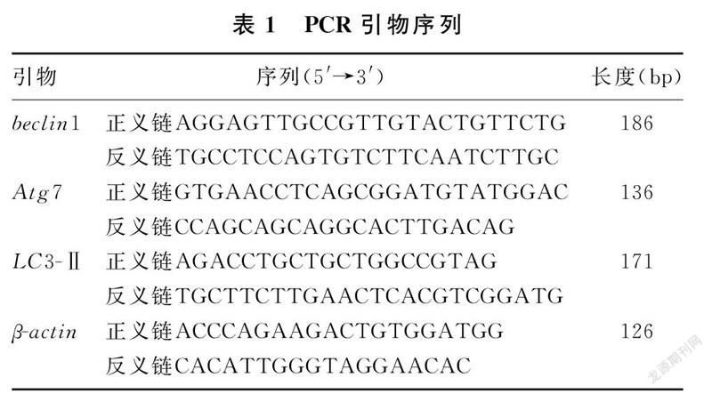
1.2.3 RT-PCR檢測beclin1、Atg7和LC3-Ⅱ的mRNA表達 采用Trizol法提取細胞總RNA,Nanodrop測定RNA含量和濃度,應用逆轉錄試劑盒將mRNA逆轉錄為cDNA。采用RT-PCR法進行擴增,擴增條件:95 ℃、600 s;95 ℃、10 s,60 ℃、10 s,72 ℃、15 s,共40個循環;95 ℃、10 s,65 ℃、60 s,97 ℃、1 s。以β-actin作為內參,用2-ΔΔCt計算目的基因相對表達量。PCR引物序列見表1。
1.3 統計學方法
應用SPSS 21.0軟件進行統計學分析,計量數據以±s表示,多組比較采用One-way ANOVA檢驗,組間兩兩比較采用LSD法。
2 結 ?果
2.1 各組心肌細胞數量比較
培養48 h后,對照組細胞數量較多,融合度達90%;H2O2組細胞數量與對照組相比明顯減少;M3K干預各組細胞數量與H2O2組相比呈劑量依賴性增加,差異均有統計學意義(F=8.685,P<0.05);H2O2+M3K高劑量組細胞數量與對照組相比差異無顯著性(P>0.05)。見表2。
4期牟婕,等. 甘露糖醛酸寡糖對H2O2誘導衰老心肌細胞的影響413
2.2 各組心肌細胞P21和P53蛋白表達比較
與對照組相比,H2O2組心肌細胞P21和P53蛋白表達顯著增加;與H2O2組相比較,M3K干預48 h后,H2O2+M3K低劑量組、H2O2+M3K中劑量組和H2O2+M3K高劑量組心肌細胞P21和P53蛋白表達隨M3K濃度的增加而降低,差異均有顯著性(F=31.507、13.111,P<0.05)。見圖1、表2。
2.3 各組細胞beclin1、Atg7和LC3-Ⅱ mRNA表達比較
與對照組相比,H2O2組心肌細胞beclin1、Atg7和LC3-Ⅱ mRNA表達水平顯著降低;與H2O2組相比,M3K干預48 h后,H2O2+M3K低劑量組、H2O2+M3K中劑量組和H2O2+M3K高劑量組心肌細胞beclin1、Atg7和LC3-Ⅱ mRNA表達呈濃度依賴性增加,差異均有統計學意義(F=10.134~61.041,P<0.05)。見表3。
3 討 ?論
衰老是導致老年人慢性疾病發生發展的重要危險因素,并且是心血管疾病發生的獨立危險因素。近年來,隨著老齡人口增加,衰老相關疾病給社會帶來了巨大的醫療和經濟負擔。因此,探討衰老相關機制、延緩衰老成為目前的研究熱點[16]。H2O2誘導的氧化損傷可以較好地模擬老年人體內氧化應激,現已被廣泛用于衰老模型的建立[17-18]。本實驗用H2O2成功誘導了心肌細胞內氧化應激,引起細胞衰老。細胞衰老后表現為細胞周期阻滯,增殖減少。M3K屬于海藻寡糖,具有多種生物學活性[11]。本研究結果顯示,用M3K干預衰老心肌細胞后,細胞周期阻滯延緩,增殖加快。
P21作為細胞周期蛋白依賴性激酶抑制因子在衰老中起重要作用,它通過與細胞周期蛋白結合,導致細胞周期阻滯,阻斷細胞增殖過程。細胞衰老后,P21蛋白表達增加,因此,P21可作為衰老標志物之一[19-20]。本實驗采用Western blot方法檢測P21蛋白的表達,觀察M3K對衰老心肌細胞的影響。結果顯示,H2O2誘導心肌細胞衰老后P21表達增加,而用M3K干預衰老細胞48 h可使P21蛋白表達下調,緩解衰老心肌細胞的細胞周期阻滯,進而延緩心肌細胞衰老。P53是氧化應激的關鍵調控分子之一[21]。P53作為“基因監護人”,可通過調節細胞周期阻滯、細胞凋亡和衰老發揮腫瘤抑制作用[22]。有研究表明,p53基因過表達小鼠可以出現早衰表型[23]。P53的表達與機體衰老呈正相關,機體衰老后,P53表達增加[24-25]。本研究結果顯示,P53在H2O2誘導的衰老心肌細胞中表達增加,而M3K干預衰老細胞可使P53蛋白表達下調,表明M3K可能延緩了H2O2誘導的心肌細胞衰老。
自噬可分為巨自噬、微自噬和分子伴侶介導的自噬3種類型,其中巨自噬是最常見的一種,其自噬過程依賴于溶酶體。LC3-Ⅱ、beclin1和Atg7是自噬溶酶體形成的關鍵蛋白,參與溶酶體膜的形成,自噬被激活后,三者的表達增加[7,26]。機體衰老后自噬調節能力也隨之下降,細胞內受損蛋白質和細胞器大量積累,進一步加速衰老[27-28]。已有大量的研究表明,激活自噬可能延緩組織和細胞衰老[29]。為了探討M3K延緩心肌細胞衰老是否與自噬的激活有關,本研究采用RT-PCR方法檢測了自噬相關基因LC3-Ⅱ、beclin1和Atg7 mRNA的表達。結果顯示,H2O2誘導的衰老細胞中LC3-Ⅱ、beclin1和Atg7 mRNA表達減少,而給予M3K處理后,細胞中LC3-Ⅱ、beclin1和Atg7 mRNA表達增加,表明M3K延緩心肌細胞衰老可能與自噬激活有關。
綜上所述,M3K可以延緩H2O2誘導的H9c2心肌細胞衰老,其作用機制可能與細胞內自噬的激活有關,但其具體的分子機制還需要進一步研究。
[參考文獻]
[1] SKIBSKA B, GORACA A. The protective effect of lipoic acid on selected cardiovascular diseases caused by age-related oxidative stress[J]. Oxidative Medicine and Cellular Longevity, 2015,2015:313021.
[2] EZZATI M, OBERMEYER Z, TZOULAKI I, et al. Contributions of risk factors and medical care to cardiovascular mortality trends[J]. Nature Reviews Cardiology, 2015,12(9):508-530.
[3] ALFARAS I, DI GERMANIO C, BERNIER M, et al. Pharmacological strategies to retard cardiovascular aging[J]. Circulation Research, 2016,118(10):1626-1642.
[4] WANG Ziling, CHEN Linbo, QIU Zhu, et al. Ginsenoside Rg1 ameliorates testicular senescence changes in Dgalinduced aging mice via antiinflammatory and antioxidative mechanisms[J]. Molecular Medicine Reports, 2018,17(5):6269-6276.
[5] CARRASCO G E, MORENO M, MORENO C L, et al. Increased Arf/p53 activity in stem cells, aging and cancer[J]. Aging Cell, 2017,16(2):219-225.
[6] JIANG C, LIU G, LUCKHARDT T, et al. Serpine 1 induces alveolar type Ⅱ cell senescence through activating p53-p21-Rb pathway in fibrotic lung disease[J]. Aging Cell, 2017,16(5):1114-1124.
[7] LAPIERRE L R, KUMSTA C, SANDRI M, et al. Transcriptional and epigenetic regulation of autophagy in aging[J]. Autophagy, 2015,11(6):867-880.
[8] KIM S N, KWON H J, AKINDEHIN S, et al. Effects of epigallocatechin-3-gallate on autophagic lipolysis in adipocytes[J]. Nutrients, 2017,9(7):680-694.
[9] SHIRAKABE A, IKEDA Y, SCIARRETTA S A, et al. Aging and autophagy in the heart[J]. Circulation Research, 2016,118(10):1563-1576.
[10] REN Jun, YANG Lifang, ZHU Li, et al. Akt2 ablation prolongs life span and improves myocardial contractile function with adaptive cardiac remodeling:role of Sirt1-mediated autophagy regulation[J]. Aging Cell, 2017,16(5):976-987.
[11] ZHU Benwei, CHEN Meijuan, YIN Heng, et al. Enzymatic hydrolysis of alginate to produce oligosaccharides by a new purified endo-type alginate lyase[J]. Marine Drugs, 2016,14(6):108-119.
[12] GUO Junjie, MA Leilei, SHI Hongtao, et al. Alginate oligosaccharide prevents acute doxorubicin cardiotoxicity by suppressing oxidative stress and endoplasmic reticulum-mediated apoptosis[J]. Marine Drugs, 2016,14(12):231-244.
[13] Guo Junjie, XU Fengqiang, LI Yonghong, et al. Alginate oligosaccharide alleviates myocardial reperfusion injury by inhibiting nitrative and oxidative stress and endoplasmic reticulum stress-mediated apoptosis[J]. Drug Des Devel Ther, 2017,11:2387-2397.
[14] ZHU Wei, WU Yan, MENG Yifang, et al. Effect of curcumin on aging retinal pigment epithelial cells[J]. Drug Des Devel Ther, 2015,9:5337-5344.
[15] CHEUNG T M, GANATRA M P, FU J J, et al. The effect of stress-induced senescence on aging human cord blood-derived endothelial cells[J]. Cardiovascular Engineering and Technology, 2013,4(2):220-230.
[16] PERRIDON B W, LEUVENINK H G, HILLEBRANDS J L, et al. The role of hydrogen sulfide in aging and age-related pathologies[J]. Aging, 2016,8(10):2264-2289.
[17] LI Ruilin, LU Zhaoyang, HUANG Jingjuan, et al. SRT1720, a SIRT1 specific activator, protected H2O2-induced senescent endothelium[J]. American Journal of Translational Research, 2016,8(7):2876-2888.
[18] SONG Zhiming, LIU Yong, HAO Baoshun, et al. Ginse-noside Rb1 prevents H2O2-induced HUVEC senescence by stimulating sirtuin-1 pathway[J]. PLoS One, 2014,9(11):e112699.
[19] HSU Y M, YIN M C. EPA or DHA enhanced oxidative stress and aging protein expression in brain of D-galactose treated mice[J]. Biomedicine (Taipei), 2016,6(3):23-30.
[20] JI Musi, SU Xiaohua, LIU Jizhen, et al. Comparison of naturally aging and D-galactose induced aging model in beagle dogs[J]. Experimental and Therapeutic Medicine, 2017,14(6):5881-5888.
[21] GAMBINO V, DE MICHELE G, VENEZIA O A, et al. Oxidative stress activates a specific p53 transcriptional response that regulates cellular senescence and aging[J]. Aging Cell, 2013,12(3):435-445.
[22] QIAN Yingjuan, CHEN Xinbin. Senescence regulation by the p53 protein family[J]. Methods Mol Biol, 2013,965:37-61.
[23] GUEST I, ILIC Z, SCRABLE H, et al. Survival of irradiated recipient mice after transplantation of bone marrow from young, old and“early aging”mice[J]. Aging, 2015,7(12):1212-1222.
[24] LESSEL D, WU D Y, TRUJILLO C, et al. Dysfunction of the MDM2/p53 axis is linked to premature aging[J]. Journal of Clinical Investigation, 2017,127(10):3598-3608.
[25] BU H, WEDEL S, CAVINATO M, et al. MicroRNA regulation of oxidative stress-induced cellular senescence[J]. Oxid Med Cell Longev, 2017,2017:1-12.
[26] WU Yingying, CHEN Chao, YU Xiao, et al. Effects of tiaozhi granule on regulation of autophagy levels in HUVECs[J]. Evidence-Based Complementary and Alternative Medicine, 2018,2018:1-10.
[27] GHOSH A K, MAU T, O’BRIEN M, et al. Impaired autophagy activity is linked to elevated ER-stress and inflammation in aging adipose tissue[J]. Aging,?2016,8(10):2525-2537.
[28] CARAMES B, OLMER M, KIOSSES W B, et al. The relationship of autophagy defects to cartilage damage during joint aging in a mouse model[J]. Arthritis Rheumatol, 2015,67(6):1568-1576.
[29] PAPP D, KOVACS T, BILLES V, et al. AUTEN-67, an autophagy-enhancing drug candidate with potent antiaging and neuroprotective effects[J]. Autophagy, 2016,12(2):273-286.
(本文編輯 馬偉平)
猜你喜歡
中國醫藥導報(2017年2期)2017-03-18 20:50:25
中國中藥雜志(2017年1期)2017-03-06 21:37:10
中國中藥雜志(2016年21期)2017-02-16 13:30:44
中國中藥雜志(2016年21期)2017-02-16 12:24:39
糖尿病新世界(2016年16期)2016-12-09 04:07:18
中西醫結合心血管病電子雜志(2016年6期)2016-11-14 12:40:03
科技視界(2016年11期)2016-05-23 08:10:09
湖北農業科學(2015年20期)2015-11-12 21:01:15
中國醫藥導報(2015年15期)2015-08-07 01:10:32
山東體育學院學報(2015年2期)2015-05-27 13:17:09
