Pre-B-cell colony-enhancing factor as a target for protecting against apoptotic neuronal death and mitochondrial damage in ischemia
2017-01-21 03:33:04XiaowanWang,ShinghuaDing
中國(guó)神經(jīng)再生研究(英文版) 2016年12期
Pre-B-cell colony-enhancing factor as a target for protecting against apoptotic neuronal death and mitochondrial damage in ischemia
Focal ischemic stroke (FIS) results from the lack of blood fow in a particular region of the brain and accounts for about 80% of all human strokes. Although tremendous eforts have been made in translational research, the treatment strategies are still limited. Tissue plasminogen activator is the only FDA-approved drug currently available for acute stroke treatment, but it is only efective within a narrow therapeutic window that is within a few hours after the onset of a stroke. Thus exploring novel molecular pathways that can reduce neuronal death and improve stroke outcomes will allow us to identify potential targets for stroke therapy. Neurons are highly metabolically active cells and especially susceptible to an ischemic insult. Energy depletion triggers rapid necrotic neuronal death in the ischemic core as well as more delayed apoptosis in the penumbral region aTher an FIS. Thus it is conceivable that rescue or compensation for energy metabolism in the neurons is an important neuronal and brain protective strategy in FIS.
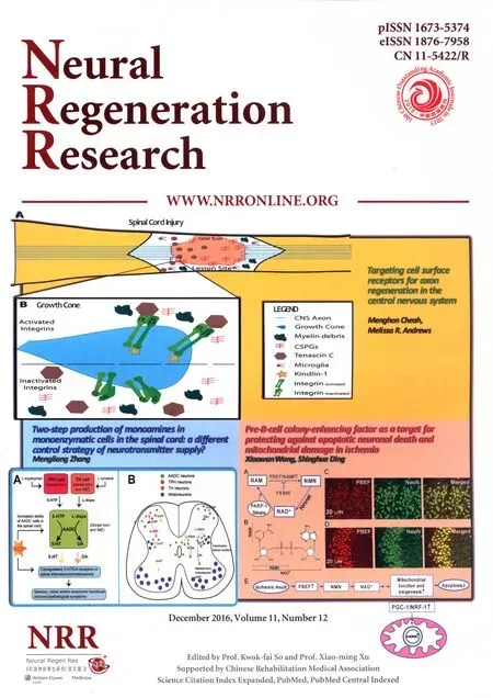
Recently, we found that pre-B-cell colony-enhancing factor (PBEF), also known as nicotinamide phosphoribosyltransferase (NAMPT), the rate-limiting enzyme in the salvage pathway of mammalian nicotinamide adenine dinucleotide (NAD+) biosynthesis through converting nicotinamide (NAM) to nicotinamide mononucleotide (NMN) (Canto et al., 2015) (Figure 1A, B), is mainly expressed in neurons in the mouse brain under physiological conditions (Figure 1C, D) (Zhang et al., 2010). NAD+is an essential coenzyme in the tricarboxylic acid (TCA) cycle and oxidative phosphorylation in the mitochondria and glycolysis in the cytoplasm. The major cellular functions of NAD+and its reduced form NADH include modulating cellular energy homeostasis. NAD+is also a substrate of some NAD+-consuming enzymes such as sirtuins (Sirt1-7), poly(ADP-Ribose) polymerases (PARP) and CD38 (Canto et al., 2015). We also found for the frst time that heterozygous knockout of PBEF increases brain infarction in a mouse model of photothrombosis (PT)-induced FIS (Zhang et al., 2010). Furthermore, we demonstrated that the enzymatic activity of NAD+synthesis is required for the neuroprotective efect of PBEF aTher glutamate and oxygen-glucose deprivation (OGD) in primary cultured cortical neurons since the overexpression of wide-type (WT) PBEF, but not mutants lacking enzymatic activity, can reduce neuronal death (Bi et al., 2012). Ischemia causes the depletion of NAD+levels and the supplementation of NAD+can protect against neuronal death in ischemia (Wang et al., 2014; Zhang et al., 2010; Bi et al., 2012). NAD+is also highly correlated with mitochondrial biogenesis (Wang et al., 2014). To further study the mechanism of PBEF in the neuronal protection in ischemia, we focused our investigation on whether the neuroprotective efect of PBEF is through regulating mitochondrial function and biogenesis.
We initially overexpressed WT and PBEF mutants H247A and H247E by DNA transfection and determined their efect on mitochondrial membrane potential (MMP) depolarization aTher excitotoxic glutamate stimulation using live cell fuorescent imaging (Bi et al., 2012). We found that neurons transfected with WT PBEF exhibited a much slower fuorescence decrease than non-transfected neurons or neurons transfected with EGFP alone; furthermore, neurons transfected with H247A and H247E mutants had the comparable rates of MMP depolarization with neurons expressing EGFP alone or non-transfected neurons (Bi et al., 2012). The results indicate that the overexpression of PBEF renders neurons more resistant to excitotoxicity-induced MMP collapse.
Ischemic stroke leads to translocation of arterial input function (AIF) from mitochondria to the nucleus. AIF release from mitochondria induces peripheral chromatin condensation, large-scale DNA fragmentation and eventually caspase-independent intrinsic apoptotic cell death (Hong et al., 2004). Next, we tested whether PBEF could inhibit AIF translocation after glutamate stimulation. We found that neurons overexpressing WT PBEF exhibited a dramatic reduction of AIF translocation rate as compared with neurons overexpressing the mutant H247A PBEF aTher glutamate excitotoxicity (Wang et al., 2016).Thus the enzymatic activity of PBEF for NAD+synthesis is required to inhibit glutamate-induced AIF translocation. Ischemia triggers intrinsic apoptosis of both caspase-3 independent pathway associated with AIF translocation and caspase-3 dependent pathway with cytochrome C release from the mitochondria. Using viral transduction approach, we found that the overexpression of PBEF can reduce cleaved caspase-3 levels after OGD, indicating that in addition to AIF translocation-involved caspase-3 independent pathway, a caspase-dependent apoptotic signaling pathway also contributes to the neuroprotective efect of PBEF in ischemia (Wang et al., 2016). Mitochondrial morphology including length, size and shape is controlled by balanced fssion and fusion processes in the neurons. Under normal conditions, new mitochondria are produced through the orchestrated action of transcriptional factors and co-activators.The balance is disrupted in injured neurons, thus causing morphological damage associated with mitochondrial dysfunction. Mitochondrial fragmentation is an early event that occurs before the release of mitochondria-dependent apoptotic proteins and is the key mechanism in glutamate excitotoxicity and AIF translocation-mediated cell death. We investigated the effect of PBEF on glutamate-induced mitochondrial fragmentation, which has been widely detected during excitotoxicity-inducedapoptosis. We found that glutamate increased the number of fragmented mitochondria with a short length and small area, but the overexpression of WT PBEF could prevent this effect, while overexpression of H247A PBEF had no effect on these parameters (Wang et al., 2016). The density of mitochondria in dendrites, however, was not affected by the overexpression of either WT or the mutant PBEF (Wang et al., 2016). Consistently, the supplementation of NAM and NAD+could also reduce glutamate-induced mitochondrial fragmentation and AIF translocation (Wang et al., 2014, 2016). Thus we conclude that PBEF prevents glutamate-stimulated mitochondrial fragmentation through its enzymatic activity of NAD+biosynthesis. It is possible that PBEF could facilitate the clearance of fragmented mitochondria via regulation of mitophagy during ischemia because PBEF signifcantly enhances autophagy (Wang et al., 2012).

Figure 1 Salvage pathway for NAD+biosynthesis and PBEF expression in neurons.
To further investigate the mechanism by which PBEF suppresses the impairment of mitochondrial biogenesis, we used AAV transduction to overexpress PBEF in a large number of neurons. We first determined the effect of PBEF on the relative amount of mitochondrial DNA (mtDNA) to nuclear DNA (nucDNA), which represents mitochondrial mass, aTher glutamate and OGD stimulations. We found a signifcant decrease of mtDNA/nucDNA ratio aTher glutamate excitotoxicity and OGD in primary cultured cortical neurons without PBEF overexpression, but the decline of mtDNA content was inhibited when neurons were overexpressed with WT PBEF. PGC-1 is a master regulator of mitochondrial biogenesis; therefore, we subsequently investigated the efect of PBEF overexpression on PGC-1 and its downstream efector NFR-1 aTher OGD. The protein levels of PGC-1 and NRF-1 reduced significantly by OGD stimulation, but PBEF overexpression signifcantly inhibited their reductions. Thus, PBEF overexpression in neurons can attenuate ischemia-induced mitochondrial damage and the impairment of mitochondrial biogenesis.
Mitochondria produce 90% of energy in mammalian cells.They are considered as the central executioner of both necrosis and apoptosis and an important therapeutic target for acute and chronic neurodegenerative diseases. Although we demonstrated that PBEF could mediate neuronal protection through the suppression of mitochondrial dysfunction and fragmentation and the impairment of mitochondrial biogenesis, some outstanding questions remain to be answered. NAD+is highly compartmentalized in cytoplasm and mitochondria of mammalian cells including neurons (Alano et al., 2007) and there is no mitochondrial localization sequence in PBEF. Accordingly it will be important to determine whether neuronal mitochondria have a distinct functional NAD+salvage pathway used to maintain the NAD+pool in the organelle. Using the subcompartment-targeted NAD+sensors (Cambronne et al., 2016), we are also able to determine the different dynamics of NAD+depletion after an ischemic insult. Using molecular biology to knockdown and overexpress PBEF in various subcompartments, we can further investigate whether mitochondrial NAD+pathway plays a predominant role in reducing neuronal death and brain damage. Sirt3 is expressed in mitochondria and plays a protective role in glutamate excitotoxicity (Kim et al., 2011). Since Sirt3 is an NAD+-consuming enzyme, further study to determine whether PBEF modulates Sirt3 activity is warranted. Moreover, PBEF may exert its neuronal and brain protective efects in ischemia through other mechanisms. A recent study reported that overexpression of PBEF in neurons in transgenic mice ameliorates white matter injury after middle cerebral artery occlusion (MCAO) through promoting extracellular PBEF levels (Jing et al., 2014), on the other hand, global PBEF overexpression in transgenic mice improves regenerative neurogenesis to promote brain recovery aTher MCAO (Zhao et al., 2015). PBEF also mediates the induction of autophagy at the early stage of ischemia, which also contributes to neuroprotection (Wang et al., 2012).
In summary, we have established that PBEF ameliorates apoptotic neuronal death after ischemia through inhibiting caspase-dependent and independent apoptotic signaling pathways; PBEF also mitigates the loss of mitochondrial mass and the impairment of mitochondrial biogenesis by inhibiting mitochondrial fragmentation and rescuing the key molecules involving mitochondrial biogenesis (Figure 1E). Our study revealed a previously unknown mechanism for the neuroprotective efect of PBEF in ischemia and indicates that inhibiting mitochondrial damage through enhancing the NAD+salvage pathway is a valuable strategy for ischemic stroke therapy.
The work was supported by NIH NS069726 and NS094539 and America Heart Association 13GRANT17020004 (to SD).
Xiaowan Wang, Shinghua Ding*
Department of Bioengineering (Wang X, Ding S), Dalton Cardiovascular Research Center (Ding S), University of Missouri, Columbia, MO, USA
*Correspondence to:Shinghua Ding, Ph.D., dings@missouri.edu.
Accepted:2016-11-28
Alano CC, Tran A, Tao R, Ying W, Karliner JS, Swanson RA, Alano CC, Tran A, Tao R, Ying W, Karliner JS, Swanson RA (2007) Diferences among cell types in NAD+compartmentalization: a comparison of neurons, astrocytes, and cardiac myocytes. J Neurosci Res 85:3378-3385.
Bi J, Li H, Ye SQ, Ding S (2012) Pre-B-cell colony-enhancing factor exerts a neuronal protection through its enzymatic activity and the reduction of mitochondrial dysfunction in in vitro ischemic models. J Neurochem 120:334-346.
Cambronne XA, Stewart ML, Kim D, Jones-Brunette AM, Morgan RK, Farrens DL, Cohen MS, Goodman RH (2016) Biosensor reveals multiple sources for mitochondrial NAD+. Science 352:1474-1477.
Canto C, Menzies K, Auwerx J (2015) NAD+metabolism and the control of energy homeostasis: a balancing act between mitochondria and the nucleus. Cell Metab 22:31-53.
Hong SJ, Dawson TM, Dawson VL (2004) Nuclear and mitochondrial conversations in cell death: PARP-1 and AIF signaling. Trends Pharmacol Sci 25:259-264.
Jing Z, Xing J, Chen X, Stetler RA, Weng Z, Gan Y, Zhang F, Gao Y, Chen J, Leak RK, Cao G (2014) Neuronal NAMPT is released aTher cerebral ischemia and protects against white matter injury. J Cereb Blood Flow Metab 34:1613-1621.
Kim SH, Lu HF, Alano CC (2011) Neuronal Sirt3 protects against excitotoxic injury in mouse cortical neuron culture. PLoS One 6:e14731.
Wang P, Guan YF, Du H, Zhai QW, Su DF, Miao CY (2012) Induction of autophagy contributes to the neuroprotection of nicotinamide phosphoribosyltransferase in cerebral ischemia. Autophagy 8:77-87.
Wang X, Li H, Ding S (2014) The effects of NAD+ on apoptotic neuronal death and mitochondrial biogenesis and function aTher glutamate excitotoxicity. Int J Mol Sci 15:1012-1022.
Wang X, Li H, Ding S (2016) Pre-B-cell colony-enhancing factor protects against apoptotic neuronal death and mitochondrial damage in ischemia. Sci Rep 6:32416.
Zhang W, Xie Y, Wang T, Bi J, Li H, Zhang LQ, Ye SQ, Ding S (2010) Neuronal protective role of PBEF in a mouse model of cerebral ischemia. J Cereb Blood Flow Metab 30:1962-1971.
Zhao Y, Guan YF, Zhou XM, Li GQ, Li ZY, Zhou CC, Wang P, Miao CY (2015) Regenerative neurogenesis aTher ischemic stroke promoted by nicotinamide phosphoribosyltransferase-nicotinamide adenine dinucleotide cascade. Stroke 46:1966-1974.
10.4103/1673-5374.195275
How to cite this article:Wang X, Ding S (2016) Pre-B-cell colony-enhancing factor as a target for protecting against apoptotic neuronal death and mitochondrial damage in ischemia. Neural Regen Res 11(12):1914-1915.
Open access statement:This is an open access article distributed under the terms of the Creative Commons Attribution-NonCommercial-ShareAlike 3.0 License, which allows others to remix, tweak, and build upon the work non-commercially, as long as the author is credited and the new creations are licensed under the identical terms.
 中國(guó)神經(jīng)再生研究(英文版)2016年12期
- 中國(guó)神經(jīng)再生研究(英文版)的其它文章
- Expression changes of nerve cell adhesion molecules L1 and semaphorin 3A aTher peripheral nerve injury
- Injury of the arcuate fasciculus in a patient with progressive bulbar palsy
- “Three Methods and Three Points” regulates p38 mitogen-activated protein kinase in the dorsal horn of the spinal cord in a rat model of sciatic nerve injury
- Biodegradable magnesium wire promotes regeneration of compressed sciatic nerves
- Electroacupuncture at Dazhui (GV14) and Mingmen (GV4) protects against spinal cord injury: the role of the Wnt/β-catenin signaling pathway
- Application of a paraplegic gait orthosis in thoracolumbar spinal cord injury