胚胎發育不良性神經上皮腫瘤
2017-03-29 06:39:35韓彤
中國現代神經疾病雜志 2017年1期
關鍵詞:信號
·臨床醫學圖像·
胚胎發育不良性神經上皮腫瘤


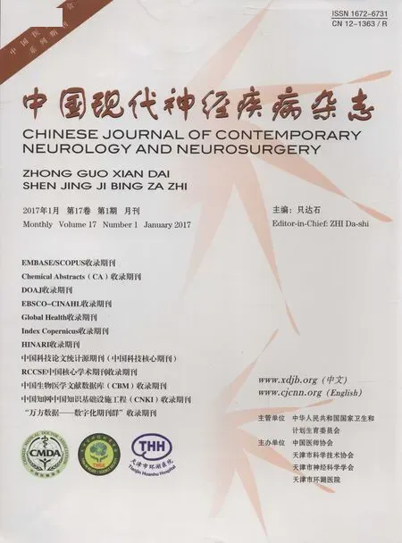
圖1 男性,18歲,主因癲發作4年、頭痛頭暈10 d入院。頭部影像學顯示左側額顳葉囊實性占位征象。手術切除左側額顳葉占位性病變。術后病理證實胚胎發育不良性神經上皮腫瘤(WHOⅠ級)1a橫斷面CT顯示,左側額顳葉低密度影,其內側壁可見斑片樣高密度結節影(箭頭所示)1b橫斷面T1WI顯示,左側顳葉囊性占位征象,囊液呈低信號,信號強度高于腦脊液(箭頭所示);其內側壁稍高信號結節影1c橫斷面T2WI顯示,病變內囊液呈高信號,附壁結節呈等信號(箭頭所示);瘤周無水腫1d橫斷面FLAIR成像顯示,鄰近病變內側壁高信號影(箭頭所示)1e橫斷面增強T1WI顯示,病變內側壁實性區斑片樣強化(箭頭所示),囊壁未見強化Figure 1 An 18-year-old male was hospitalized because of epilepsy for 4 years and headache for 10 d.MRI showed a cystic solid mass located in left fronto-temporal lobe.The lesion was totally resected and postoperative pathological diagnosis was dysembryoplastic neuroepithelial tumor(DNT, WHOⅠ).Axial CT showed hypodensity shadow in left fronto-temporal lobe with patchy hyperdensity nodule located in its medial wall(arrow indicates,Panel 1a).Axial T1WI revealed a cystic mass with hypointensity,which was slightly higher than cerebrospinal fluid,located in left temporal lobe(arrow indicates).A mural nodule with slightly high-intensity was found in the medial wall(Panel 1b).Axial T2WI showed high-intensity cystic fluid with an isointensity mural nodule(arrow indicates).No edema was found in the surrounding tissue(Panel 1c).Axial fat suppression FLAIR showed high-intensity signal adjacent to medial wall of lesion(arrow indicates,Panel 1d).Axial enhanced T1WI showed patchy enhancement in solid component of medial wall(arrow indicates)and no enhancement was found in the cystic wall(Panel 1e).
胚胎發育不良性神經上皮腫瘤(DNT,WHOⅠ級)是中樞神經系統少見良性腫瘤,屬神經元和混合性神經元-膠質腫瘤范疇。1988年由Daumas-Duport等首次命名。多見于兒童和青年,好發于大腦皮質,顳葉最為多見,其次為額葉,基底節、腦室、腦干、小腦、透明隔和胼胝體等亦有報道,腫瘤生長緩慢,臨床主要表現為難治性癲,預后良好,術前明確診斷十分重要。典型胚胎發育不良性神經上皮腫瘤呈底部位于大腦皮質、尖部朝向腦深部的楔形或腦回樣結構,囊性或囊實性,邊界清晰,瘤周無水腫,無明顯占位征象,鄰近大腦皮質可并存皮質發育不良。CT表現為皮質和皮質下界限清晰的低密度影(圖1a),20%病灶可見斑片樣鈣化;位于大腦凸面的腫瘤因生長緩慢致顱骨內板受壓變薄。MRI顯示病灶內多發結節樣和假囊性結構,T1WI呈低信號,信號強度略高于腦脊液(圖1b);T2WI可見囊性或多囊性“肥皂泡”樣結構,呈高信號(圖1c),部分病變內有分隔;FLAIR成像呈略低或高信號,病變邊緣可見線樣、斑片樣或環形更高信號帶,即“環形征”(圖1d),具有診斷特異性,可能與腫瘤邊緣圍繞含膠質-神經元成分的疏松組織有關。部分病變可見附壁結節,信號強度略高于大腦皮質。增強掃描可見少部分病變內或邊緣線樣、斑片樣、結節樣或環形強化(圖1e),系增生的神經膠質細胞伴血管增生所致。應注意與位于皮質和皮質下的囊性腫瘤、帶附壁結節的腫瘤(如節細胞膠質瘤、多形性黃色星形細胞瘤、毛細胞型星形細胞瘤、少突膠質細胞瘤)相鑒別。
(天津市環湖醫院神經放射科韓彤供稿)
Dysembryoplastic neuroepithelial tumor
HAN Tong
Department of Neuroradiology,Tianjin Huanhu Hospital,Tianjin 300350,China(Email:mrbold@163.com)
10.3969/j.issn.1672-6731.2017.01.015
猜你喜歡
鴨綠江(2021年35期)2021-04-19 12:24:18
考試與評價·高一版(2020年6期)2020-11-02 02:45:24
媽媽寶寶(2019年10期)2019-10-26 02:45:34
中國生殖健康(2019年3期)2019-02-01 06:12:26
鐵道通信信號(2018年11期)2019-01-19 01:15:08
電子制作(2018年11期)2018-08-04 03:25:42
鐵道通信信號(2018年2期)2018-04-18 12:18:10
鐵道通信信號(2016年11期)2016-06-01 12:11:32
鑿巖機械氣動工具(2016年3期)2016-03-01 04:00:25
中國病理生理雜志(2015年8期)2015-12-21 12:38:06