Vestigial like family member 3 is a novel prognostic biomarker for gastric cancer
2019-08-14 07:31:12LiHuaZhangZhuoWangLongHaiLiYanKuiLiuLinFangJinXiaoWeiQiChunZhangTengWangDongHua
World Journal of Clinical Cases 2019年15期
Li-Hua Zhang,Zhuo Wang,Long-Hai Li,Yan-Kui Liu,Lin-Fang Jin,Xiao-Wei Qi,Chun Zhang,Teng Wang,Dong Hua
Abstract
Key words: Vestigial like family member 3; Stomach adenocarcinoma; HER2 mutation;Gastric cancer; Bioinformatics analysis
INTRODUCTION
Gastric cancer (GC) is a frequently occurring malignancy of the digestive tract[1],and it was the fifth most commonly diagnosed and the third most common cause of cancerrelated mortality worldwide in 2018[2].Lack of early diagnosis and ineffective treatment options for the advanced stages are the primary reasons for the dismal 5-year survival rate of GC[3].Therefore,it is imperative to identify novel biomarkers for early diagnosis and accurate prediction of prognosis.Vestigial like family member 3(VGLL3) is a member of the vestigial like family of proteins[4],and is associated with epithelial ovarian cancer[5]and soft tissue sarcoma[6].The aim of this study was to determine the expression status and prognostic utility of VGL33 in GC.To this end,we mined the transcriptomic data of GC from the ONCOMINE and GEPIA databases,and determined its correlation with the survival data from GEPIA and ONCOLINC.VGLL3 levels were upregulated in GC samples and associated with a poor prognosis.The bioinformatics data were successfully validated on the tumor and normal gastric tissues resected from GC patients.Based on the VGLL3 levels,the patients were stratified into the high and low expression groups,and a prognosis prediction model was established on the basis of VGLL3 status and clinical data.Our findings show a strong prognostic role of VGLL3 in GC,which can potentially translate to clinical applications.
MATERIALS AND METHODS
Patients and tissue samples
A total of 172 tissue samples were obtained from gastric adenocarcinoma patients who underwent major surgery at the Affiliated Hospital of Jiangnan University between 2009 and 2012.The tissues were immediately fixed in formalin and embedded in paraffin for further analysis.In addition,fresh samples were dissected from 30 patients at the Affiliated Hospital of Jiangnan University in November 2018,and immediately stored in liquid nitrogen for molecular analysis.The mean follow-up duration was 43.1 mo and ranged from 0.2 to 86 mo.None of the patients received chemotherapy or radiotherapy before surgery[7].The tumor stage classification was determined by three pathologists who were blinded to the patient data according to the guidelines of the American Joint Committee on Cancer (AJCC).Freshly resected tissues were obtained from 202 patients,and were fixed in formalin and embedded in paraffin.In addition,tissue specimens from 30 patients were flash frozen in liquid nitrogen.All participating clinicians and patients provided written informed consent.
Bioinformatics analysis
The VGLL3 mRNA levels in the GC and normal gastric tissues were determined by integrated analysis of the Oncomine database (www.oncomine.org)[8]using Coexpedia (www.coexpedia.org)[9].ONCOLINC (http://www.oncolnc.org/cancer/)and Coexpedia were used to analyze the prognostic value of WISP1 in STAD.The STRING database (http://www.string-db.org) was utilized to construct the proteinprotein interaction (PPI) network[10].
前列腺癌是男性生殖系統(tǒng)中常見的腫瘤,發(fā)病率隨年齡而增長(zhǎng),前列腺癌的發(fā)病率和死亡率僅次于肺癌,位居癌癥死亡的第二位。目前治療前列腺癌的主要方法是內(nèi)分泌治療,但大部分患者經(jīng)過一段時(shí)間的治療后進(jìn)展為去勢(shì)抵抗性前列腺癌[5-7]。當(dāng)患者發(fā)展為去勢(shì)抵抗性前列腺癌時(shí)對(duì)內(nèi)分泌治療藥物就會(huì)失效,對(duì)前列腺腫瘤的發(fā)展失去控制,患者的病情愈加惡劣,加速了死亡。因此,尋找新的有效的治療方法已成為當(dāng)務(wù)之急[8-9]。
RNA isolation and RT-qPCR
Total RNA was extracted from frozen tissue samples using Trizol reagent (Invitrogen,Carlsbad,CA) according to the manufacturer’s instructions,and reverse transcribed using the PrimeScript RT-PCR kit (Takara,Japan)[11].RT-qPCR was performed on the ABI 7500 RealTime PCR System (Applied Biosystems,Inc.USA) using SYBR Green Master Mix (Takara,Japan),and the VGLL3 levels were normalized to β-actin.The following primers were used:Forward primer,5’-CCAACTACAGTCACCTCTGCTAC-3’ and reverse primer,5’-ACCACGGTGATTCCTTACTCTTG-3’ for VGLL3; forward primer,5’-CCTGTGGCATCCACGAAACT-3’ and reverse primer,5’-GAAGCATTTGCG GTGGACGAT-3’ for β-actin.All reactions were performed in triplicates,and the 2-??Ctmethod[12]was used to quantify the relative expression levels of VGLL3.
Western blot analysis
Total protein was extracted from GC and para-cancerous tissues using the RIPA lysis buffer (Pierce,Thermo Scientifc,Cramlington,United Kingdom)[13],and quantified with the enhanced BCA protein assay kit (KeyGEN BioTECH,Jiangsu,China).Equal amount (40 mg) of proteins per sample were separated by sodium dodecyl sulfatepolyacrylamide gel electrophoresis (SD-PAGE),and then transferred to polyvinylidene difluoride membranes (Bio-Rad,Hercules,CA,United States).After blocking with 5% non-fat milk for 1 h at room temperature (RT),the membranes were incubated overnight with anti-VGLL3 (ab83555,Abcam) and β-actin (ab8226,Abcam)primary antibodies at 4 °C.After washing thrice with TBST,the membranes were incubated with HRP-conjugated secondary antibody (1:1000) for 1 h at RT.The protein bands were visualized with an ECL chemiluminescence system after short exposure to X-ray films (Kodak,Japan).Densitometric analysis was performed with Image Pro-Plus software,and the relative expression levels of VGLL3 were normalized to tubulin.
Immunohistochemistry (IHC)
The formalin-fixed tissues were dehydrated,embedded in paraffin,and cut into 5 μm thick sections.IHC was performed according to the manufacturer’s protocol[14].Briefly,the sections were heated in citrate buffer in a microwave,cooled,and incubated overnight with anti-VGLL3 antibody (1:50,clone PA5-68441,Invitrogen) at 4 °C.After labeling with the secondary antibody for 1 hour at RT,color was developed using liquid DAB substrate.Three pathologists blinded to the patient identity observed and graded VGLL3 positivity as 0,1 (1%-29% positively-stained cells),2 (30%-69%),or 3 (70%-100%),and the staining intensity as 0 (negative),1(weak staining),2 (moderate staining),or 3 (strong staining).The immunoreactive score (IRS) was then calculated for each sample by multiplying the staining intensity and positivity scores,and graded as:0-1,“-”; 2-3,“+”; 4-6,“++”; and 6-9,“+++”[15].Based on the IRS,the samples were stratified into the high (4-9) and low (0-3) VGLL3 expression groups.
Fluorescence in situ hybridization (FISH)
The tissue sections were treated with denaturing solution to denature the DNA into single strands,and hybridized with the PathVysion probes (No.36-161060,PathVysion HER-2DNA Probe Kit).After washing the unbound probe with DNA,the sections were counterstained with the nuclear dye DAPI (4’,6 diamidino-2-phenylindole).Positive LSI HER-2/neu and CEP 17 signals were counted under a fluorescence microscope,and the ratio of the copy number of HER-2/neu gene to that of chromosome 17 was calculated.
Statistical analysis
Statistical analyses were performed with R*64 version 3.5.2 software and Graphpad 6.01.HER2 status (positive or negative) was designated on the basis of the combined FISH and IHC results as per the American Society of Clinical Oncology/College of American Pathologists Clinical Practice Guidelines.VGLL3 expression levels in the tumor and normal samples were compared by the Student'st-test,and the association between VGLL3 and clinico-pathological features was assessed by the chi-square test.The overall survival (OS) curve was plotted by the Kaplan-Meier method and analyzed by the log-rank test.Univariate and multivariate analyses of the prognostic factors were performed using the Cox proportional hazard regression model.A nomogram was formulated based on the results of the multivariate analysis with the package of rms in R version 3.5.2 (http://www.r-project.org/)[16].The receiver operating characteristic (ROC) curve was plotted to determine the sensitivity and specificity of the VGLL3-based prognostic score[17],and the area under the curve(AUC) was calculated.AllP-values were two-tailed and considered statistically significant when less than 0.05.
RESULTS
VGLL3 mRNA and protein levels are upregulated in GC samples
GEPIA database profiling showed that although VGLL3 was significantly overexpressed in GC samples of advanced TNM stages relative to those at the lower stages (P =0.0163),there was no significant difference between the GC and normal tissues (P> 0.05).Integrated analysis of multiple VGLL3 transcriptome datasets from the ONCOMINE database,however,showed that VGLL3 was significantly overexpressed in GC (P =0.003),and notably associated with a poor prognosis according to the ONCOLINC database analysis (P =0.01,n= 378) (Figure 1).In addition,GEPIA online survival analysis further confirmed that VGLL3 was a marker of poor prognosis in GC (P =0.0062,n= 384).To validate thein silicodata,we analyzed the expression levels of VGLL3 in the tumor and normal gastric tissues resected from 30 GC patients,and observed significantly higher levels of VGLL3 mRNA (P< 0.001) and protein (P< 0.001) in the GC tissues compared to normal tissues (Figure 2).Taken together,VGLL3 is upregulated in GC and possibly associated with a worse prognosis.
VGLL3 is a potential prognostic factor for GC
The relationships between VGLL3 levels and the clinico-pathological characteristics of the patients are summarized in Table 1.High levels of VGLL3 were positively correlated with tumor lymph node metastasis (P= 0.028) and TNM stage (P= 0.041).In addition,the median survival time of the GC patients was significantly lower in the high VGLL3 expression group than in the low expression group (35 movs49 mo,P=0.019; Figure 3C).Univariate analysis showed that the VGLL3 expression level (P=0.019) was the only significant prognostic factor of GC,whereas age,gender,differentiation,T stage,lymph node status,TNM stage,and HER2 status did not show any significant results (P> 0.05).After including the significant factors in the Cox proportional hazards regression model for multivariate prognostic analysis,we found that VGLL3 expression (P= 0.019) was an independent risk factor for GC.ROC curves were plotted to determine the power of the VGLL3 prognostic score in predicting the 3- and 5-year survival,and the AUCs were 0.613 and 0.706,respectively (Figure 3I).We constructed a PPI network of VGLL3 using the STRING database,and identified YAP1,TEAD1,WWTR1,and VGLL4 as upstream of VGLL3,and POU1F1,TEAD3,AKAP11,SYT17,CHMP28,TEAD4,and ERBBE2 (HER2) as downstream proteins(Table 2).
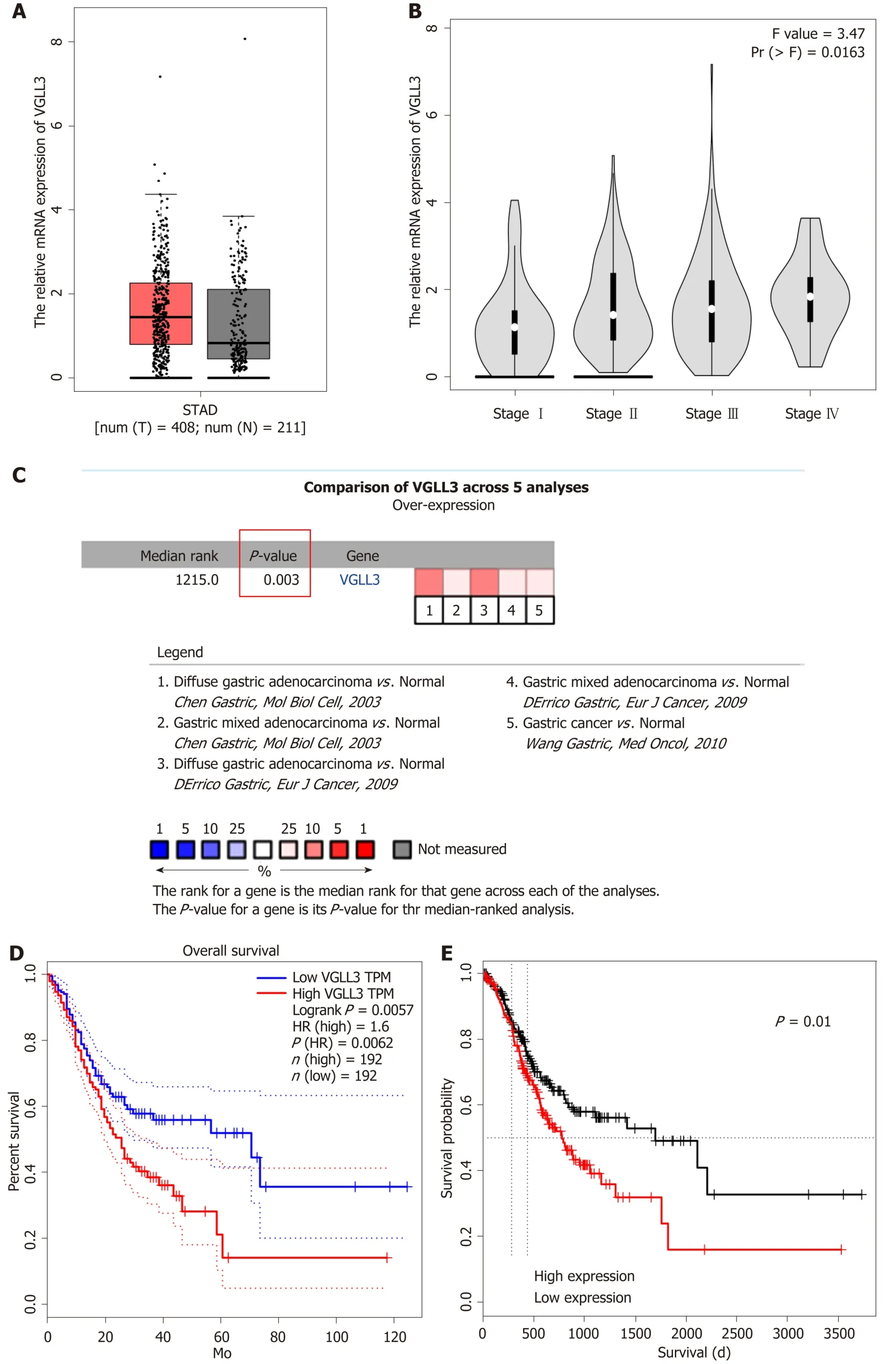
Figure 1 Vestigial like family member 3 mRNA expression levels in gastric cancer tissues determined by integrated analysis of different databases.
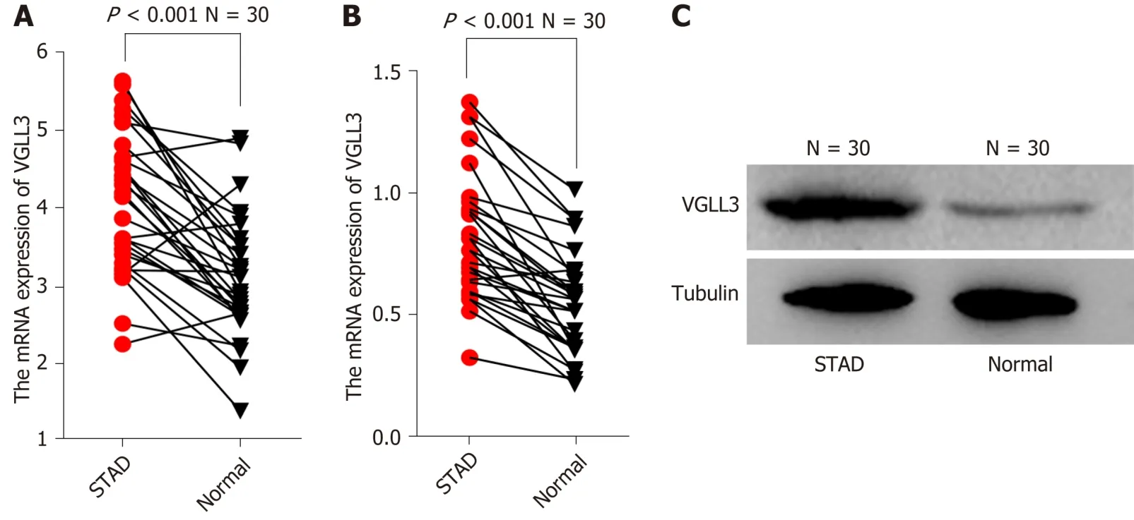
Figure 2 Vestigial like family member 3 mRNA and protein expression levels in gastric cancer.
DISCUSSION
GC is the third most common cause of cancer-related deaths worldwide,mainly due to ineffective diagnostic and therapeutic options.It is often detected at the advanced stage,and is highly heterogeneous.Therefore,novel molecular biomarkers for the early diagnosis and treatment of GC are urgently needed.VGLL3 is a member of the vestigial-like family proteins that are related to sex and maturation[18-20],and is reportedly associated with epithelial ovarian cancer and soft tissue sarcoma.Through bioinformatics data mining and integrated analysis,we found that VGLL3 mRNA was not only upregulated in the GC tissues relative to normal gastric tissues,but also significantly associated with advanced TNM stage GC and poor survival outcomes.To validate thein silicodata,we analyzed the gastric tissues of GC patients,and found abnormally high levels of VGLL3 in the tumor relative to normal tissues.In addition,highin situexpression of VGLL3 was correlated with tumor lymph node metastasis,TNM stage,and HER2 mutation,but not with age,gender,differentiation,T stage,or M stage.The patients were stratified into the high and low VGLL3 expression groups,and the former showed a significantly poor survival.Univariate and multivariate analyses further indicated that VGLL3 expression and TNM stage were independent risk factors for the prognosis of GC.Finally,the respective AUCs of the 3- and 5-year survival of the VGLL3-based prognostic model were 0.613 and 0.706,indicating a somewhat imperfect predictive ability.This could be due to the insufficient number of samples and other confounding factors such as the patient's state of mind,economic status,and family environment.Taken together,we identified VGLL3 as a novel prognostic biomarker for GC.
To further elucidate the underlying molecular mechanisms,we analyzed the PPI network of VGLL3 through the STRING database,and found that the Hippo pathway was significantly enriched.In addition,the identified upstream molecules of VGLL3 are YAP1[6],TEAD1,WWTR1,VGLL4,and the downstream molecules are POU1F1,TEAD3,AKAP11,SYT17,CHMP28,and TEAD4.Interestingly,the expression level of VGLL3 was related to ERBBE2 (HER2) mutation,although the relevant mechanistic connection is unclear.
To conclude,VGLL3 is highly expressed in GC and an independent risk factor,and further studies are needed to determine its underlying mechanism.Nevertheless,VGLL3 is a novel biomarker for GC prognosis and a potential therapeutic target for this malignancy.
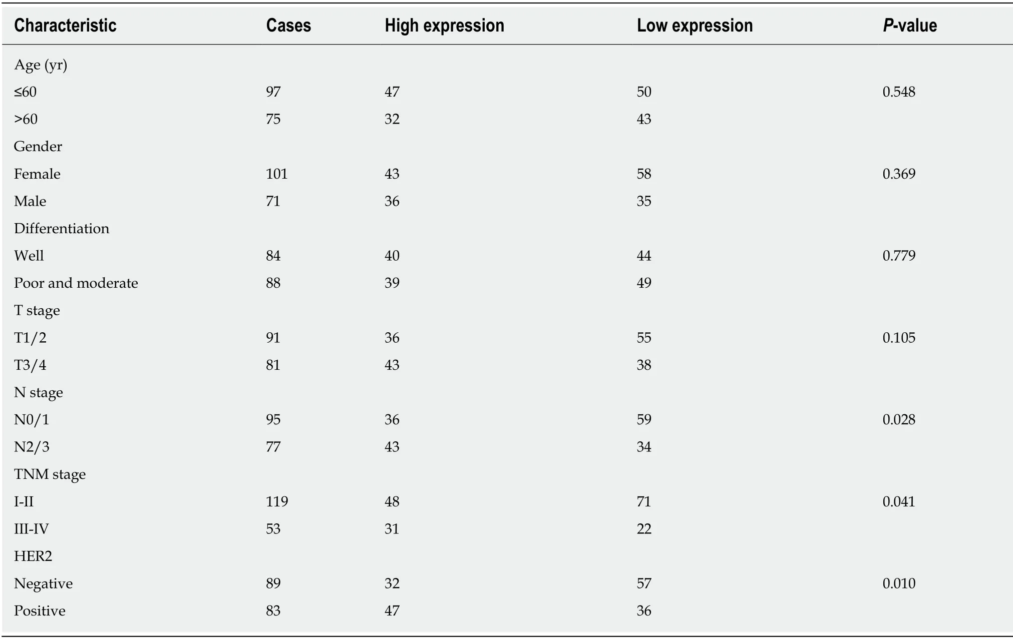
Table 1 Characteristics of gastric cancer patients according to vestigial like family member 3 expression status
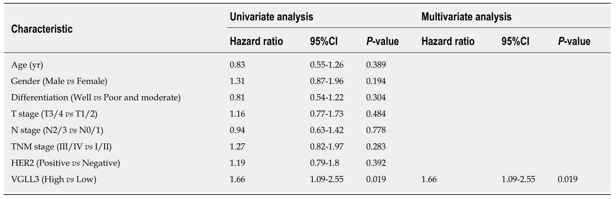
Table 2 Univariate and multivariate analyses of factors associated with overall survival
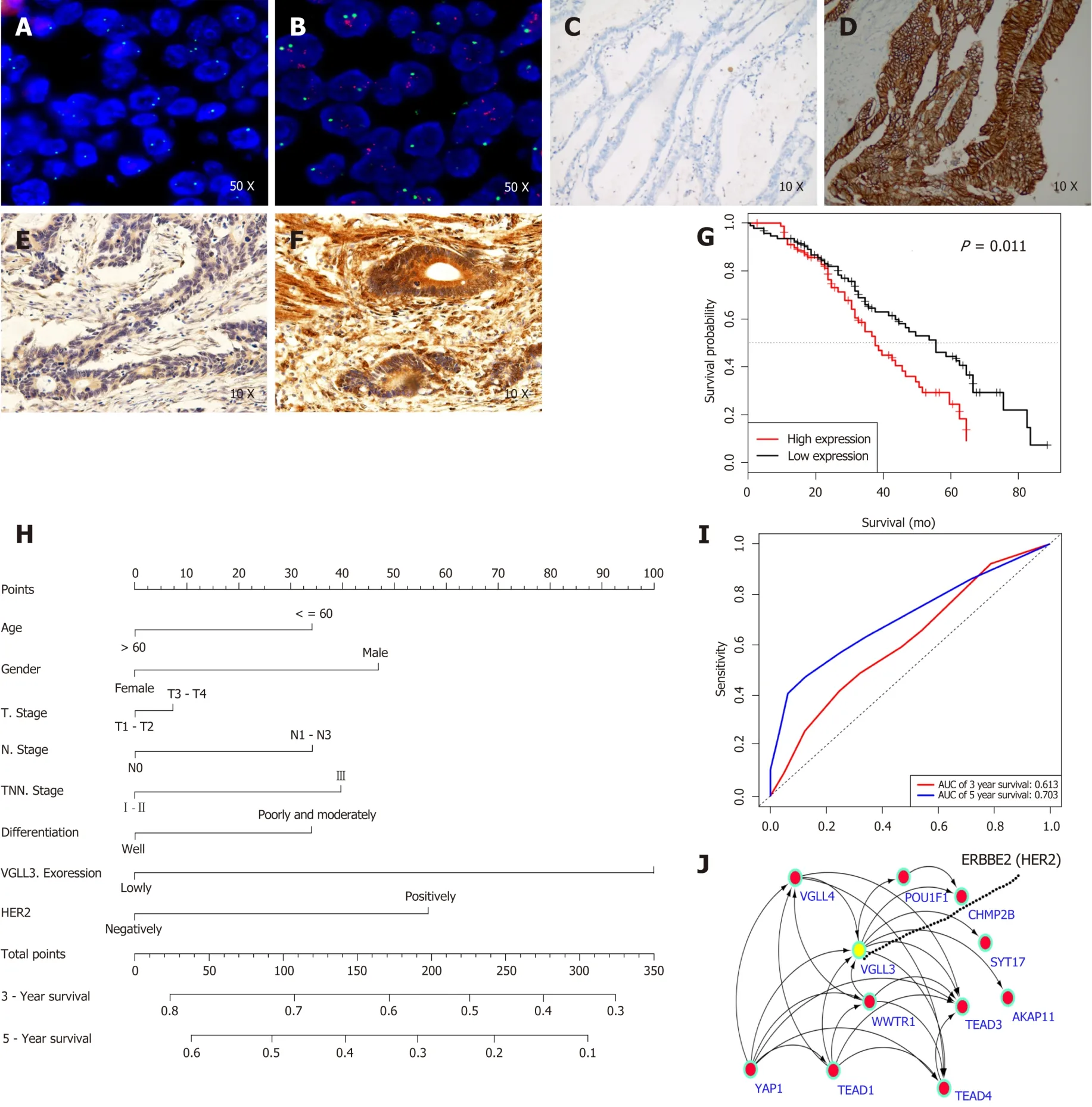
Figure 3 Prognostic value and putative molecular mechanism of vestigial like family member 3 in gastric cancer.
ARTICLE HIGHLIGHTS
Research background
Gastric cancer (GC) is the most prevalent gastrointestinal tract malignancy.The prognosis of GC patients remains relatively poor.It is urgent to explore prognostic markers for GC.
Research motivation
There are insufficient reports about the correlation between VGLL3 and GC.
Research objectives
The aim of the present study was to explore the expression pattern and clinical significance of VGLL3 in GC.
Research methods
It was found that VGLL3 would be a potential prognostic marker by bioinformatics analysis.To validate thein silicodata,the authors identified the expression of VGLL3 in GC patient samples by immunohistochemistry and evaluated clinical outcomes.
Research results
Analysis of the ONCOMINE and GEPIA databases showed that VGLL3 was significantly upregulated in GC tissues,and associated with the tumor TNM stage.In addition,the high VGLL3 expression group had a significantly worse prognosis compared to the low expression group,as per both GEPIA and ONCOLNC.The bioinformatics results were validated by the significantly higher VGLL3 mRNA and protein levels in the GC tissues compared to the adjacent normal tissues in a cohort of 30 GC patients.Furthermore,highin situexpression of VGLL3 protein was associated with more advanced N and TNM stages and HER2 mutation in a cohort of 172 patients.Kaplan-Meier analysis showed that the high VGLL3 expression group had a worse prognosis compared to the low VGLL3 expression group.Multivariate analysis showed that VGLL3 expression status was an independent risk factor for prognosis.In addition,the prognostic risk model nomogram showed that VGLL3 was the most important indicator,with an AUC of 0.613 for 3-year survival and 0.706 for 5-year survival.Finally,the protein interaction network analysis revealed that VGLL3 is likely involved in the Hippo signaling pathway.
Research conclusions
VGLL3 is overexpressed in GC tissues and associated with a poor prognosis,indicating its potential as a novel prognosis biomarker and therapeutic target for GC.
Research perspectives
The present study suggested that VGLL3 is a novel prognostic biomarker for GC,and the significance of VGLL3 as a promising therapeutic target for GC is highlighted.
猜你喜歡
保健醫(yī)苑(2023年2期)2023-03-15 09:03:04
中國(guó)臨床醫(yī)學(xué)影像雜志(2022年2期)2022-05-25 13:24:34
家庭醫(yī)學(xué)(下半月)(2020年3期)2020-05-30 12:42:02
家庭醫(yī)學(xué)(下半月)(2020年3期)2020-05-30 12:42:00
家庭醫(yī)學(xué)(下半月)(2020年3期)2020-05-30 12:42:00
中國(guó)生殖健康(2019年7期)2019-01-06 09:27:34
Coco薇(2016年2期)2016-03-22 02:42:52
Coco薇(2015年1期)2015-08-13 02:47:34
醫(yī)學(xué)研究雜志(2015年12期)2015-06-10 06:57:46
小雪花·成長(zhǎng)指南(2015年4期)2015-05-19 14:47:56
 World Journal of Clinical Cases2019年15期
- World Journal of Clinical Cases的其它文章
- Percutaneous coronary intervention for ostial lesions of the left main stem in a patient with congenital single left coronary artery: A case report
- Common iliac artery occlusion with small intestinal transection caused by blunt abdominal trauma: A case report and review of the literature
- c.753_754delAG,a novel CFTR mutation found in a Chinese patient with cystic fibrosis: A case report and review of the literature
- Fever and neck pain after pacemaker lead extraction: A case report
- Endometriosis of the duplex appendix: A case report and review of the literature
- Gastric duplication cyst mimicking large cystic lymphangioma in an adult: A rare case report and review of the literature