Effect of GSH and niacin combination on protein oxidation,ER stress,glycation and aggregation in HLE cells under high glucose condition
2020-01-08 03:13:08NinaHandayani1NurPermatasari2HidayatSujuti3AchmadRudijanto
國際眼科雜志 2020年1期
Nina Handayani1,Nur Permatasari2,Hidayat Sujuti3,Achmad Rudijanto
Abstract
·KEYWORDS:PCC;GRP78;AGEs;αβ crystalline;diabetic cataracts
INTRODUCTION
Studies have shown that cataracts are still the leading cause of preventable blindness worldwide.In fact,cataracts were responsible for one out of every three cases of blindness and one out of six cases of visual impairment at the end of 2010[1-3].Furthermore,patients with diabetes often experience cataract maturity ten years earlier than those without diabetes[4].This is mainly due to increases in protein glycosylation and the formation of free radicals related to hyperglycemia.Continual increases in free radical production,combined with the inability of biological systems to detoxify these,lead to oxidative stress.
Crystalline,which is the main protein in the lens and constitutes around 90% of the lens,undergoes changes that occur due to oxidation which form carbonyl proteins.The level of carbonyl proteins,derived from amino acids during metal-catalyzed oxidation of proteinsinvitroandinvivo,represents a direct measure of the oxidative injury to these molecules[5].There are many forms of measurements of protein oxidation and one of these is protein carbonyl content (PCC).
In the process of synthesis and modification of proteins in the lens,they experience unfolding,misfolding,or incomplete folding processes.These imperfect proteins can accumulate in the endoplasmic reticulum (ER)and cause ER stress.As a result of this condition,a cellular stress signal is sent as a reaction to ER stress,known as the unfolding protein response (UPR).UPR triggers a broad transcription response that adjusts the protein folding capacity in the ER.In addition,chronic hyperglycemia in diabetes can worsen ER stress due to increases in glycation and oxidative stress.One of the markers in ER stress conditions is glucose reactive protein (GRP)78 which functions as a chaperone.In addition,its expression is also obtained in lens fiber cells[6].
The study by Mulhern on senile cataracts and diabetic cataracts shows that increased levels of glycation of crystalline proteins can also cause Maillard reactions that play an important role in cataract formation[6].Accordingly,non-enzymatic glycation causes advanced glycation end products (AGEs)formation.The accumulation of AGEs on the crystalline lens is one of the pathogeneses related to cataracts in patients with diabetes mellitus.The lens is also susceptible to protein glycation due to limited or no recycling processes in the lens proteins.This makes it easier to aggregate and form high molecular weight particles that cause disruption of lens clarity and cataract formation[4,7].
In chronic hyperglycemia conditions such as diabetes,there is a decrease of reduced glutathione (GSH)levels caused by a reduction of oxidized glutathione (GSSG)synthesis and regeneration due to the used of nicotinamide adenine dinucleotide phosphate hydrogen (NADPH)co-factor in the conversion of glucose to sorbitol.As a result,decreased GSH levels are considered to increase oxidative stress which can also trigger cataract formation.Therefore,in chronic hyperglycemia conditions,it is necessary to consider GSH levels in the cell to overcome the decrease of GSH levels.Niacin,which is needed in NADPH synthesis,is also important in GSSG regeneration process so that it prevents GSSG from becoming prooxidant.
Based on all of the above,it is hypothesized that GSH and niacin combinations can delay the onset of diabetic cataracts.This study examines the possible protective effect of these combinations against cataract formation in high glucose treated lens epithelial cell cultures by assessing protein oxidation,ER stress,glycation and aggregation of αβ crystalline.
MATERIALS AND METHODS
MaterialSuppliesThe human lens epithelial (HLE-B3)cell line was purchased from ATCC (Manassas,VA).The PCC ELISA kit,GRP78 ELISA kit,and AGEs ELISA kit,were purchased from MyBioSource (San Diego,USA).The rabbit αβ-crystalin primary antibody (BS 124 63R)was purchased from Bioss (Bioss Antibodies Inc.,Massachusetts,USA),and the secondary antibody,anti-rabbit biotin,was purchased from Santa Cruz Biotechnology (Gene Company Ltd.,Shanghai,China).
CellTreatmentThe human lens epithelial (HLE-B3)cell line and stock cultures were maintained in MEM medium (Gibco,Thermo-Fisher Sc,USA)supplemented with 20% FBS,penicillin (50 U/mL)and streptomycin (50 U/mL)at 37°C in the presence of 5% CO2.Overnight,5×104cells were seeded into 24 well plates in MEM medium containing 10% FBS,treated with 25 mmol/L glucose and co-treated with three different doses combination of GSH (10,30,100 μmol/L)and niacin 25 μmol/L.These then were harvested after 72h and analyzed for PCC and glucose reactive protein (GRP78)measurement.Another 21 well plates were harvested after two weeks for AGEs measurement and Western Blot analysis.
ProteinCarbonylContentandGRP78AnalysisAfter 72h exposure to high glucose (25 mmol/L)and co-treated with three different doses of combination GSH (10,30,100 μmol/L)and niasin 25 μmol/L,cells were harvested and lysed and then centrifuged at 10 000 rpm for 2-3min.The proteins were isolated from cultured cells and protein concentrations were measured using the nanodrop protein methods.The cells were then measured using PCC and GRP ELISA kits according to the manufacturer’s instructions (MyBioSource).The absorbance was measured at 532 nm using a spectrophotometer (Model 550 Microplate Reader Bio-Rad,Singapore).
AdvancedGlycationEndProductsAnalysisIn a separate experiment,cells were harvested after two weeks’ exposure in high glucose,and the cell lysate was collected for ELISA AGEs analysis using commercial assay kits according to the manufacturer’s instructions (MyBioSource).Before the analysis,protein concentrations were measured using the nanodrop protein methods.The absorbance was measured at 532 nm using the spectrophotometer.
WesternBlotAnalysisTotal cellular proteins were extracted using a cell lysis buffer containing 2% SDS,100 mmol/L tris HCl,30 gliserol,and 10 mmol/L PMSF.Protein concentration was measured using nanodrop protein methods.Proteins were resolved by 4-12% SDS-polyacrylamide gel electrophoresis (SDS-PAGE)and transferred to trans-blot semi-dry transfer membranes (BioRad Lab Pte.Ltd,Singapore).The membranes were blocked with 0.5% skim milk,before being incubated overnight at 4°C.These were then,washed with 0.05% TBS-tween 3×5min and incubated with primary αβ-crystalline antibody BS 12463R 1∶1000 for 2h.Then,repeated wash with 0.05% TBS-tween 3×5min and incubated with secondary antibodies,anti-rabbit-biotin for 1h at room temperature.After complete five washes,and incubated with SAHRP 1∶1000 (Dako,Santa Clara USA)and TMB Membrane substrate,the proteins were detected and analysis of protein bands was conducted using Corel Draw software.
StatisticalAnalysisOne-way ANOVA or Kruskal Wallis was used for testing the statistical significance between groups.Normality of data was tested with Saphiro Wilk.Levene’s test was used for testing the homogeneity of the data between groups.A value ofP<0.05 was considered to be significant.The SPSS 18.0 statistical package was used for all data analyses.
RESULTS
EffectofGSHandNiacinCombinationonPCCLevelsFigure 1 shows the highest PCC rate was in the K+ groups (1.49±0.138)and the lowest average was in the group treated with GSH 10 μmol/L and niacin 25 μmol/L (1.4±0.267).After administration of GSH 10 μmol/L combined with niacin 25 μmol/L,the average of PCC level was lower than control group (K+).In contrast,administration of GSH 30 μmol/L and 100 μmol/L combined with Niacin 25 μmol/L resulted in a higher PCC level than the control group.
EffectofGSHandNiacinCombinationonGRP78LevelsAmong the groups treated with GSH 100 μmol/L and Niacin 25 μmol/L (0.08±0.267)there was a small reduction in the GRP78 level.However,this could not be statistically proven.It is interesting to note that in the other three groups this reduction did not happen (Figure 2).
EffectofGSHandNiacinCombinationonAGEsLevelsAs described in Figure 3,AGEs levels were significantly reduced in all groups.The lowest average AGEs level was in the group treated with GSH 10 μM and niacin 25 μM (P1,0.86±0.267).This implies that this combination was the most effective in decreasing AGEs levels.
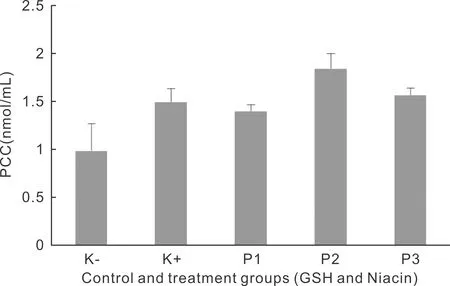
Figure 1 Mean of PCC levels in lens epithelial cell cultures with reduced glutathione (GSH)and niacin administrations.
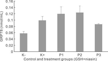
Figure 2 Mean of GRP78 levels in lens epithelial cell culture with reduced glutathione (GSH)and niacin administrations.

Figure 3 Mean of AGEs level in lens epithelial cells culture with reduced glutathione (GSH)and niacin administrations.
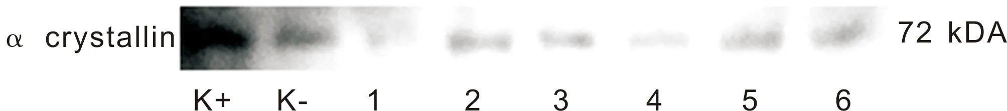
Figure 4 α crystalline expression in HLE-B3 cells treated with GSH and combination of GSH and niacin.
AlphaβCrystallineExpressioninHumanLensEpithelialCellsUsing the Western Blot analysis,it revealed that αβ crystalline was found at 72 kDa molecular weight.This high molecular weight suggested that there had been aggregation of protein.The positive control group had the highest blotting density of α crystalline (K+).On the other hand,there was a reduction of blotting density in the groups treated with GSH and those treated with a combination of GSH and niacin (Figure 4).
DISCUSSION
The aims of this study are to determine the effect of a glutathione and niacin combination onprotein carbonyl content (PCC),glucose reactive protein (GRP78/BiP),AGEs levels and the expression of αβ crystalline in lens epithelial cell cultures treated with high levels of glucose.This research was conducted using three different combinations of glutathione and niacin to investigate their efficacy.
PCC is a relatively stable marker of oxidative stress that occurs at the beginning of oxidation.The three different combinations of glutathione and niacin in this experiment did not decrease PCC levels in all groups.The results contradict previous study conducted by Zhengetal[8]involving the mechanism of carbonylation of proteins in brains of rats which showed a relationship between a decrease in reduced glutathione (GSH)and the occurrence of carbonylation of proteins.Furthermore,Sinbadetal[9]observed in glycated Human Serum Albumin (HSA)samples a decrease in carbonyl content level after administration of niacin using the dosages 50,100,200 and 500 μmol/L.Niacin is considered to have the ability to break down free carbonyl groups formed as a result of reduced carbonyl content and act as an antioxidant.
Some studies conducted by Zheng and Sinbad[8-9]regarding the administration of GSH and niacin separately showed a decrease in PCC formation.In contrast,in this study a combination of GSH and niacin did not reduce PCC levels significantly.In reviewing the literature,it appears that no research related these combinations have been done before.As a result,the cause of this condition is not clearly understood.However,it is reasonable to assume that it is probably caused by the excessive GSH administration that affects the GSH:GSSH ratio,leading to an accumulation of GSSG which is a prooxidant.
The results of the combination of GSH and niacin on GRP78 showed that there was an increase in GRP78 levels in the positive controls compared to negative controls.Surprisingly,administrating a combination of GSH and niacin failed to show a decrease of GRP78 levels in all groups.
Based on the literature,administration of GSH or niacin separately were able to improve the ER stress.One of the potential functions of GSH on ER is considered to be reduced non-native disulphide bonds formed in protein oxidative stress processes and use enzymes that catalyze oxidation[10].Two studies conducted by Aquilanoetal[11]and Wangetal[12]suggested GSH has an important role in the isomerization of non-native disulfide bonds.Both studies showed an increase in non-native disulfide bond formation when total GSH levels decrease[11-12].In addition,nicotinic acid is estimated to reduce ER stress by protecting the protein against the malfolding process and preventing Ca2+depletion.Another study by Vargasetal[13]showed that 6-amino nicotinamide administration,which causes niacin deficiency,can induce GRP78 activity.
Before entering the secretory pathway,proteins and protein membranes have to undergo complete folding in the ER,it is a basic protein folding factory which controls the quality of its products.Only proteins that are assembled perfectly and function properly are sent to the appropriate destination[14].During the process of cell development,the cell experiences a situation which causes excess folding capacity in the ER.This causes ER stress that changes or disrupts the folding process.In addition,during ER stress conditions,there is imbalance of protein folding with their capacity[15]and this condition can trigger UPR[16].
The administration of three combinations of GSH and niacin were not able to reduce GRP78 levels as expected.A possible explanation for this might be that the process of oxidative stress in the ER is a complex process which involves a lot of mechanisms and require a precise combination dosage of GSH and niacin to reduce stress.In addition,the recovery ability in the ER may require a longer time-span when compared to oxidative stress that occurs in the cell membrane.
Research on combinations of GSH and niacin against levels of AGEs shows an increase in the level of AGEs in the positive control compared to negative controls with significant differences.By administrating three combinations of GSH and niacin,there was a significant decrease in AGEs levels in all groups.
Glycation of the lens protein occurs when glucose or other reduced sugars react with the e amino group of lysine residues or the amino part of the protein.This results in the formation of schiff base (SB)[17].The SB then performs amadori rearrangements through Maillard reactions and produces more stable ketoamine or amadori products,referred to as early glycation products.In an advanced stage,amadori products dehydrate and rearrange themselves to form cross-links between adjacent proteins[18-21].As a result,protein aggregates or AGEs are formed.The condition of long-standing hyperglycemia is believed to cause the formation of AGEs that are irreversible glycation products and this process causes cataract formation.This study proves that there is an elevated level of AGEs in lens epithelial cells exposed to high glucose and combination of GSH and niacin decrease the levels of AGEs significantly in all groups.Therefore,GSH and niacin are believed to have potential effect of preventing glycation.GSH fulfills the function of co-factors in methylglyoxal elimination (AGEs)by using two glyoxalase enzymes known as the glyoxalase I and glyoxalase II.In addition,niacin prevents the formation of methylglyoxal and also protects the protein in its original form[9].These results show that the combination of GSH and niacin are effective in preventing cataracts predominantly through the AGEs pathway.
The results of αβ crystalline expression using the western blot examination showed increased blotting density,which represented an increase of protein aggregation in positive control group.In contrast,there was a decrease in blotting density in the groups treated with GSH or a combination of GSH and niacin.Evidence of protein aggregation was indicated by an increase in molecular weight to 72 kDa from 20 kDa.Furthermore,this result also found that GSH or combination of GSH and niacin prevented protein aggregation.The presence of protein aggregation indicates that the process of cataract formation has begun,even though it cannot be seen in a clinical examination of the lens.
In summary,the results showed that GSH and niacin combination prevented the cataract formation by decreasing the AGEs levels and preventing protein aggregation.Therefore,it appears the AGEs pathway seems to be dominant to prevent cataracts in diabetic patients.We suggest that further research should be undertaken to investigate the most effective dose combination of GSH and niacin needed to prevent cataracts from all pathways.
