Giant ventral hernia simultaneously containing the spleen, a portion of the pancreas and the left hepatic lobe:A case report
2020-05-13 07:34:44XiaGangLuoChenLuWuLinWangFeiZhouChunZhaoYu
World Journal of Clinical Cases 2020年9期
Xia-Gang Luo, Chen Lu, Wu-Lin Wang, Fei Zhou, Chun-Zhao Yu
Xia-Gang Luo, Chen Lu, Wu-Lin Wang, Fei Zhou, Chun-Zhao Yu, Department of General Surgery,the Second Affiliated Hospital of Nanjing Medical University, Nanjing 2l0011, Jiangsu Province, China
Abstract
Key words: Giant ventral hernia;Spleen;Pancreas;Liver;Mesh;Case report
INTRODUCTION
Ventral hernia is a common complication of abdominal surgery, with an incidence ranging from 9% to 20%[1].The risk of ventral hernia is about 10% for elective abdominal surgery, while it is significantly increased to 33% following emergency abdominal operation[2].The contents of ventral hernia may include omentum,preperitoneal adipose tissue, small intestine or colon.Ventral hernia with protrusion of more than two parenchymal organs simultaneously is extremely rare.The development of giant ventral hernia has a negative impact on the physiology of patients, such as pain, and inability to carry out daily activities.More importantly,significantly increased negative psychological effects, such as disturbances to mood,sleep, relationships with others and pleasure of life, lead to a poor quality of life[3].
We here report a rare case of asymptomatic giant ventral hernia with the protrusion of total spleen, partial pancreas and left hepatic lobe simultaneously through abdominal incision for 15 years in a 21-year-old female.The patient underwent a ventral hernia repair with Mesh at our hospital and had a good recovery.
CASE PRESENTATION
Chief complaints
A 21-year-old female patient was admitted to the Department of General Surgery in the Second Affiliated Hospital of Nanjing Medical University (Nanjing, China) on July 1, 2019, with chief complaints of giant ventral hernia without any other special discomfort for 15 years.
Personal and family history
The patient underwent umbilical hernia repair at the age of 1 year.Approximately 5 years later, ventral hernia recurred and repair with Mesh was performed, but the operation failed due to postoperative infection, and a huge mass appeared in the left abdominal wall.The mass increased gradually with the development and maturity of the body.
The patient denied fever, cough, expectoration, nausea, vomiting, defecation difficulties, melena and hematochezia.There was no other significant surgery and special medications in her past medical and family histories.
Physical examination upon admission
No abnormality was found in her vital signs.Physical examination showed no obvious cardiovascular or respiratory abnormalities.Her abdomen was soft and there were no obvious signs of tenderness or rebound pain.Murphy’s sign was negative.Many irregular and obsolete surgical scars could be seen on the abdominal wall.There was a giant ventral hernia about 10 cm × 10 cm with clear boundaries,tenderness and poor mobility (Figure 1A).Auscultation revealed no abnormal bowel sounds.
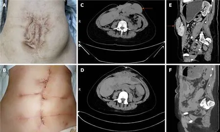
Figure 1 Patient-related image.
Laboratory examinations
Electrocardiogram was unremarkable and laboratory examination results, including blood, urine, stool, liver and kidney function, and coagulation tests, were within normal ranges.
Imaging examinations
Chest X-ray examination was unremarkable and computed tomography (CT) scan of the abdomen confirmed a giant ventral hernia containing liver, pancreas, spleen and blood vessels (Figure 1C and E).
FINAL DIAGNOSIS
Based on the examination results, together with the patient's history and symptoms, a giant ventral hernia was diagnosed.
TREATMENT
As the patient was unable to endure the inconvenience of life and the potential risk of spleen or liver rupture, she underwent a ventral hernia repair with Mesh at our hospital.Adequate pre-operative assessment and discussion were carried out due to the absence of indications for emergency surgery.During the laparotomy, between the adipose layer of the abdominal wall and the anterior sheath of the rectus abdominis, herniation of the spleen, part of the left lobe of the liver and the pancreas was observed, and the herniation was closely adhered to each other (Figure 2).The spleen, part of the left liver lobe and pancreas were safely and thoroughly separated from severe and extensive adhesions and reverted into the abdominal cavity.The hernia cystic ring was about 10 cm × 10 cm in size, and approximately 3/4 of the hernia sac was located under the left abdominal wall and 1/4 under the right abdominal wall (Figure 3).When the intra-abdominal adhesions were separated at approximately 5 cm around the defect, a COOK 15 cm × 13 cm biological Mesh was fixed to the retroperitoneum (Figure 4).Silica gel drainage tube was inserted into the left hernia sac cavity and then negative pressure ball was connected.The original skin scar was removed and tension reduction suture was made.The postoperative recovery was uneventful.Postoperative abdominal CT scan revealed that the contents of ventral hernia, including liver, pancreas, spleen and blood vessels, had returned to the abdominal cavity (Figure 1D and F).
OUTCOME AND FOLLOW-UP
She had a good recovery and was discharged 18 d after the operation.During the 3-mo follow-up, the patient remained asymptomatic and the appearance of the surgical incision was greatly improved (Figure 1B).
DISCUSSION
Ventral hernia is divided into congenital and acquired hernia.The latter is mainly due to previously abdominal surgery or unsuccessful hernia repair.Therefore, its repair is very complex and difficult[4].The incidence of ventral hernia is rising rapidly due to the increase of elderly, obese and diabetic patients[5]and other factors, such as poor nutrition, surgical site infection and increased intra-abdominal pressure[6].In the vast majority of cases, the contents of ventral hernia include small intestine, colon, fat or fibrous tissue with the most serious complication of impaired intestinal blood supply which usually leads to intestinal incarceration and necrosis[4].Abdominal parenchymal organs such as liver, spleen and pancreas are rarely found within the ventral hernia either separately or simultaneously.The clinical cases were searched through PubMed between January 2000 and January 2019 using the following search terms, single or in combination:Hernia, liver, spleen and pancreas.Reports of hiatal hernia, diaphragmatic hernia, para-esophageal hernia, and inguinal hernia were excluded.Twenty cases were identified (Table 1).The case we present here, to our knowledge, is the first case of giant ventral hernia which simultaneously contained the spleen, a portion of the pancreas and the left hepatic lobe.
Ventral herniation of spleen is extremely rare and almost all spleen-related hernias have been found to be associated with various types of diaphragmatic hernia, with only one case of congenital umbilical hernia.CT confirmed that spleen, small intestine, colon and pancreas protruded through umbilical hernia in an 82-year-old female patient who presented with left abdominal pain and weakness.Splenectomy and abdominal wall reconstruction were carried out without delay to avoid the rupture of the spleen with hemorrhage[7].Few studies have reported that pancreas exists in umbilical or paraumbilical hernia.According to the previous literature, in 3 of the 12 patients, pancreas protruded through umbilical hernia (n= 2) and paraumbilical hernia (n= 1), rather than ventral hernia.In addition to pancreas,herniation of small intestine, colon (n= 3) and spleen (n= 1) could also be found.Although pancreatic herniation is rare, its clinical manifestations and complications are serious as the incarceration of the head of pancreas could easily lead to acute pancreatitis.The most serious clinical symptoms were found in a 67-year-old female patient who suffered from severe epigastric pain radiating to the back, nausea and vomiting caused by acute pancreatitis and intestinal obstruction.The patient died of multiple organ failure 20 d later although emergency operation was performed[8].Another 56-year-old female patient with pancreatic herniation had a similar outcome and died of multiple organ failure 24 h after surgery[9].The occurrence of hepatic herniation in 9 patients was all related to incisional hernia.The most frequent symptom of hepatic herniation was abdominal pain (n= 6) and swelling (n= 5).Among the 9 patients, 5 received observation and conservative treatment for the primary disease, but one of these patients died 9 d after admission due toPseudomonassepsis[10].Open incisional hernia repair was performed in 2 patients[6,11]and in another case, the same surgery was performed simultaneously with the common bile duct exploration[12].Although hepatic herniation usually protrudes through a large area of abdominal wall defect, it rarely leads to serious clinical manifestations.Hepatic encephalopathy or hepatic dysfunction occurred in a patient when the herniated left hepatic lobe was present[11].In another patient, herniation of the right hepatic lobe might contribute to the clinical consequence of vascular compromise, which resulted in Budd-Chiari syndrome[13].At present, there are no relevant epidemiological data due to the rarity of hepatic herniation[14].
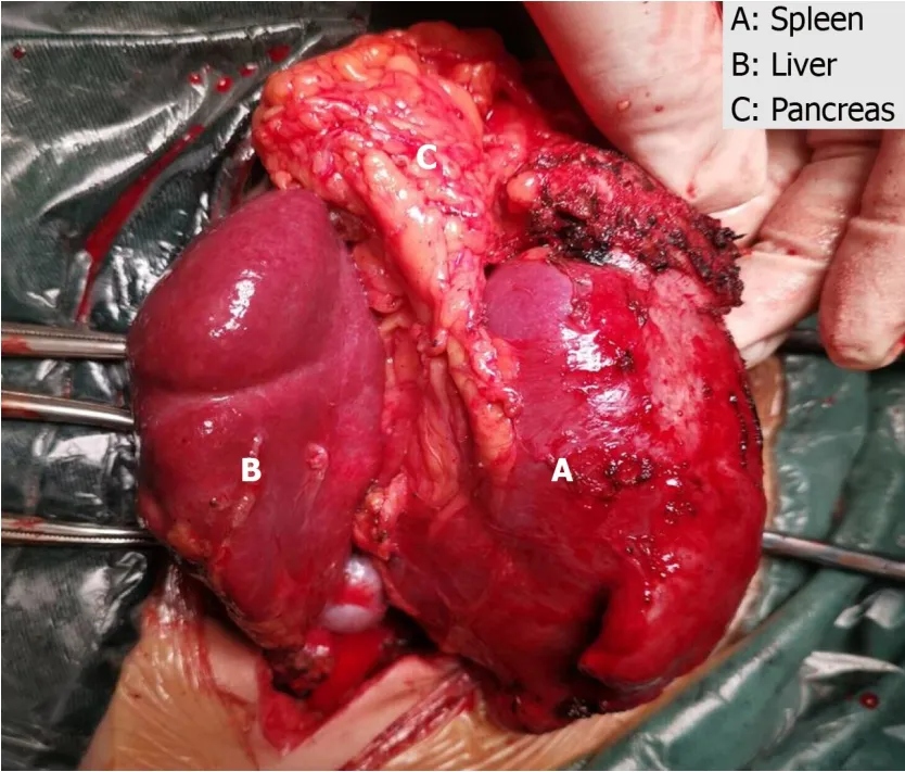
Figure 2 The spleen, part of the left lobe of the liver and the pancreas were observed between the adipose layer of the abdominal wall and the anterior sheath of the rectus abdominis.
All of the 12 cases were diagnosed by CT scan.Although it is not a routine imaging examination, CT scan is recommended for the diagnosis, especially for the content of hernia and the choice of treatment strategy[6].Surgeons can evaluate and determine the safest and desirable Mesh placement method based on CT scan.Enhanced abdominal CT scan can assess the relationship between abdominal viscera and defects, especially in obese patients and those with a history of recurrent ventral hernia.Although enhanced abdominal CT may not be helpful in assessing whether or not there is adhesion, or the degree of adhesion, it can discover hidden signs of inflammation or infection, which helps determine what surgical approach will be adopted and whether Mesh will be used or not[15].As such, in this case, although the patient was asymptomatic and liver function was normal, enhanced abdominal CT scan was performed to evaluate whether there was acute pancreatitis or the hepatic vein was compressed.
The type of herniated parenchymal organs is important for the choice of treatment strategy.When the spleen or pancreas protrudes through the hernia ring, patients often need to receive surgical treatment (elective or emergency operation) because of the serious complications caused by this pathological condition[8,9].However, hepatic herniation alone rarely causes serious clinical manifestations, conservative treatment might be the first choice.Surgical treatment must be comprehensively evaluated only when conservative treatment is ineffective, and the benefits from the operation outweigh the risks[6].In clinical practice, surgeons should make a comprehensive assessment based on their own experience and the individual characteristics of the hernia, such as the location, size and different organs of the herniation.In the present case, intraperitoneal Onlay Mesh repair of the open ventral hernia was performed using biological Mesh.Considering that the patient was a 21-year-old young woman,we chose an absorbable biological Mesh to avoid the possible pain and influence on body growth that might be caused by the non-retractable Mesh.
The development of giant ventral hernia has a negative impact on the physiology of patients, such as pain, and inability to carry out daily activities.The skin of abdominal wall in many patients with ventral hernia has not been repaired satisfactorily although the ventral hernia was successfully repaired, consequently declining the patient's sense of self-worth[16].More importantly, significantly increased negative psychological effects, such as disturbances to the mood, sleep, relationships with others and pleasure of life, lead to a poor quality of life[3].In our case, the young woman suffered from a giant ventral hernia for nearly 15 years.Many serpentine scars on her abdominal wall made her almost autistic.In addition, she was advised not to take exercise like other students because of the potential risk of liver and spleen rupture during school time.These unusual pressures caused the patient to be selfabased.During the treatment, multiple serpentine scars on the abdominal wall were removed simultaneously, and psychological comfort and encouragement were provided.At the time of discharge, the patient had almost got rid of the psychological shadow.
CONCLUSION
We report an extremely rare case of giant ventral hernia simultaneously containing the spleen, part of the pancreas and liver, which has been successfully treated.We highlight that surgeons should avoid abdominal hernia when performing abdominal surgery in obese, elderly and diabetic patients.In addition, we recommend that surgeons should pay more attention to the psychological treatment while restoring the abdominal physiological function in ventral hernia patients.
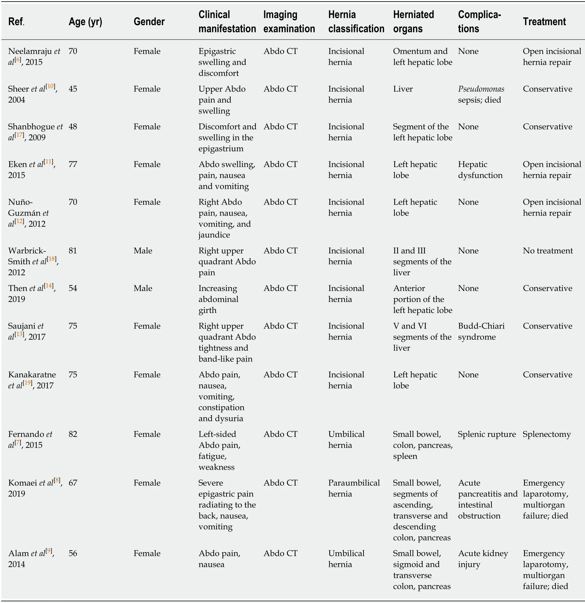
Table 1 Clinical data of 12 published cases
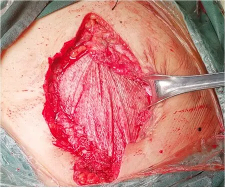
Figure 4 A COOK 15 cm × 13 cm biological Mesh was fixed to the retroperitoneum.
 World Journal of Clinical Cases2020年9期
- World Journal of Clinical Cases的其它文章
- Rare primary lymphoepithelioma-like carcinoma of the renal pelvis
- Portal hypertension in a patient with biliary hamartomas:A case report
- Mesonephric adenocarcinoma of the uterine cervix with rare lung metastases:A case report and review of the literature
- Endoscopic ultrasonography elastography in the diagnosis of intrapancreatic ectopic spleen:A case report
- Application of curved ablation in liver cancer with special morphology or location:Report of two cases
- COVlD-19 managed with early non-invasive ventilation and a bundle pharmacotherapy:A case report