Laparoscopic umbilical trocar port site endometriosis: A case report
2020-05-14 01:51:14
World Journal of Clinical Cases 2020年8期
Xue Ao, Wei Xiong, Shi-Qiao Tan, Department of Obstetrics and Gynecology, West China Second University Hospital, Key Laboratory of Birth Defects and Related Diseases of Women and Children (Sichuan University), Ministry of Education, Chengdu 610041, Sichuan Province, China
Abstract
BACKGROUND
Abdominal wall endometriosis can occur secondary to gynecological and obstetric pelvic laparotomy; however, this is a rare clinical event. There are few cases of endometriosis involving the incision site of a laparoscopic surgery,especially for those of the endometrial nodule at the umbilical trocar port site where the camera is placed.
CASE SUMMARY
We describe the case of a 37-year-old woman who presented with a 2-year history of a tough swelling below the umbilicus, which presented periodical pain during menstruation. The patient had undergone laparoscopic ovarian cystectomy 4 years prior, and we theorized that the umbilical nodule was a complication of that laparoscopic surgery. Histological analysis confirmed the diagnosis of abdominal umbilical scar endometriosis secondary to previous laparoscopic surgery. Surgical removal of the nodule followed by three cycles of leuprorelin was curative.
CONCLUSION
Abdominal mass and pain in women of childbearing age with a previous history of pelvic surgery should support consideration of endometriosis at the surgical site.
Key words: Endometriosis; Laparoscopy; Abdominal wall; Scar endometriosis; Ovarian cystectomy; Case report
INTRODUCTION
Endometriosis occurs mostly in pelvic organs and is rarely detected in extrapelvic locations, such as the abdominal wall, central nervous system, lungs, kidneys, ureters,limbs, gallbladder, nasal cavity, brain, and distant skin[1,2]. Abdominal wall endometriosis (AWE) is the most common site of extrapelvic endometriosis. AWE can be spontaneous, but it can also occur after gynecological and obstetric pelvic surgery,with approximately 57% of cases developing secondary to cesarean section[3]. In addition, AWE can also be detected in the abdominal wall incision after ovarian cyst removal, appendectomy, and tubal ligation, being clinically defined as scar endometriosis or incision endometriosis.
Herein, we report a case of AWE detected in the umbilicus, which served as the trocar port site where the laparoscope body and camera were placed during previous laparoscopic surgery.
CASE PRESENTATION
Chief complaints
A 37-year-old woman found a slow-growing nodule under the skin of the surgical scar on the umbilicus which caused periodic pain and presented to the Dermatology Department of West China Hospital, Sichuan University (Chengdu, China) on March 29, 2019 for treatment. Histological analysis of a fine needle aspiration biopsy of the nodule suggested endometriosis.
History of present illness
Umbilical nodules appeared at the incision site of the original laparoscopic cannula at the lower edge of the umbilicus about 4 years prior, which had gradually increased from 0.5 cm to approximately 1.5 cm in diameter and were accompanied by pain during menstruation.
History of past illness
The patient had undergone laparoscopic ovarian cyst removal and bilateral salpingoplasty at the Department of Reproductive Endocrinology of our hospital 6 years prior. After cyst removal, pathological examination had led to the diagnosis of endometrial cyst.
Physical examination
The patient’s vital signs were normal. Abdominal examination revealed no tenderness or rebound tenderness. At the incision of the original laparoscopic cannula at the lower edge of the umbilicus, three brown hard nodules of different sizes were detected, with the maximum diameter of about 1.5 cm. No redness or ulceration was found on the surface of the nodules (Figure 1). The boundary between the nodules and surrounding tissues was clear.
Laboratory testing
Fine needle biopsy of the umbilical nodule confirmed the diagnosis of umbilical endometriosis. Positive expression of the immunohistochemical markers’ estrogen receptor, progesterone receptor, and p63 was found in the infiltrated glands of the dermis; in addition, progesterone receptor and cluster of differentiation 10 were expressed in the periglandular interstitial space. Ki67 was about 10% positive in the gland and interstitium. Serum level of tumor marker carbohydrate antigen 19-9 was 39.1 U/mL and that of carbohydrate antigen 12-5 was 19.7 U/mL.

Figure 1 Endometriosis at the laparoscopic umbilical trocar port site (orange arrow).
Imaging examination
No superficial ultrasound examination of the abdominal wall was performed because the nodules could be seen clearly by the naked eye. Transvaginal Doppler showed that the endometrial echo was uneven, with a slightly strong echo (endometrial polyp could not be excluded). A cyst about 1.5 to 2.9 cm in diameter was present on both ovaries (chocolate cyst of the ovary is likely).
FINAL DIAGNOSIS
Abdominal umbilical scar endometriosis secondary to previous laparoscopic surgery.
TREATMENT
The nodule was surgically removed, and leuprorelin was administered for three cycles.
OUTCOME AND FOLLOW-UP
Postoperative anatomy showed the cut surface of the mass to be gray-white, grayyellow, and solid. Both intraoperative and postoperative histological analyses confirmed umbilical endometriosis (Figure 2), and the wound recovered well after operation, without any recurrence to date.
DISCUSSION
This is a case report of an AWE that appeared in the abdominal wall incision after minimally invasive laparoscopic surgery. With the extensive development of laparoscopic surgery, there have been few reports of endometriosis at the cannula incision. Laparoscopic incision endometriosis was first reported by Dentonet al[4]in 1990, as umbilical endometriosis after laparoscopic sterilization in a 37-year-old woman. According to Akbarzadeh-Jahromiet al[5], there have been 17 cases reported in the English literature. However, according to our literature review, the total number of cases reported in either English or Chinese is 71 to date, including 32 cases in English and 39 cases in Chinese. Overall, prospective studies are required to increase the quality of the existing literature. Most scholars agree that the etiology of scar endometriosis is endometrial cell planting during surgical treatment[1,5].Endometriosis of the abdominal wall most commonly involves tissues around incisions following surgery, such as from cesarean section, abdominal wall incision after hysterectomy, and laparoscopic cannula incision for specimen collection. When taking out the tissue containing endometrial cells, the abdominal incision is most likely to contact endometrial cells. In our case, incision endometriosis occurred in the umbilical cannula incision. This incision is only used to place the laparoscopic camera,so the chance of contact with endometrial cells is small. We suspect that gloves or instruments contaminated with endometrial cells were responsible for the endometrial cells or debris present at the abdominal incision closed (by suture).
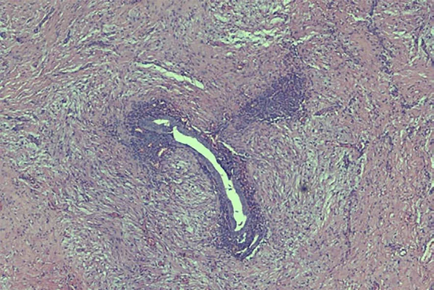
Figure 2 Histopathology of the umbilical nodules: Endometrial tissue (×400).
Abdominal wall mass (82%-96%) and pain (41%-87%) are the most common symptoms of AWE[6,7]. Only 57% of patients report periodic symptoms[7], and 3.08%(2/65) are asymptomatic[8]. Therefore, the periodicity of lump pain related to menstruation is not a necessary condition to diagnose AWE. In a study by Hortonet al[7]that reviewed 29 articles, the average age of patients was 31.4 years (95%confidence interval: 29.1-33.8 years) and the average time of symptom onset was 3.6 years (95% confidence interval: 2.5-4.8 years)[7]. Anandet al[9]described the appearance of ectopic umbilical scar nodules being usually above the incision, with brown skin changes on the surface of the nodules, consistent with the appearance in our case(Figure 1).
AWE should be differentiated from incisional hernia, granulation tissue,hematoma, abscess, sediment, desmoid fibromatosis, lipoma, infection, soft tissue sarcoma, and metastatic malignant tumors[7,10]. Guptaet al[11]described cases of sporadic desmoid fibromatosis in the umbilical cord 8 mo after laparoscopic cholecystectomy. Desmoid fibromatosis is a rare benign soft tissue tumor originating from muscle-aponeurosis, without malignant potential; however, it can be locally invasive and is similar to AWE. It has been reported that many cases are misdiagnosed as hernia[5,12], and one case was misdiagnosed as umbilical granuloma[13].
Most AWEs have typical clinical symptoms, including palpable masses and pain without compression, combined with abdominal surgery history, which indicates a preliminary diagnosis of AWE. Nevertheless, there is no gold standard for preoperative diagnosis. Nodules on ultrasound (a modality characterized by low cost,safety, and noninvasiveness) are usually pear-shaped, with a solid hypoechoic or cystic appearance. Ultrasound, magnetic resonance imaging, or computed tomography can assist in determining the extent of lesions. Ultrasound-guided fine needle puncture can confirm the diagnosis and exclude the diagnosis of malignancies before operation. Surgical resection not only treats patients but can also confirm the diagnosis.
It is widely accepted that surgical excision is the most appropriate approach, and surgical excision of normal tissue 5-10 mm away from the edge of the lesion is required to prevent recurrence, according to the literature[3,5]. In the case of extensive lesions, computed tomography or magnetic resonance imaging can assess the location and size of AWE more accurately than ultrasound; moreover, they can prevail for the determination of whether to accumulate fascia and rectus abdominis muscles and if preoperative surgical evaluation is needed for reticular repair of the abdominal wall[3].Large lesion area and rectus muscle involvement often indicate a higher recurrence rate. Apart from surgical excision, therapeutic percutaneous cryoablation can reduce the volume of lesions, as shown by preliminary studies but not yet confirmed by highquality literature. Due to the histological characteristics of scars, it is difficult to achieve therapeutic effects around the lesion by oral medication alone. Oral contraceptives and progesterone can attenuate symptoms only slightly but cannot eliminate the lesion[3]. However, in a study conducted by Alborziet al[14], who performed laparoscopic exploration after resection of abdominal wall mass in 30 AWE patients, it was found that 28 (93.3%) patients had pelvic endometriosis. Our case report is likely to support this conclusion. Therefore, after surgical resection of AWE,we believe that the postoperative treatment should be supplemented with medication.
A Japanese group, Tsurugaet al[15], previously reported the case of a 49-year-old woman with mixed endometrioid and clear cell carcinoma caused by endometriosis in the laparoscopic cannula during laparoscopic oophorocystectomy. Nevertheless,malignant transformation of peritoneal endometriosis is extremely rare. Clear cell carcinoma of abdominal incision is the most common histological type, followed by endometrioid carcinoma[16]. Other types include mixed adenocarcinoma, serous carcinoma, clear cell carcinoma, and carcinosarcoma. There is no authoritative consensus or guideline on the treatment of AWE malignancies. Despite the lack of direct evidence to support the benefit of chemotherapy at present, adjuvant chemotherapy or radiotherapy is typically administered after extensive surgical operation. The malignant transformation of AWE emphasizes the importance of surgical excision and pathological biopsy.
In consideration of the etiology of AWE iatrogenic implantation, prevention is the fundamental precaution to eliminate AWE. Clinically, the dissemination of endometrial cells should be avoided as much as possible in the first pelvic surgery for patients with ovarian function. In addition, preventive measures can be taken, such as using specimen bags to collect specimens. Saline irrigation should be carefully used when closing abdominal wall incisions, and potentially contaminated cannula incisions should also be flushed out. In addition, instruments contacting the specimens should be rinsed, hands and stitches should be flushed, or the stitches should be replaced. Laparoscopic surgery should not be performed during the menstrual period, and the postoperative complications should be reduced as much as possible for patients.
CONCLUSION
In summary, incisional endometriosis after laparoscopic surgery is rare, and the biological characteristics of the endometriosis are the appearance, growth, infiltration,and formation of nodules and masses in tissues outside the endometrium. Hence,abdominal mass and pain in women of childbearing age with a previous history of pelvic surgery should support consideration of AWE.
 World Journal of Clinical Cases2020年8期
- World Journal of Clinical Cases的其它文章
- Clinicopathological differences and correlations between right and left colon cancer
- Bedside score predicting retained common bile duct stone in acute biliary pancreatitis
- Stability and infectivity of coronaviruses in inanimate environments
- Status, challenges, and future prospects of stem cell therapy in pelvic floor disorders
- Unusual presentation of congenital radioulnar synostosis with osteoporosis, fragility fracture and nonunion: A case report and review of literature
- Predictive factors for central lymph node metastases in papillary thyroid microcarcinoma