Phosphatidylinositol-3,4,5-trisphosphate dependent Rac exchange factor 1 is a diagnostic and prognostic biomarker for hepatocellular carcinoma
2020-09-18 08:03:24YiCaiQiaoZhengDeJiaoYao
World Journal of Clinical Cases 2020年17期
Yi Cai,Qiao Zheng,De-Jiao Yao
Yi Cai,Qiao Zheng,De-Jiao Yao,Department of Oncology,Hospital of Chengdu University of Traditional Chinese Medicine,Chengdu 610000,Sichuan Province,China
Abstract
Key words:Phosphatidylinositol-3,4,5-trisphosphate dependent Rac exchange factor 1;Overexpression;Prognosis;Hepatocellular carcinoma;Hepatitis B virus infection;Diagnosis
INTRODUCTION
Hepatocellular carcinoma (HCC) is a major malignant tumor,with the 5thhighest cancer-related mortality[1].Currently,the treatments for HCC include surgery,ablation,liver transplantation,chemotherapy,targeted therapy and some novel theranostic approaches[2].According to the Barcelona Clinic Liver Cancer staging,surgical resection,ablation,and liver transplantation are performed in patients at an early stage,and for those patients at an intermediate or advanced stage,palliative treatments are recommended,which include sorafenib targeted therapy and transarterial chemoembolization[2,3].In addition,some novel treatments have been reported.However,immune checkpoint blocking therapy (pembrolizumab) has no significant impact on the overall survival (OS) or progression-free survival (PFS)compared with chemotherapy[4,5].Some studies have reported novel theranostics based on the tumor microenvironment,which have a significant effect on liver tumor inhibition,but this was only found in the laboratory research stage[6-8].Drug resistance is a common challenge in HCC treatment,which contributes to a poor prognosis.Furthermore,the diagnosis of HCC in the clinic is also poor,as due to insignificant early symptoms,many HCC patients are diagnosed at an intermediate or advanced stage,which contributes to fewer opportunities for surgical resection[9,10].Thus,the study of novel molecular biomarkers or pharmaceutic targets is critical for HCC therapy.
Phosphatidylinositol-3,4,5-trisphosphate dependent Rac exchange factor 1 (P-Rex1)is a Rho guanine nucleotide exchange factor,which activates Rac1 and plays a critical role in neutrophil and macrophage function[11,12].P-Rex1 was first purified from the cytoplasm of neutrophils,and P-Rex1 deficiency in a mouse model resulted in impaired Rac1 activation,reactive oxygen species formation and chemotaxis[13-15].In cancer,P-Rex1 was reported to be involved in regulation of the PI3K/AKT,mTOR and MEK/ERK pathways[16,17].P-Rex1 could interact with Gβγ and PIP3 to mediate the process of Rac-GDP to Rac-GTP in some cancer types[18,19].Furthermore,P-Rex1 is also involved in the progression of melanoma through the regulation of melanoblasts migration[20-22].However,its expression and role in HCC remain unclear.This study aimed to examine the expression of P-Rex1 in HCC and analyze the potential association between P-Rex1 and the development of HCC,and further evaluate the potential value of P-Rex1 in the clinical diagnosis and prognosis of HCC.
MATERIALS AND METHODS
Reagents
The total ribonucleic acid (RNA) extraction reagent Trizol was purchased from Thermo Fisher Scientific Co.,Ltd (Waltham,United States).The first-strand complementary DNA (cDNA) synthesis kit (with genomic deoxyribonucleic acid digester) and the Hieff quantitative polymerase chain reaction (qPCR) SYBR Green Master Mix (with high Rox) were obtained from Yeasen Technology (Shanghai,China).The specific qPCR primer was synthesized by Sangon Technology (Shanghai,China).The detailed sequence for P-Rex1 is as follows:Forward:5’- TGG AGT ATT GTT TAC ACC CGGA-3’;Reverse:5’-CTC GTA CAC GCA GAA CTT GTC-3’.
Clinical samples
Ninety resected liver tumor tissues and adjacent normal liver tissues were obtained from the Department of Oncology,Hospital of Chengdu University of Traditional Chinese Medicine.Tissue collection was undertaken during surgery,and the whole resected tissues including tumor and surrounding normal tissues were washed with cold saline solution and then preserved in the refrigerator (-80°C).All patients provided informed consent,and the study was approved by the Ethics Committee of the Hospital of Chengdu University of Traditional Chinese Medicine.Patient information is as follows:Malevsfemale (69vs21),age ≤ 50vsage >50 (24vs66),hepatitis B virus (HBV) infection positivevsnegative (38vs52),lymph node invasion positivevsnegative (48vs42),distant metastasis positivevsnegative (24vs66),alphafetoprotein (AFP) <20 ng/mLvs20 ng/mL ≤ AFP ≤ 200 ng/mLvsAFP >200 ng/mL(16vs28vs46).
Real-time qPCR analysis
The resected tissues were homogenized and then lysed with Trizol for 30 min at room temperature.The supernatant was collected and treated with isopropyl alcohol.Total RNA was washed with 75% ethanol and then used as a template to synthesize the first-strand cDNA according to the first-strand cDNA synthesis kit manual.The acquired cDNA was amplified with the Hieff qPCR SYBR kit as previously reported[23].High Rox was used as an internal control.
Validation with bioinformatic analysis
The messenger RNA expression of P-Rex1 in liver tumor and adjacent normal liver tissues was validated with the cancer genome atlas (TCGA,https://www.cancer.gov/tcga).The protein expression in liver tumor and normal liver tissues was obtained from the human protein atlas as reported previously[24,25].
The receiver operating characteristic analysis
Receiver operating characteristic (ROC) analysis was used to confirm the diagnostic value of P-Rex1 in liver cancer.The adjacent normal liver was used as the control group,and the corresponding tumor tissues were set as the disease group.The area under the curve (AUC) value was used to assess the diagnostic value of the indicated marker.
Survival analysis using the Kaplan-Meier method
Survival analysis (including OS,PFS,and relapse-free survival (RFS)) was conducted with the Kaplan-Meier plotter.The median expression level of P-Rex1 was applied as the cut-off of the high or low expression patient group.
Statistical analysis
Statistical difference was determined by Graphpad Prism 8.0,and the difference between the two groups was calculated with the Student’st-test.Pearson analysis was used to assess the correlation between AFP and P-Rex1.The survival analysis was conducted with the logrankPmethod.
RESULTS
The overexpression of P-Rex1 in HCC
To evaluate the potential role of P-Rex1 in HCC,we firstly examined the expression of P-Rex1 using the real-time quantitative PCR assay in 90 resected HCC tumor tissuesand corresponding adjacent normal liver tissues.As shown in Figure 1A,the expression level of P-Rex1 was significantly upregulated in liver tumor tissues.The details of the expression difference of P-Rex1 in liver tumor and normal tissues are shown in Figure 1B,the T/N ratio >5 was considered statistically different (23/90,25.6%),and the number of patients with a ratio between 1 and 5 was 56 cases (62.2%),with only 11 patients showing lower expression in tumor tissues than in adjacent tissues (12.2%).These results demonstrated that P-Rex1 was frequently overexpressed in liver tumor tissues.
P-Rex1 expression was closely associated with clinical features of HCC
The upregulation of P-Rex1 in liver tumor tissues suggested that P-Rex1 might be involved in the development of HCC.To further demonstrate the potential role of PRex1 in HCC,we evaluated the association between P-Rex1 and the following clinical features,HBV infection,lymph node invasion,distant metastasis,and serum AFP level.HBV infection is an important risk factor for HCC,and results in a marked difference in the choice of treatment[26,27].Figure 2A shows that P-Rex1 expression was significantly higher in patients with HBV infection than in those without HBV infection.Metastasis is a critical factor contributing to the poor prognosis of HCC patients in the clinic[28];thus,we conducted a further analysis of lymph node invasion and distant metastasis.The data revealed that patients with lymph node invasion or distant metastasis had increased P-Rex1 expression in tumors (Figure 2B and C).AFP is the gold standard biomarker for HCC diagnosis,and some studies have also revealed that AFP can predict prognosis during HCC treatment[29,30].Thus,we also evaluated the correlation between P-Rex1 and AFP.As shown in Figure 2D,patients with an AFP level more than 200 ng/mL had higher expression of P-Rex1.The detailed correlation analysis is also shown in Table 1,where the 90 HCC patients were divided into the high expression group and the low expression group according to the median value of P-Rex1 expression in liver tumor tissues.Compared with HCC patients with low P-Rex1 expression,the P-Rex1 high expression group included more patients with HBV infection (33 casesvs5 cases),lymph node invasion (37 casesvs11 cases),distant metastasis (22 casesvs2 cases) and increased AFP concentration (31 casesvs15 cases).It is noteworthy that the patients with high P-Rex1 expression had lower false negative AFP results (14/45,31%),and patients with low P-Rex1 expression had higher false-negative AFP results (35/45,67%).These findings revealed that P-Rex1 expression was closely associated with the clinical features of HCC and may participate in the development of HCC.
P-Rex1 could act as a good diagnostic biomarker for HCC
The marked difference in P-Rex1 between HCC tumor and liver normal tissues as mentioned above was closely associated with some pathological features of HCC.Considering the significant upregulation of P-Rex1 in liver tumor,and poor diagnosis in the clinic,we hypothesized that the upregulation of P-Rex1 may be used as a diagnostic biomarker for HCC.To confirm this hypothesis,ROC analysis was conducted on all 90 HCC patients.The results are shown in Figure 3A,and the AUC value was high at 0.758 (P<0.001,95%CI = 0.688-0.827).Based on this,P-Rex1 could be a potential biomarker for the diagnosis of HCC.It is necessary to fully understand the role of P-Rex1 in the diagnosis of HCC,and the importance and shortcomings of AFP in clinical practice.In the clinic,a concentration of AFP over 200 ng/mL is highly suspicious of liver cancer[31].To evaluate the diagnostic potential of P-Rex1 in HCC,we also examined the ROC analysis in patients with AFP >200 ng/mL and AFP <200 ng/mL,respectively.As shown in Figure 3B,the ROC analysis in patients with higher serum AFP showed a higher AUC value (0.878,P<0.001) than patients with low AFP levels (0.639,P<0.05).Combined with the results of Figure 2D,P-Rex1 expression was closely associated with serum AFP concentration.We further tested the correlation between serum AFP level and P-Rex1 expression in liver tumor,and a positive correlation was observed between P-Rex1 and AFP (Figure 3C);therefore,P-Rex1 may be a new AFP-related factor in HCC.Considering the significance of HBV on P-Rex1 expression (Figure 2A),we further examined HCC patients with and without HBV infection,respectively,using ROC analysis.The ROC analysis is shown in Figure 3D,and the AUC value in HBV positive patients was higher than that in HBV negative patients (0.857vs0.666).Thus,P-Rex1 as a diagnostic biomarker could be more effective for HBV-related HCC patients.
Validation of P-Rex1 overexpression in HCC
In the above-mentioned results,we revealed the overexpression of P-Rex1 in livertumor,and the significant difference in P-Rex1 could be a diagnostic biomarker for HCC patients,especially in patients with HBV infection.Here,we further validated the overexpression of P-Rex1 by bioinformatics analysis.As shown in Figure 4A,samples from TCGA liver cancer were included,and the GTEx value of the liver tumor and adjacent normal liver were also included.The results showed that P-Rex1 was significantly increased in liver tumor tissues,which was consistent with our clinical HCC samples.Moreover,we also evaluated the protein expression of P-Rex1 in the human protein atlas database.Immunohistochemistry staining of P-Rex1 was increased in liver tumor cells compared with normal liver tissues,and the main expression of P-Rex1 was in the cytoplasm (Figure 4B and C).The bioinformatics analysis validated the overexpression of P-Rex1 in liver tumor,which confirmed the results of the clinical HCC samples.
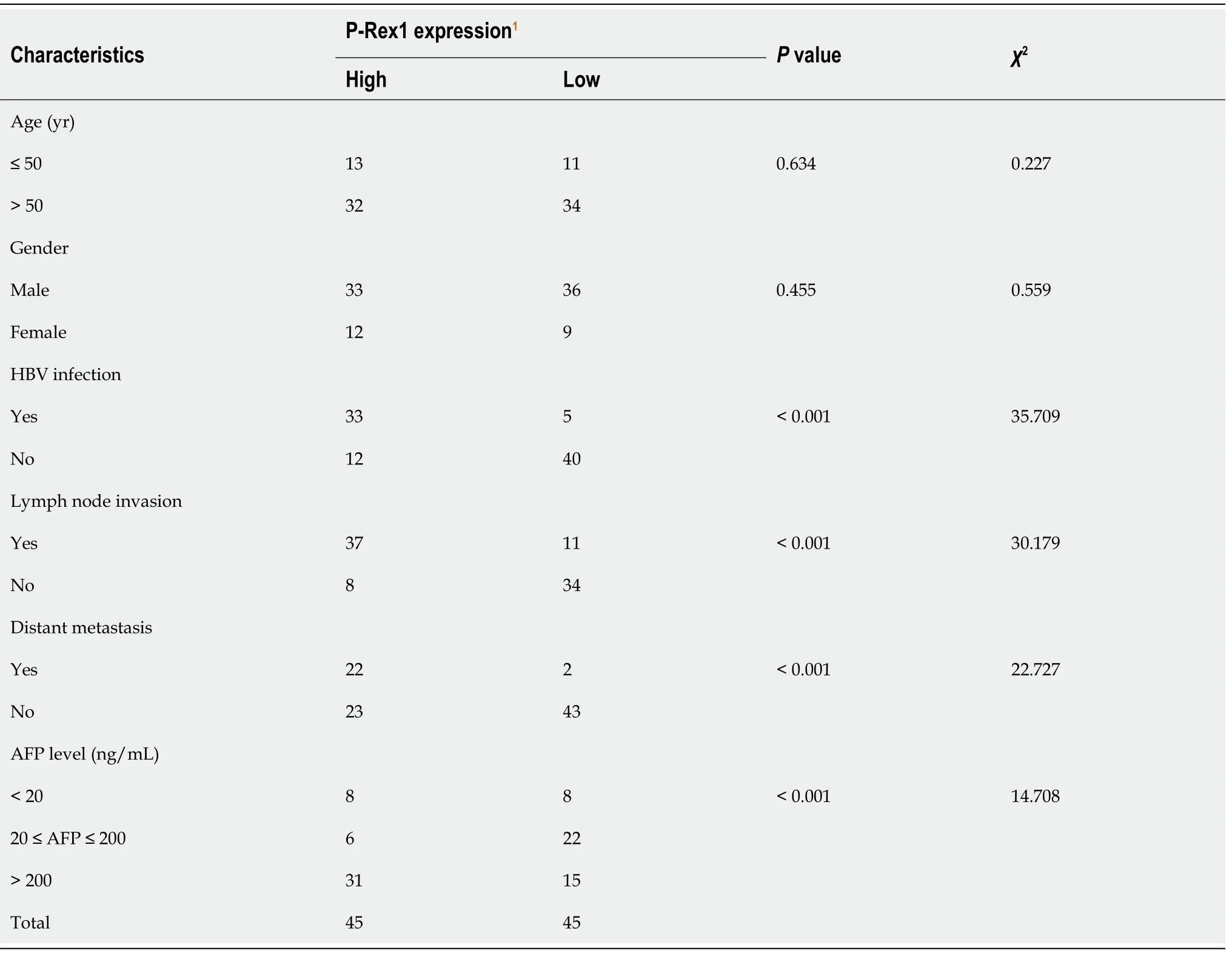
Table 1 The association between phosphatidylinositol-3,4,5-trisphosphate dependent Rac exchange factor 1 and clinical features in 90 hepatocellular carcinoma patients
P-Rex1 is a prognostic biomarker for HCC
Our data showed the upregulation of P-Rex1 in liver tumor tissues,and P-Rex1 expression was further enhanced in patients with HBV infection,lymph node invasion or distant metastasis,suggesting that P-Rex1 was closely associated with the clinicalpathological features of HCC.The ROC analysis revealed the potential of P-Rex1 as a diagnostic biomarker for HCC patients,especially for HBV positive patients.P-Rex1 expression was closely associated with the development of HCC.To understand the role of P-Rex1 in HCC,the survival analysis was applied in this study.As shown in Figure 5,HCC patients with high P-Rex1 had a higher OS,PFS and RFS.Table 2 shows that the median survival time of the P-Rex1 high expression group was significantly longer than patients with low expression of P-Rex1 (OS 25.5 movs14 mo),PFS (40.97movs18.7 mo),RFS (37.23 movs16.83 mo).
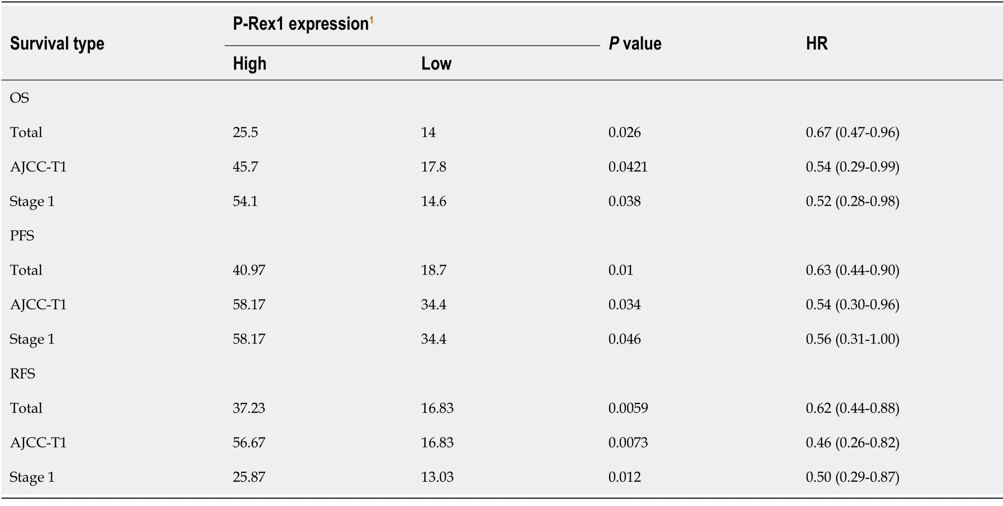
Table 2 The association of phosphatidylinositol-3,4,5-trisphosphate dependent Rac exchange factor 1 expression with median survival time in the 5-yr survival analysis
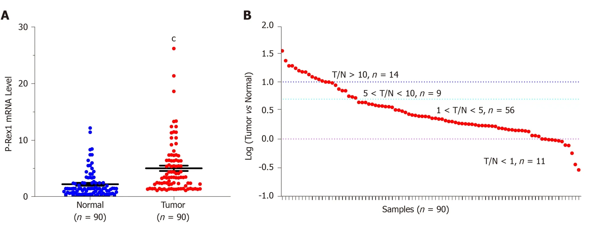
Figure 1 Phosphatidylinositol-3,4,5-trisphosphate dependent Rac exchange factor 1 messenger ribonucleic acid expression was increased in liver cancer tissues compared with adjacent normal tissues.A:Ninety resected liver tumor tissues and adjacent normal liver tissues were subjected to real-time quantitative polymerase chain reaction to determine the messenger ribonucleic acid level of P-Rex1;and B:The expression ratio between tumor tissues and normal liver tissues in 90 resected samples,the ratio was assessed by log treatment.cP <0.001 was considered significantly different.P-Rex1:Phosphatidylinositol-3,4,5-trisphosphate dependent Rac exchange factor 1;T:Liver tumor;N:Normal liver.
P-Rex1 is a good prognostic biomarker even in early stage HCC patients
Higher P-Rex1 expression was revealed as a favorable factor in the analysis of OS,PFS,and RFS,suggesting that P-Rex1 could serve as a new prognostic biomarker for HCC patients.In clinical practice,HCC therapy showed that prognostic evaluation was poor in the early stage of HCC.Considering the prolonged survival time in patients with high P-Rex1 expression,we further examined the OS,PFS,and RFS in early stage HCC patients.In this study,the AJCC-T1 or stage 1 was considered early stage.As shown in Figure 6A-C,the higher P-Rex1 group of HCC patients with AJCC-T1 showedprolonged OS [logrankP= 0.042,HR = 0.54 (0.29-0.99)],PFS [logrankP= 0.034,HR =0.54 (0.3-0.96)],and RFS [logrankP= 0.0073,HR = 0.46 (0.26-0.82)].Similar results were also observed in HCC patients with stage 1 (Figure 6D and F).The analysis of median survival time in HCC patients with AJCC-T1 or stage 1 is shown in Table 2.The median survival time in the higher P-Rex1 expression group was longer than in patients with lower P-Rex1 expression [OS (P<0.05),PFS (P<0.05),RFS (P<0.05)].

Figure 2 P-Rex1 levels were closely associated with the pathological features of hepatocellular carcinoma.A:The P-Rex1 expression level in patients with HBV infection;B:lymph node invasion;C:distant metastasis;and D:alpha-fetoprotein levels were determined,respectively.cP <0.001 was considered significantly different compared with the negative groups,respectively.P-Rex1:Phosphatidylinositol-3,4,5-trisphosphate dependent Rac exchange factor 1;HBV:Hepatitis B virus;AFP:Alpha-fetoprotein.
DISCUSSION
HCC is one of the most lethal human cancers,especially in East Asia.Many risk factors contribute to the high incidence,such as HBV infection,aflatoxin exposure,alcoholic l iver injury and non-alcoholic fatty liver disease[32].Multiple methods are applied in current HCC clinical practice,but the 5-year survival rate is not satisfactory.Poor diagnosis and drugresistance are the two major challenges in the treatment of HCC.These challenges have attracted considerable attention in order to overcome these problems,and the identification of novel biomarkers and pharmaceutic targets is critical in the management of HCC.
In this study,we identified that P-Rex1 was overexpressed in liver tumor tissues(Figures 1 and 4),which was consistent with previous reports in other cancers.Bakeret al[33]reported hyperactivation of P-Rex1 in prostate cancer[33].P-Rex1 amplification is also involved in the progression of melanomaviathe PAK1/P38 pathways[34].We hypothesized that a close association between P-Rex1 and risk factors of HCC exists,such as HBV infection,lymph node invasion and distant metastasis.Our results confirmed this hypothesis.Interestingly,we also found the P-Rex1 was furtherenhanced in the AFP-positive HCC patients (Figure 2D),and this positive correlation is demonstrated in Figure 3C.Considering the diagnostic value of AFP in HCC,we then tried to evaluate the potential application of P-Rex1 as a diagnostic biomarker.Our findings showed that P-Rex1 was a diagnostic biomarker with a higher AUC value (Figure 3A).We also determined the effect of HBV infection on the upregulation of P-Rex1 levels,and conducted ROC analysis in HCC patients with and without HBV infection.A significant increase in the AUC value in HBV positive infection was discovered (Figure 3D).Therefore,P-Rex1 is a novel diagnostic biomarker for HBVrelated HCC patients.

Figure 3 Phosphatidylinositol-3,4,5-trisphosphate dependent Rac exchange factor 1 was a good diagnostic biomarker for hepatocellular carcinoma,especially in patients with hepatitis B virus infection.A:A total of 90 hepatocellular carcinoma tissues were subjected to receiver operating characteristic (ROC) analysis.The phosphatidylinositol-3,4,5-trisphosphate dependent Rac exchange factor 1 (P-Rex1) expression in the liver tumor was set as the patient group,and adjacent normal tissues was set as the control group;B:The patients with different alpha-fetoprotein levels were subjected to ROC analysis based on the P-Rex1 expression;C:The correlation between alpha-fetoprotein and P-Rex1 expression was analyzed in 90 liver tumor tissues;and D:Patients with and without hepatitis B virus infection were subjected to ROC analysis based on P-Rex1 expression.ROC:Receiver operating characteristic;95%CI:95% confidence interval;AUC:Area under the curve;AFP:Alpha-fetoprotein.P-Rex1:Phosphatidylinositol-3,4,5-trisphosphate dependent Rac exchange factor 1.
The effect of the overexpression of P-Rex1 in HCC is unknown.To determine the potential function of P-Rex1 in HCC,the effect of P-Rex1 expression on survival time was assessed.Analysis of OS,PFS,and RFS demonstrated that patients with higher PRex1 expression had prolonged survival time (Figure 5 and Table 2).P-Rex1 was reported to be an important factor which regulates immune signaling pathways,and the complex regulatory mechanism of tumor immunity is also considered the most important factor in the development of liver cancer.Thus,high P-Rex1 correlated with the development of liver cancer,but high expression of P-Rex1 also improved survival time,suggesting that high expression of P-Rex1 might be a compensatory phenomenon.We also evaluated early stage HCC patients,as prognosis is poorly studied in these patients.Our findings revealed that even in early stage HCC,P-Rex1 was a favorable prognostic biomarker (Figure 6 and Table 2).Other researchers previously reported that P-Rex1 expression could be a marker for sensitivity to PI3K inhibitors in breast cancer[35];thus,we believe that P-Rex1 is a critical biomarker in thediagnosis and prognosis of HCC.The overexpression of P-Rex1 in HCC will be an encouraging pharmaceutic target in the treatment of HCC.
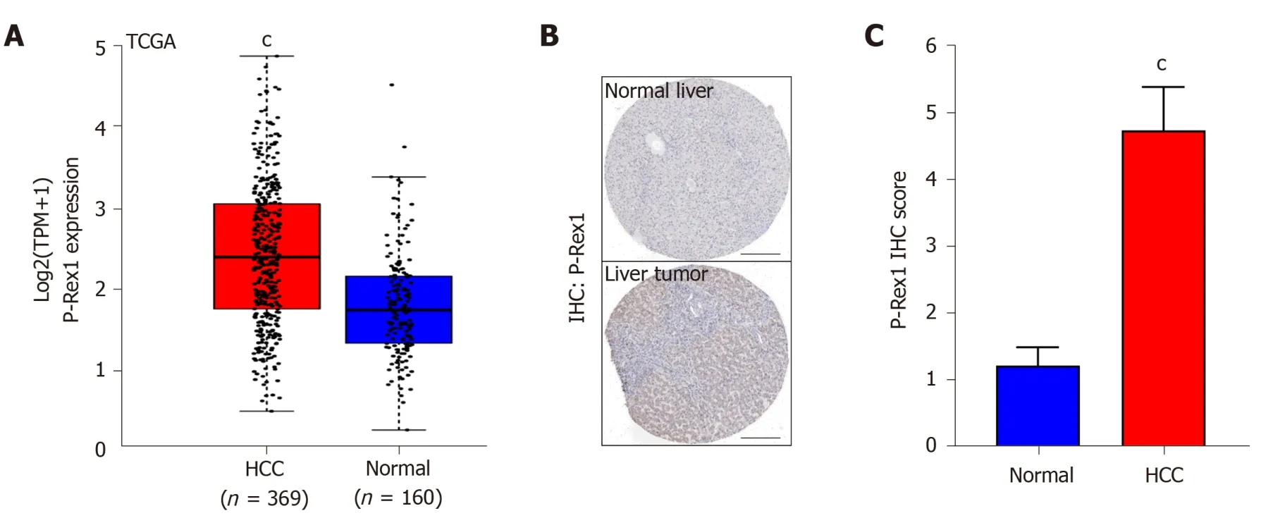
Figure 4 Validation of phosphatidylinositol-3,4,5-trisphosphate dependent Rac exchange factor 1 expression in the cancer genome atlas and human protein atlas database.A:The expression of phosphatidylinositol-3,4,5-trisphosphate dependent Rac exchange factor 1 (P-Rex1) in hepatocellular carcinoma liver tumor tissues and adjacent normal tissues in the cancer genome atlas were subjected to analysis;B:The protein expression of P-Rex1 in liver tumor tissue and normal liver tissue was determined by the human protein atlas database;and C:The immunohistochemistry score of P-Rex1 in panel B was quantified by Quantity One software.cP <0.001 was considered significantly different compared with normal liver tissues.TCGA:The cancer genome atlas;HCC:Hepatocellular carcinoma.
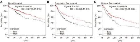
Figure 5 Survival analysis was conducted in hepatocellular carcinoma patients with high or low phosphatidylinositol-3,4,5-trisphosphate dependent Rac exchange factor 1 expression.A:Using the cancer genome atlas,hepatocellular carcinoma patients were subjected to overall survival analysis;B:Progression-free survival;and C:Relapse-free survival in those with high or low phosphatidylinositol-3,4,5-trisphosphate dependent Rac exchange factor 1 expression.The median level was applied to patient clustering.HR:Hazard ratio.

Figure 6 The overall survival,progression-free survival,relapse-free survival analysis of phosphatidylinositol-3,4,5-trisphosphate dependent Rac exchange factor 1 in early-stage hepatocellular carcinoma patients.A:Hepatocellular carcinoma patients with T1 stage based on the American Joint Committee on Cancer were included in the overall survival analysis,B:Progression-free survival analysis,C:Relapse-free survival analysis according to the phosphatidylinositol-3,4,5-trisphosphate dependent Rac exchange factor 1 expression;D:The overall survival analysis,E:Progression-free survival analysis,and F:Relapse-free survival analysis of phosphatidylinositol-3,4,5-trisphosphate dependent Rac exchange factor 1 was assessed in hepatocellular carcinoma patients with stage 1.OS:Overall survival;PFS:Progression-free survival;RFS:Relapse-free survival;AJCC-T1:T1 stage based on the American Joint Committee on Cancer;HR:Hazard ratio.
ARTICLE HIGHLIGHTS
Research background
Phosphatidylinositol-3,4,5-trisphosphate dependent Rac exchange factor 1 (P-Rex1) is regarded a critical regulator in the inflammation response,and a recent study showed the significance of P-Rex1 in cancers.
Research motivation
P-Rex1 functions as a tumor promoter in many cancers,but the expression and the clinical significance of P-Rex1 in hepatocellular carcinoma (HCC) are still unknown.
Research objectives
The study aimed to evaluate the potential value of P-Rex1 in the diagnosis and prognosis of HCC.
2.3.4 精密度與準(zhǔn)確度試驗(yàn) 按“2.3.3”項(xiàng)下方法配制ZG02低、中、高質(zhì)量濃度(2、100、200 μg/L)的質(zhì)控樣品各5份,按“2.2.4”項(xiàng)下方法孵育2 h后,進(jìn)樣分析,考察日內(nèi)精密度;連續(xù)測(cè)定3 d,考察日間精密度。將實(shí)測(cè)質(zhì)量濃度與理論質(zhì)量濃度進(jìn)行比較,考察準(zhǔn)確度。結(jié)果,各質(zhì)控樣品的日內(nèi)、日間RSD均小于10%,準(zhǔn)確度為87.26%~94.58%,詳見表1。
Research methods
Ninety resected HCC tissues were examined by real-time quantitative polymerase chain reaction to analyze P-Rex1 expression.The correlation between P-Rex1 expression and pathological features was determined.Bioinformatics analysis was applied to validate the results.
Research results
P-Rex1 was highly expressed in liver tumor,and P-Rex1 expression was closely correlated with lymph node invasion,hepatitis B virus (HBV) infection,distant metastasis,and alpha-fetoprotein levels.P-Rex1 acts as a diagnostic biomarker with a higher area under the curve value,especially in patients with HBV infection.P-Rex1could also act as a favorable prognostic factor in HCC,even in patients with early stage HCC.
Research conclusions
Our data suggest that P-Rex1 is overexpressed in HCC and is associated with HCC progression.P-Rex1 is a new diagnostic biomarker for HBV-related HCC and is also a favorable prognosis factor for HCC.
Research perspectives
The overexpression of P-Rex1 was confirmed in this study,and P-Rex1 was closely associated with the pathological features of HCC.A positive correlation between PRex1 and the risk factors of HCC was also observed,suggesting that P-Rex1 is an important mediator in the development of HCC.Survival analysis showed that P-Rex1 is a favorable prognosis biomarker.Therefore,the overexpression of P-Rex1 in the development of HCC and the effect on cell proliferation,migration or differentiation will be further demonstrated in a future study.
猜你喜歡
當(dāng)代陜西(2022年5期)2022-04-19 12:10:18
當(dāng)代陜西(2022年5期)2022-04-19 12:10:12
新世紀(jì)智能(數(shù)學(xué)備考)(2021年9期)2021-11-24 01:14:28
湘潮(上半月)(2021年4期)2021-07-20 08:05:28
當(dāng)代陜西(2021年1期)2021-02-01 07:18:02
汕頭大學(xué)學(xué)報(bào)(自然科學(xué)版)(2020年4期)2020-12-14 07:05:00
當(dāng)代陜西(2020年20期)2020-11-27 01:43:10
福建基礎(chǔ)教育研究(2019年3期)2019-05-28 23:47:21
Coco薇(2016年2期)2016-03-22 02:42:52
Coco薇(2015年1期)2015-08-13 02:47:34
 World Journal of Clinical Cases2020年17期
- World Journal of Clinical Cases的其它文章
- Diagnosis and treatment of an elderly patient with 2019-nCoV pneumonia and acute exacerbation of chronic obstructive pulmonary disease in Gansu Province:A case report
- Active surveillance in metastatic pancreatic neuroendocrine tumors:A 20-year single-institutional experience
- Shear wave elastography may be sensitive and more precise than transient elastography in predicting significant fibrosis
- Diagnosis and treatment of mixed infection of hepatic cystic and alveolar echinococcosis:Four case reports
- Surgical strategy used in multilevel cervical disc replacement and cervical hybrid surgery:Four case reports
- Gallbladder sarcomatoid carcinoma:Seven case reports