BDNF對缺糖缺氧小鼠mPFC腦區神經元突觸形成的影響
2020-10-20 04:48:18喻曉路傅慧
醫學信息
2020年17期
喻曉路 傅慧
摘要:目的 ?探討腦源性神經生長因子(BDNF)在缺糖缺氧復灌條件下對mPFC腦區突觸前膜蛋白Synapsin-1的保護作用。方法 ?將18只小鼠隨機分為正常組、缺糖缺氧復灌組、缺糖缺氧復灌給藥組,每組6只。正常組mPFC腦區低糖DMEM培養基培養,缺糖缺氧復灌組(OGD/R)mPFC腦區EBSS培養基培養15 min后復灌1 h,缺糖缺氧復灌給藥組(OGD/R+BDNF)mPFC腦區EBSS培養基中加入BDNF培養15 min后復灌1 h,采用免疫印跡分析和RT-qPCR檢測分析突觸前膜蛋白Synapsin-1的表達量和mRNA表達量。結果 ?缺糖缺氧復灌組Synapsin-1蛋白和mRNA表達量低于正常組,差異有統計學意義(P<0.05);缺糖缺氧復灌給藥組Synapsin-1蛋白表達量高于缺糖缺氧復灌組,差異有統計學意義(P<0.05);缺糖缺氧復灌給藥組Synapsin-1蛋白和mRNA表達量與正常組比較,差異無統計學意義(P>0.05)。結論 ?缺糖缺氧對mPFC腦區神經元突觸有損傷作用,BDNF對神經元突觸起到保護作用。
關鍵詞:腦源性神經生長因子;mPFC;缺糖缺氧;突觸前膜蛋白
中圖分類號:R614 ? ? ? ? ? ? ? ? ? ? ? ? ? ? ? ? 文獻標識碼:A ? ? ? ? ? ? ? ? ? ? ? ? ? ? ? ? ? DOI:10.3969/j.issn.1006-1959.2020.17.018
文章編號:1006-1959(2020)17-0063-04
Abstract:Objective ?To investigate the protective effect of brain-derived nerve growth factor (BDNF) on the presynaptic membrane protein Synapsin-1 in mPFC brain under the condition of hypoglycemia and hypoxia reperfusion.Methods ?18 mice were randomly divided into normal group, hypoglycemia and hypoxia reperfusion group, hypoglycemia and hypoxia reperfusion administration group, 6 mice in each group. Normal group mPFC brain area was cultured in low-sugar DMEM medium, hypoglycemia hypoxia reperfusion group (OGD/R) mPFC brain area EBSS medium was cultured for 15 min and then reperfused for 1 h, hypoglycemia hypoxia reperfusion administration group (OGD/R+BDNF) mPFC brain area EBSS medium was cultured with BDNF for 15 min and reperfused for 1 h.Western blot analysis and RT-qPCR were used to analyze the expression and mRNA expression of the presynaptic membrane protein Synapsin-1.Results ?The expression of Synapsin-1 protein and mRNA in the hypoglycemia and hypoxia reperfusion group was lower than that of the normal group,the difference was statistically significant (P<0.05);Synapsin-1 protein expression in the hypoglycemia and hypoxia reperfusion administration group was higher than that of the hypoglycemia hypoxia reperfusion group, the difference was statistically significant (P<0.05); Synapsin-1 protein and Compared with the normal group, the mRNA expression level was not statistically different (P>0.05).Conclusion ?Glucose and hypoxia could damage neuronal synapses in the mPFC brain region, and BDNF could protect neuronal synapses.
Key words:Brain-derived nerve growth factor;mPFC;Hypoglycemia and hypoxia;Presynaptic membrane protein
1.3統計學處理 ?采用統計學軟件SPSS 13.0進行處理,計量資料采用(x±s)表示,采用單因素方差分析,one-way ANOVE進行組間比較,P<0.05 為差異有統計學意義。
2結果
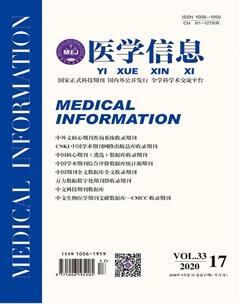
2.1三組小鼠mPFC腦區病理形態 ?鏡檢結果顯示,三組腦片中細胞核形態清晰、完整,提示模型制作成功,短時間的缺塘缺氧沒有造成大面積的組織壞死和細胞凋亡,完全符合后續實驗要求,見圖1。
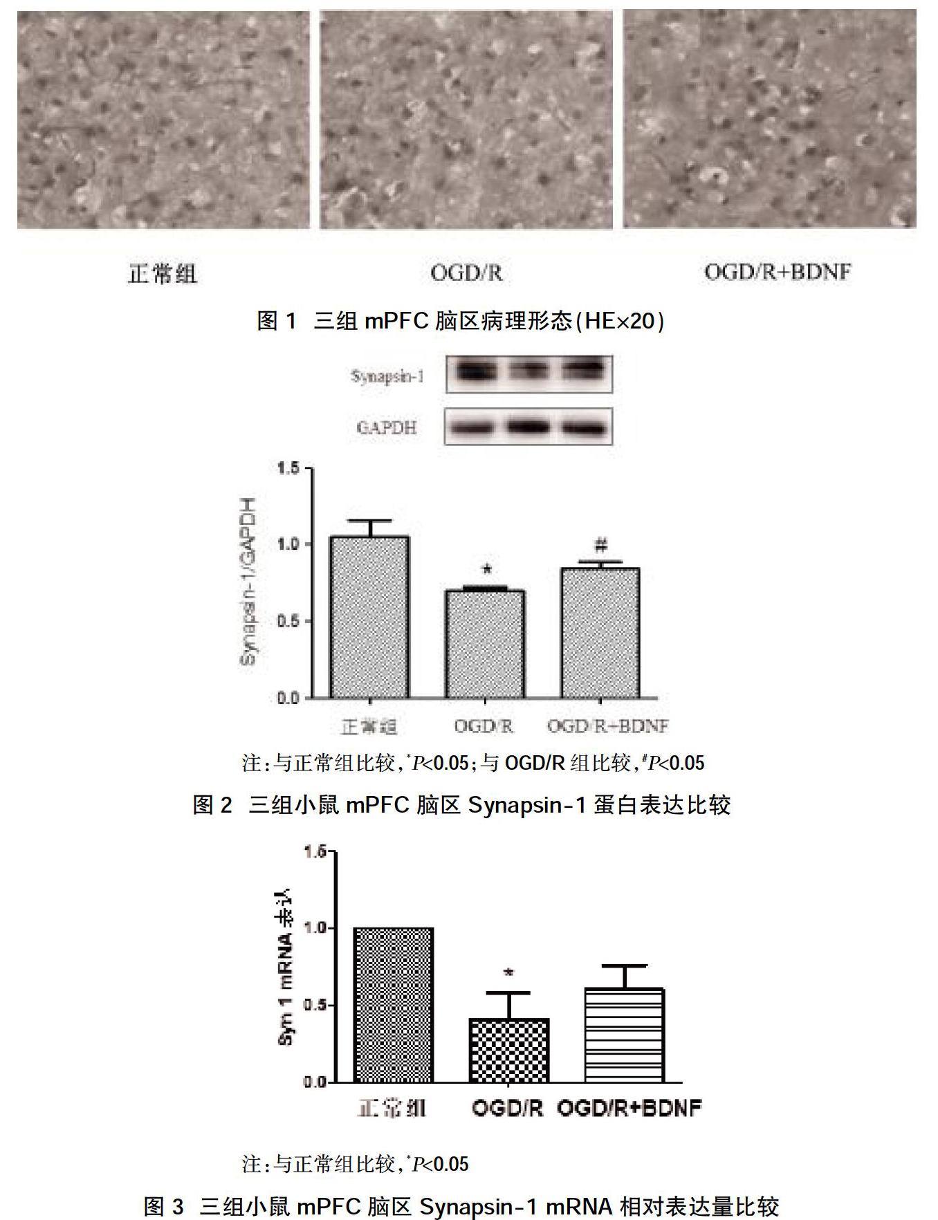
2.2三組小鼠mPFC腦區Synapsin-1蛋白表達比較 ?WB檢測結果顯示,正常組Synapsin-1蛋白的相對表達量為(1.0573436±0.171485464),缺糖缺氧復灌組Synapsin-1蛋白的相對表達量為(0.702825767±0.03653467),缺糖缺氧復灌給藥組Synapsin-1蛋白的相對表達量為(0.845764±0.073608716);缺糖缺氧復灌組Synapsin-1蛋白的表達量低于正常組、缺糖缺氧復灌給藥組,差異有統計學意義(P<0.05);缺糖缺氧復灌給藥組與正常組Synapsin-1蛋白表達量比較,差異無統計學意義(P>0.05),見圖2。
2.3三組小鼠mPFC腦區Synapsin-1 mRNA相對表達量比較 ?正常組Synapsin-1 mRNA的相對表達量為(1.00000±1.00000),缺糖缺氧復灌組Synapsin-1 mRNA的相對表達量為(0.4126427±0.299804),缺糖缺氧復灌給藥組Synapsin-1 mRNA的相對表達量為(0.604788±0.271342)。……
登錄APP查看全文