Malignant solitary fibrous tumor in the central nervous system treated with surgery,radiotherapy and anlotinib:A case report
2022-01-24 09:24:46DongYongZhangLanSuYiWeiWang
World Journal of Clinical Cases 2022年2期
INTRODUCTION
Solitary fibrous tumor (SFT) is a rare soft tissue tumor of mesenchymal cell origin.An SFT mainly develops in the pleural cavity,but it can arise in a variety of non-pleural soft tissue sites throughout the body.The incidence of SFT in the central nervous system (CNS) is very low and usually originates from the cranial meninges.The NGFIA-binding protein (NAB2) and signal transducer and activator of transcription(STAT6) gene fusion was identified as a driver mutation of SFT.Based on SFT and hemangiopericytoma (HPC) containing identical genetic abnormalities,they were considered a new combined entity in the 2016 CNS classification[1].This classification described three grades of SFT/HPC,grade I,grade II and grade III.The first case of intracranial SFT was described by Carneiro[2] in 1966.This tumor most commonly affects adults and is generally benign in the CNS[3].Malignant SFT of the CNS is exceedingly rare.
No standardized treatment guideline is available for malignant intracranial SFTs.The main treatments are surgical resection and postoperative radiotherapy.Despite the combination of surgery and adjuvant radiotherapy,the control rates of malignant SFT have been disappointing.Targeted therapy for the treatment of the soft tissue tumors has been developed recently.Anlotinib (Chia-tai Tianqing Pharmaceutical Co.,Ltd,China) is a newly multitargeted tyrosine kinase inhibitor with anti-neoplastic and anti-angiogenic activities.It inhibits tumor angiogenesis and proliferation.There are ongoing phase I/II/III clinical trials of anlotinib for different carcinomas and sarcomas in China and other countries.To our knowledge,there are no studies on anlotinib for the treatment of intracranial SFT.In this report,we present a girl with intracranial SFT who was effectively treated by surgery,radiotherapy and anlotinib.We discuss the histopathological features,next-generation sequencing results and anlotinib treatment of cancer,together with a brief review of the literature on SFT treatment by monotherapy.
Yes, I believe it sounds hopeful, he nodded. And I have a hunch8 we might do well to have him share a bit of his world with us. I just received a list of the poor families in our school who most need help through the Christmas collection. Here, take a look at it.
CASE PRESENTATION
Chief complaints
A 9-year-old girl presented to our hospital emergency department with a 3-wk history of ineffective right limb movement.
Imaging examinations
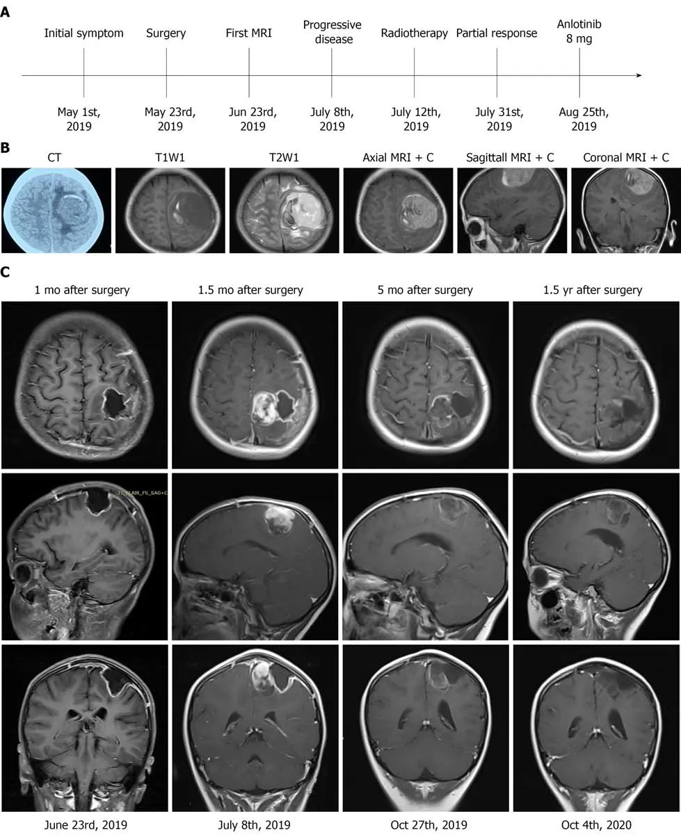
The treatment timeline is shown in Figure 1A.A head computed tomography (CT)scan revealed a quasi-circular mass in the left frontal-parietal region with high-density and associated hemorrhage (Figure 1B).Brain magnetic resonance imaging (MRI)revealed low signals on T1 weighted imaging with high surrounding signals.High signals on T2 weighted imaging with low surrounding signals were observed,with marked enhancement on contrast measuring 4.8 cm × 5.0 cm × 4.5 cm in the left motor area of the frontal-parietal lobes (Figure 1B).The imaging characteristics were similar to meningioma.An unenhanced chest CT scan revealed no nodules in the chest.
And then I came to my dormitory 303. I considered that I would spend four years here (in fact I moved to another one year later) and my dorm mates5 are all there. Most of them came from Sichuan and they were chatting with a happy voice, but I can t understand them! Again, I felt myself isolated! I hated that kind of feeling, and then I said to hello to them! To my surprise they are very friendly to me and warm-hearted! I no longer felt afraid. And I got along well with them. But at the first night here, I burst out to tears for that I was missing7 my family. I don t know why. Everyday when I was at home, I was just eager to go to school, to experience the wonderful college life but when coming here, I am just eager to go back! It s quite strange though, you must know this kind of feeling!
Laboratory examinations
The patient was diagnosed with malignant SFT.
Physical examination
Her temperature was 36.6 ℃,resting respiratory rate was 16 breaths/min,heart rate was 90 bpm and blood pressure was 120/75 mmHg.Neurological examination showed that her Glasgow Coma Scale score was 15 (E4V5M6),and muscle strength was grade 2 in the right limbs.She did not have any other neurological deficits.
Personal and family history
We present the first known case of malignant intracranial SFT treated with surgery,adjuvant radiotherapy and anlotinib,leading to a good prognosis.SFT is a rare soft tissue tumor in the CNS.The first case of SFT in the CNS was described by Carneiro[2]in 1966.SFT most commonly affects adults and is generally benign.A few cases have been described in children.An MRI scan 1 mo after surgery showed that the tumor had been successfully resected in our case.However,2 wk after the first MRI examination,the tumor rapidly progressed as shown by head MRI in this 9-year-old patient.The immunohistochemical features of intracranial SFT and HPC overlap.They share an inversion atfused withandgenes,and the combined term is “SFT/HPC”[1].This classification described three grades of SFT/HPC,grade IIII.Grade I was previously diagnosed as SFT.Grade II was previously diagnosed as CNS HPC.Grade III was previously termed anaplastic HPC and malignant SFT.Our patient had grade III CNS SFT and had a poor prognosis.However,disease progression was controlled following her treatment regimen.
So the Princess and the baby floated and drifted in the chest on the sea all day and night, but the baby was not afraid of the waves nor of the wind, for he did not know that they could hurt him, and he slept quite soundly
History of past illness
As our patient was young,a diagnosis of synovial sarcoma was considered.The diagnosis of synovial sarcoma depends on the cytogenetic change(;18) (p11;q11),which is detected by fluorescence in situ hybridization assay[14].In our case,fluorescence in situ hybridization assay showed that the(;18) was negative.Thus,the diagnosis in our patient was not synovial sarcoma.Another differential diagnosis we considered was schwannoma.However,the tumor was located in the frontalparietal lobe,not the area where the cranial nerves are located.Therefore,a schwannoma was ruled out.
So Cola-Mattheo rose at cock-crow, took a large basket under his arm, and carefully collected all the broken fragments of pots and pans, and jugs13 and lamps, and other trash of that sort
Timmy remains13 forever in my heart as a constant reminder14 of the possibility of miracles. From him, I have learned to challenge the thought of failure as it comes into my mind. To this day, Timmy inspires me to reach beyond the accepted knowledge of the times, and to remember the kupuna wisdom that teaches all things are possible if you truly believe.
History of present illness
The nuclear expression of STAT6 on immunohistochemistry is very important in the pathological diagnosis of SFT[4,5].Although-fusion seems to be important in the diagnosis of SFT,its detection may be difficult unless highthroughput sequencing is performed to detect breakpoints.Most SFTs have shown-fusion,and the remaining 11% of cases lacked an identifiable-fusion[5].Our case did not show-fusion following highthroughput sequencing,and no nuclear STAT6 expression was observed on immunohistochemical analysis.Hematoxylin and eosin staining showed a high-grade component mimicking a spindle cell sarcoma,small-cell sarcoma and other entities that have no morphological resemblance to SFT.The dedifferentiation of SFT can induce the loss of STAT6 expression on immunohistochemistry[6-8].We speculated that Ki-67 proliferation index was 80%,and the tumor was highly dedifferentiated,which suggest the reasons for negative STAT6 on immunohistochemistry.Although STAT6 is a sensitive marker in the diagnosis of SFT,other immunohistochemical indices,such as CD99,Bcl-2 and vimentin are also sensitive markers for the diagnosis of intracranial SFT[9-11].Han[12] analyzed 53 cases using immunohistochemistry and found that CD99 was positive in 94.3% of cases and Bcl-2 was positive in 96.2% of cases.
MULTIDISCIPLINARY EXPERT CONSULTATION
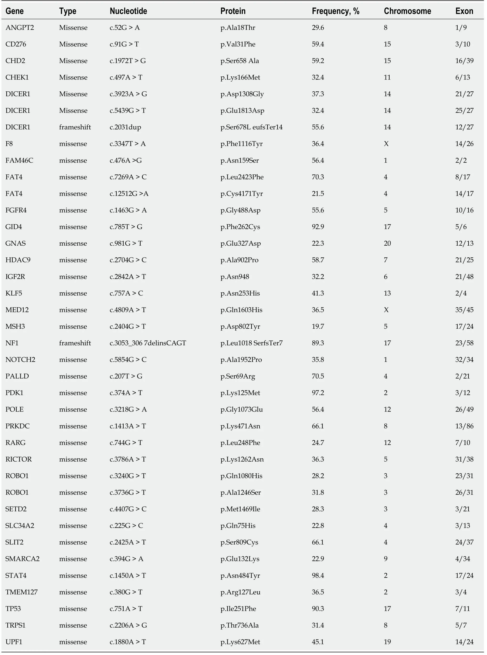
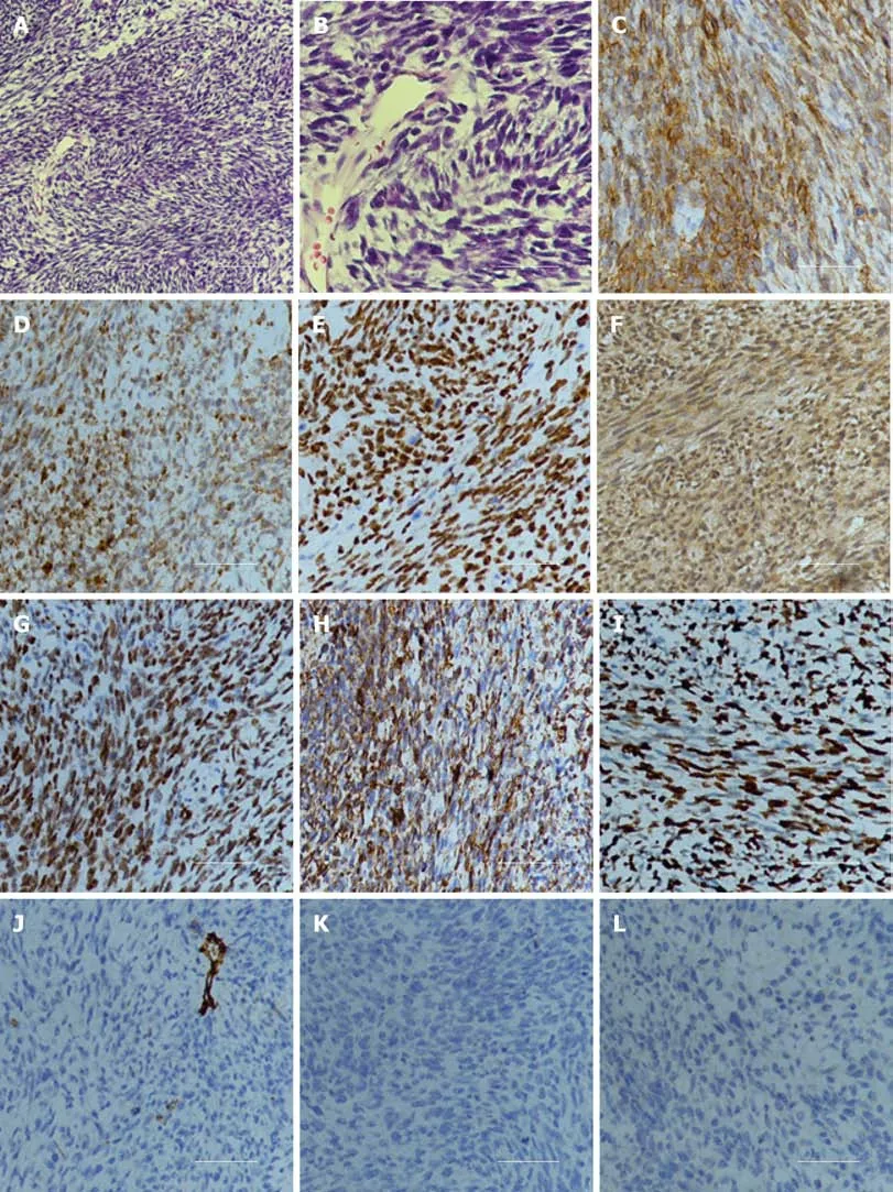
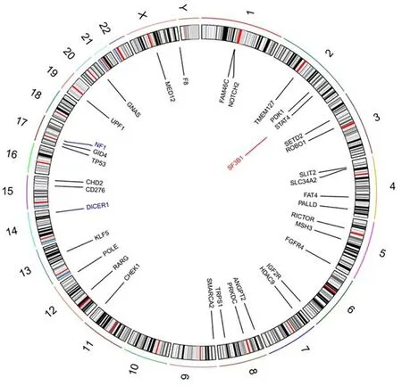
Examination of a frozen section of the biopsy revealed features of malignant tumor.Hematoxylin and eosin staining showed that a large number of spindle or oval cells were diffusely distributed,with deep staining of a null,“staghorn” vascular pattern,hypercellularity and increased mitotic activity were observed in the tumor (>4 mitosis/10 high-power fields) (Figures 2A and B).Immunohistochemistry examination of the specimen showed positivity for CD99,Bcl-2,TP53,IDH1,TLE-1 and vimentin(Figures 2C-2H) and negativity for CD34,STAT6,CK,EMA,Olig2,PR,SSTR2,CD68,S-100 and GFAP (Figures 2J-2L).Ki-67 proliferation index staining was highly positive in 80% of tumor cells (Figure 2I).Based on the above findings,the pathological diagnosis of malignant SFT was confirmed.The patient’s parents sent the specimen to the Department of Neuropathology,Beijing Neurosurgical Institute to confirm the diagnosis.The pathological diagnosis concurred with that at our hospital.The primary surgical tissue was subjected to whole-exome sequencing by next-generation sequencing (Genetron Health Co.,Ltd,Beijing,China).A global landscape of gene mutations was generated from the whole exome sequencing data (Figure 3).A total of 38 somatic gene mutations,including 36 missense mutations,2 frameshift mutations and 1 gene amplification were detected.These gene alterations were divided into:genes related to chemotherapy and targeted drug-related genes.In targeted drugrelated genes,fibroblast growth factor receptor ()(>A,)and(>,) were detected (Table 1).However,this test did not reveal any gene fusion,especially-gene fusion.
FINAL DIAGNOSIS
Laboratory tests including complete blood cell counts,bleeding time,activated partial thromboplastin time,prothrombin time,liver and renal function and blood glucose level were within normal ranges.
TREATMENT
The patient underwent left craniotomy under general anesthesia,and the mass was resected.After surgery,her motor examination showed improvement in the right limb,and she could walk unaided.Head MRI was performed 1 mo after surgery,and the results showed no residual tumor (Figure 1C).The patient was advised to undergo adjuvant radiotherapy due to high Ki-67,and MRI (1.5 mo after surgery) before radiotherapy planning showed a solid mass,suggesting a progressive tumor(Figure 1C).The patient was unable to walk by herself,and physical examination showed muscle strength was grade 3 in the right limbs.The patient received 60 Gy and 30 fractions of intensity modulated radiotherapy.Mannitol was administered to relieve her symptoms by reducing intracranial pressure.After 19 d of radiotherapy,the patient could walk unaided.Considering her pathological diagnosis,andmutation and progression of the disease,the oncologist at Beijing Cancer Hospital advised the patient to undergo anlotinib treatment.She received anlotinib 8 mg po qd for 1-14 d of a 21 d cycle at home 3 mo after surgery.During anlotinib treatment,the patient occasionally suffered from loss of appetite,and her laboratory results were within the normal range.MRI performed 5 mo after surgery showed that the tumor had not progressed (Figure 1C).The patient took anlotinib orally for 17 mo.
The Ogre s wife, who believed she could conceal26 them from her husband till morning, let them come in, and brought them to warm themselves at a very good fire; for there was a whole sheep upon the spit, roasting for the Ogre s supper.
OUTCOME AND FOLLOW-UP
Follow-up MRI showed that the tumor had not progressed 1.5 years after surgery(Figure 1C).An unenhanced chest CT scan revealed no nodules in the chest.
DISCUSSION
She had no personal or family history of benign or malignant tumors.
The patient had ineffective right limb movement for 3 wk.She also had headaches,accompanied by nausea and vomiting and excess sleep.A head CT scan revealed a quasi-circular mass in the left frontal-parietal region with high-density and associated hemorrhage.
The differential diagnosis of SFT in the CNS includes meningioma.MRI initially showed that the mass was broadly attached to the dura matter in our patient.However,the dural tail sign,which is the most important characteristic of meningioma on MRI,was absent.Secondly,EMA and SSTR2 were negative in our case by immunohistochemistry,as opposed to meningioma in which EMA and SSTR2 are both positive[13].Thirdly,meningioma is usually a benign tumor,and SFT tends to be aggressive.In our case,Ki-67 proliferation index was 80% and showed that the tumor was very malignant.These features can be used to differentiate SFT from meningioma.
The patient had no history of prior illness.
A malignant SFT has been shown to be hypercellular,mitotically active (>4 mitosis/10 high-power fields),with cytological atypia,tumor necrosis and /or infiltrative margins[15].Although our case did not show-fusion following high-throughput sequencing and STAT6 expression on immunohistochemistry,CT scanning,MRI imaging,morphologic examination,conventional immunohistochemistry (positive for CD99,Bcl-2 and vimentin),high Ki-67 proliferation index and the exclusion of other tumors resulted in the diagnosis of malignant SFT.
SFT is frequently benign and if gross total resection is performed,it will not recur.HPC tends to be an aggressive tumor with the potential for local recurrence and metastases[16].Although gross total resection offers a potential cure,improvement or preservation of motor function is crucial for patients.During surgery,we found that the tumor invaded the arachnoid,and gross total resection could cause the damage to the underlying cortex.Thus,gross total resection was not performed in our patient.Her muscle strength after surgery was better than that before surgery,and she could walk unaided.However,the disadvantage of this surgical method is that the tumor can recur.MRI (1.5 mo after surgery) showed a solid mass on the cut margin.SFT after subtotal resection has a high risk of local recurrence[17],and adjuvant radiation may be beneficial in some cases[18].Rana[19] analyzed 155 intracranial SFT patients and found that adjuvant radiation did not prolong overall survival time compared with the surgery group.However,Bishop[20] found that the treatment of SFT with combined surgery and radiotherapy led to excellent local control.Therefore,the role of radiotherapy in SFT is still unclear.Due to the low number of patients,it is difficult to conduct high-quality clinical trials on this treatment regimen.
The tumor location in our case was in the motor area,and gross total resection of the tumor while preserving neurological function was relatively difficult.Head MRI 1 mo after surgery showed that the tumor had been successfully resected.However,2 wk after the first MRI examination,brain MRI revealed tumor recurrence.The patient was advised to undergo adjuvant radiotherapy due to a high Ki-67 index and rapid recurrence.The patient received 60 Gy and 30 fractions of intensity modulated radiotherapy.After 20 d of radiotherapy,her limb weakness improved.We speculated that there were three main reasons for rapid alleviation of the patient’s symptoms.The first is that the time for the tumor to press against the motor zone of the cortex was relatively short and did not cause necrosis of cortical cells.Radiotherapy induces tumor cell necrosis,and the functions of these cells are then restored.The second is that we administered mannitol to relieve symptoms by reducing intracranial pressure.The third is that the tumor was more sensitive to radiotherapy,and it was effectively controlled.Our patient showed that radiotherapy resulted in excellent local control of intracranial malignant SFT.
The troll then showed him the three bushels of money which he had earned during the past year; they stood beside the other three, and all the six now belonged to him
Activation of oncogenes is a key mechanism of tumorigenesis and generally arises from a genetic mutation or amplification.As the therapeutic value of the genetic mutation depends heavily on the clinical prognosis,proving their direct involvement in personal targeted therapy has become a task for doctors.However,the extent of genetic intratumoral heterogeneity in SFTs is largely unknown.In our study,nextgeneration sequencing analysis showed significant germline aberrations and somatic point mutations were identified in hotspot cancer-related genes in this patient,such as FGFR4 and TP53.
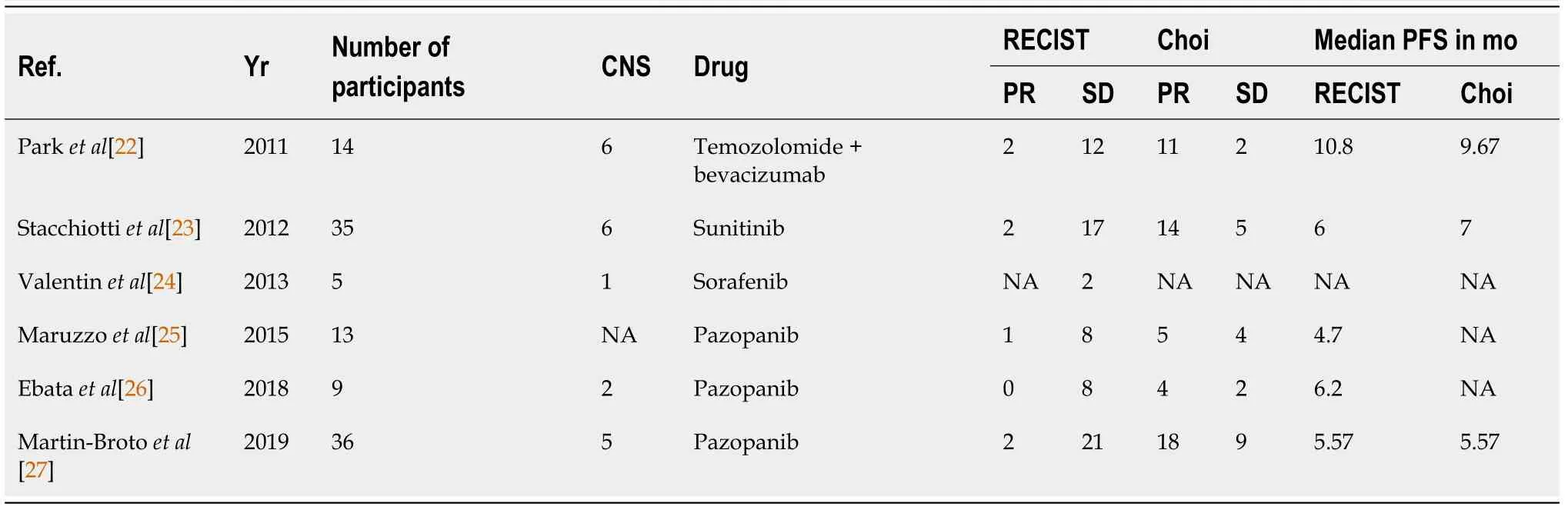
Anlotinib,a newly designed oral small-molecule receptor tyrosine kinase inhibitor,was developed independently by Chia-Tai Tianqing Pharmaceutical Co.,Ltd.in China.This drug was approved by the China Food and Drug Administration for the treatment of non-small cell lung cancer,soft tissue sarcoma,metastatic renal cell carcinoma and medullary thyroid cancer[21].Anlotinib inhibits tumor angiogenesis and proliferation,targeting vascular endothelial growth factor,FGFR,platelet-derived growth factor α/β and c-Kit[21].Several prospective studies suggest the potential efficacy of antiangiogenic treatments in SFT[22-27] (Table 2).Our patient received anlotinib 8 mg po qd for 1-14 d of a 21 d cycle after adjuvant radiotherapy for 17 mo.The follow-up MRI showed that the tumor had not progressed.
Oncogene alterations are present in all FGFR family members in human cancers.FGFR is a receptor tyrosine kinase consisting of an intracellular tyrosine-kinase domain and an extracellular ligand-binding domain.FGFR 1-3 often occur in amplifications or fusions in some cancers.However,FGFR 4 is infrequently mutated in cancers[28].FGFR 4 mutations are present in 6% of melanomas[29].One recent report found that 7% of cancers had FGFR aberrations,and FGFR 4 mutation was found in 0.5% of 4853 tumors[30].Y367C mutation of the FGFR 4 gene in the breast cancer cell line MDA-MB453 promoted tumor growth[31].Futami[32] identified FGFR 4 mutation in one of 83 gastric tumor specimens,and cells expressing this mutation showed a malignant phenotype.Multi-targeted tyrosine kinase inhibitors can be used to inactivate FGFR 4 by disrupting ATP binding in its tyrosine kinase domains[33].Potential mechanisms of FGFR 4 activation include FGFR 4 overexpression and somatic mutations[34].Therefore,we speculated that FGFR 4 mutation was likely to be the “driver” mutation and resulted in increased FGFR signaling in this patient.FGFR 4 mutation might be a key anticancer target for anlotinib in the treatment of malignant intracranial SFT.
TP53 is a tumor suppressor gene and plays a crucial role in malignant tumor progression.There is mounting basic and clinical evidence to show that tumors with TP53 gene mutations have a better response to antiangiogenic drugs than TP53 wildtype tumors[35].Recent research found that anlotinib induced apoptosis in TP53 D259Y and R248G mutants,which were able to induce apoptosis through their transcription-independent function[36].Fang[37] identified three cases with TP53 mutations (p.S183X on exon 5,p.S241F on exon 7,p.R175H on exon 5,K320fs on exon 9) that might represent biomarkers for predicting the effects of anlotinib in non-small cell lung cancer.Wu[38] reported a patient with pulmonary artery sarcoma harboring a TP53 mutation (p.R110P in exon 4) who had a favorable response to anlotinib.Kurisaki-Arakawa[39] found dedifferentiated SFTs in the pelvis with a TP53 mutation (p.A158H in exon 5).Morimitsu[40] found the TP53 mutation p.A116T in 1 of 17 cases with solitary extrapleural fibrous tumors.These findings suggest that various mutations of TP53 in SFTs are common,and tumors with TP53 mutations are more likely to respond to anlotinib.Based on the next-generation sequencing analysis,we speculated that TP53 mutation also plays a very important role in malignant SFT of the CNS treated with anlotinib monotherapy.
CONCLUSION
There is currently no standard treatment regimen for malignant SFT of the CNS.There is no effective targeted drug that can improve the prognosis of malignant intracranial SFT.This is the first report in the world of a patient with malignant intracranial SFT treated with surgery,radiotherapy and anlotinib monotherapy.Based on preliminary data,we speculated that FGFR 4 and TP53 mutations might be beneficial in the treatment of malignant intracranial SFT with anlotinib.Basic research and larger,randomized controlled trial are needed to confirm the results of the present study.
 World Journal of Clinical Cases2022年2期
- World Journal of Clinical Cases的其它文章
- Successful management of delirium with dexmedetomidine in a patient with haloperidol-induced neuroleptic malignant syndrome:A case report
- Using a fretsaw in treating chronic penial incarceration:A case report
- Occupational fibrotic hypersensitivity pneumonia in a halogen dishes manufacturer:A case report
- Accelerated Infliximab Induction for Severe Lower Gastrointestinal Bleeding in a Young Patient with Crohn’s Disease:A Case Report
- Tension pneumocephalus following endoscopic resection of a mediastinal thoracic spinal tumor:A case report
- Primary adrenal diffuse large B-cell lymphoma with normal adrenal cortex function:A case report