Vitrectomy with residual internal limiting membrane covering and autologous blood for a secondary macular hole:A case report
2022-01-24 09:24:50HuangFangYingShuangQingWuWeiPingHuLiYangNiZiLongZhangYongGenXu
World Journal of Clinical Cases 2022年2期
INTRODUCTION
Myopic foveoschisis (MF) is a common complication of pathological myopia and is characterised by intraretinal splitting in the macular region,which can eventually develop into macular hole (MH) or even retinal detachment[1,2].It can result from the combined action of tangential and longitudinal vitreoretinal tractions,and surgery can release these two types of traction[3].
Vitrectomy is the mainstay of treatment for MF,but it is still disputable whether internal limiting membrane (ILM) peeling should be performed[4].A recent metaanalysis concluded that vitrectomy with ILM peeling may lead to a better anatomic outcome of MF than that without peeling.However,vitrectomy with ILM peeling did not significantly improve the best corrected visual acuity (BCVA) or lead to fewer complications[5].Qi[6] reported that vitrectomy with fovea-sparing ILM peeling may lead to a better anatomic outcome of MF and fewer complications and similar visual function than that without peeling.A meta-analysis by Azuma[3] suggested that vitrectomy with fovea-sparing ILM peeling could achieve similar anatomic outcomes in the treatment of MF,with greater visual benefits and a lower risk of MH than complete ILM peeling.Furthermore,Lai[7] reported that vitrectomy combined with ILM repositioning and autologous blood clot was significantly effective for closing MHs,reattaching the retina and improving BCVA in patients with MH retinal detachment.
In the present case,we found that fovea-sparing ILM peeling was advantageous for providing residual ILM to treat MH secondary to vitrectomy for MF.Vitrectomy combined with residual ILM covering and autologous blood was effective for closing secondary MH and improving BCVA.
CASE PRESENTATION
Chief complaints
A 52-year-old woman presented to our hospital with a complaint of blurred vision in the right eye for 7 years.
The Ant-King, with his thousands and thousands of followers31, had come during the night, and the grateful creatures had industriously32 gathered all the millet together and put it in the sacks
History of present illness
The patient had no other positive symptoms.
History of past illness
The patient had no previous medical history.
Personal and family history
A recent meta-analysis reported that compared with complete ILM peeling,foveasparing ILM peeling may lead to similar morphological effects and may contribute to greater visual benefits and a lower risk of postoperative MH formation[3].Foveasparing ILM peeling could release tangential traction force and preserve the integrity of Müller cells in the macular fovea.Thus,the traction force over the extremely thinned foveal tissue can be minimised,and less irritation and injury are helpful to maintain the stability of the macular area and avoid postoperative MH formation.
Physical examination
The messenger took back this answer, but a second time returned with the request that Don Giovanni would present them with his picture, so that they might know what sort of a person to expect
Laboratory examinations
Five months after the initial vitrectomy,postoperative OCT (Figure 2) demonstrated that the split range of the outer nuclear layer gradually reduced,a new split of the inner nuclear layer appeared,and the MH gradually expanded.The MH diameter was 406 μm at 1 wk after the initial vitrectomy and expanded to 617 μm at 5 mo after the initial vitrectomy.The epiretinal membrane proliferated on the residual ILM,wrinkled and caused the disordered shape of the nasal retinal nerve fibre layer.
So they dug a hole, and then the little hare said, The next thing is to make a fire in the hole, and they set to work to collect wood, and lit quite a large fire
In a narrow creek28 she found a whole troop of little human children, quite naked, and sporting about in the water; she wanted to play with them, but they fled in a great fright; and then a little black animal came to the water; it was a dog, but she did not know that, for she had never before seen one
At the initial examination,her BCVA was 20/100 in the right eye and 20/60 in the left eye.The standard equivalent refractive error was-10.5 dioptres in the right eye and-12.5 dioptres in the left eye.The intraocular pressures were 12.2 and 12.3 mmHg and axial lengths were 25.79 and 27.1 mm in the right and left eyes,respectively.In both eyes,the crystalline lens demonstrated opacification,and the anterior chamber was deep and clear,with tessellated fundus.
She was beautiful, said her mother; she was quite differentfrom the beauties they call antiques, for they are so damaged. Abeauty ought to be perfect, and Kaela was a perfect beauty. Alfred wept, and mamma wept, and they both wore mourning. Theblack dress suited mamma very well, and she wore mourning the longest.
Imaging examinations
The secondary MH was detected after 1 wk,and second vitrectomy combined with residual ILM covering and autologous blood was performed 5 mo after the first surgery.Standard 25-G three-port pars plana vitrectomy was performed in the right eye under retrobulbar anaesthesia.The residual vitreous was removed,and the residual ILM was stained with indocyanine green (0.5 mg/mL).The residual ILM flap was inverted and covered the MH.Fluid-gas exchange was conducted.Then,fresh blood obtained from the patient’s vein was injected gently to cover the macula.The patient was instructed to remain in the supine position for 30 min and then change to the prone position for 3 d and avoid the supine position thereafter until the gas was absorbed.
She wove one month, she wove two months-all the winter Vasilissa sat weaving, weaving her fine thread, till the whole piece of linen46 was done, of a texture47 so fine that it could be passed, like thread, through the eye of a needle. When the spring came she bleached48 it, so white that no snow could be compared with it. Then she said to the old woman: Take thou the linen to the market, grandmothers and sell it, and the money shall suffice to pay for my food and lodging49. When the old woman examined the linen, however, she said:

FINAL DIAGNOSIS
Initial vitrectomy with fovea-sparing ILM peeling for MF was performed on 10 September 2020.Standard 25-G three-port pars plana vitrectomy combined with phacoemulsification and intraocular lens implantation was performed in the right eye under retrobulbar anaesthesia.After cataract surgery,the central vitreous core was removed,and the posterior hyaloid membrane was removed from the macular surface.The ILM was stained with indocyanine green (0.5 mg/mL).The ILM was removed in the macular area (fovea-sparing,2 PD).Fluid-gas exchange was conducted with gas tamponade by room air at the end of the surgery.The patient was instructed to remain in the prone position for 3 d and avoid the supine position during the follow-up period until the gas was absorbed.
TREATMENT
The final diagnosis of the case was binocular cataract,binocular MF,binocular high myopia and pulmonary nodules.
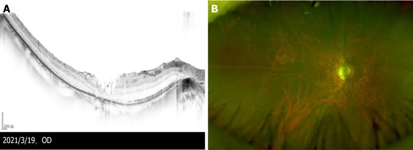
Preoperative optical coherence tomography (OCT) (Figure 1A) showed that vitreomacular traction was present in the right eye,the retinal nerve fibre layer was split on the temporal side and the outer nuclear layer was split extensively on the entire macula.Preoperative optomap (Figure 1B) revealed tessellated fundus in the right eye.
OUTCOME AND FOLLOW-UP
Her prothrombin time and blood analysis,blood chemistry and urinalysis results were normal.The electrocardiographic findings were normal,and chest computed tomography showed pulmonary nodules.

Postoperative OCT 3 wk after the second vitrectomy revealed that the MH closed,covered by flocculent ILM (Figure 3A).The split of each layer was significantly reduced,and photoreceptor cells were absent in the macular area.Optomap(Figure 3B) revealed tessellated fundus only.The patient’s BCVA was 25/100 in the right eye,but she complained of central scotoma.
DISCUSSION
The patient was married and had a son.There was no family history of ocular disease.
The residual ILM may contract 2-10 mo postoperatively.Although it does not affect vision in the short term,it can still cause pathological changes similar to that of epiretinal membrane formation.The ILM is composed of type IV,type VI and type VIII collagen,which is the basement membrane of Müller cells[2].Removal of the ILM can affect the adhesion of Müller cells,leading to its dysfunction[8].Therefore,the size of the preserved ILM in the macular region warrants further study.
The secondary MH was detected 1 wk after the initial vitrectomy.MH usually results from the natural progression of MF and is a common complication of vitrectomy[4].The stiffness of Müller cells might considerably increase in MF,which might translate to higher mechanical force transmission to photoreceptors[9].The inner retinal surface of the MF was vulnerable to developing a break,and tangential and anteroposterior traction between the vitreous and retina might induce MH formation[10].Gao[11] investigated the possible mechanisms and risk factors for the development of secondary MH in MF based on preoperative OCT findings and found that a preoperative inner segment/outer segment (IS/OS) junction defect can be a risk factor for MH development.Kumar[12] found that vitrectomy with intraoperative OCT (I-OCT)-guided fovea-sparing ILM peeling helps in complete removal of traction,resolution of retinoschisis and good functional recovery,with fewer intraoperative and postoperative complications.Therefore,if preoperative OCT reveals an IS/OS junction defect,we should pay more attention to MH formation.Vitrectomy should be performed centripetal rather than centrifugal near the macula,and anteroposterior traction should be avoided to protect Müller cell processes and photoreceptor axons from damage.I-OCT may be used as it is associated with few intraoperative and postoperative complications.
In the present case,5 mo after the initial vitrectomy,we observed a gradual reduction in the split range of the outer nuclear layer,a new split of the inner nuclear layer and gradual enlargement of the MH.The repair process of MF may be centripetal,gradually moving from the peripheral retina to macula.However,due to the expansion of the sclera in high myopia,the MH could not be closed.A new split was formed in the inner nuclear layer due to the longitudinal traction caused by the repair process of the outer nuclear layer and tangential traction caused by the epiretinal membrane in the residual ILM against retinal extension.
Vitrectomy combined with various auxiliary methods have been used to treat MH[7,13].Lai[7] repaired MH with retinal detachment in patients with high myopia by vitrectomy combined with ILM repositioning and autologous blood.Moreover,96% of the MH was closed with the retina reattached after the first surgery,and 100% of the MH was closed with the retina reattached after the second surgery.Morizane[14]used a free autologous flap of the ILM to fill the MH and then covered it with lowmolecular-weight viscoelastic material to repair the refractory MH,and the hole closure rate was 90%.Faria[15] reported that the external limiting membrane and ellipsoid zone were better repaired with inverted ILM covering in the MH than with inverted ILM filling the MH.
We treated the secondary MH successfully using 25-G vitrectomy combined with residual ILM inversion and covering of the MH and autologous blood.Fresh blood forms a blood clot within minutes,keeping the residual ILM flap inverted and covering the MH,thereby reducing the risk of dislocation after surgery[7].The inverted ILM flap and blood clot mixture covering on the MH might seal the hole and provide a smooth surface for glial cell proliferation.The components and growth factors present in the blood could facilitate the healing processes of the MH.Additionally,the serum may reduce the toxic effects of indocyanine green staining of the ILM[16].
Our case report suggests that fovea-sparing ILM peeling provides a better chance to repair the secondary MH after initial vitrectomy.Additional cases need to be evaluated.
CONCLUSION
We consider that fovea-sparing ILM peeling can treat MF with fewer surgical complications effectively.When secondary MH develops after initial vitrectomy,the residual ILM in the macula can also provide better repair of the MH.
 World Journal of Clinical Cases2022年2期
- World Journal of Clinical Cases的其它文章
- Successful management of delirium with dexmedetomidine in a patient with haloperidol-induced neuroleptic malignant syndrome:A case report
- Using a fretsaw in treating chronic penial incarceration:A case report
- Occupational fibrotic hypersensitivity pneumonia in a halogen dishes manufacturer:A case report
- Accelerated Infliximab Induction for Severe Lower Gastrointestinal Bleeding in a Young Patient with Crohn’s Disease:A Case Report
- Tension pneumocephalus following endoscopic resection of a mediastinal thoracic spinal tumor:A case report
- Primary adrenal diffuse large B-cell lymphoma with normal adrenal cortex function:A case report