Langerhans cell histiocytosis presenting as an isolated brain tumour:A case report
2022-03-15 07:12:32HanXiangLiangYueLongYangQingZhangZhiXieEnTaoLiuShuXiaWang
World Journal of Clinical Cases 2022年4期
INTRODUCTION
Langerhans cell histiocytosis (LCH) is an uncommon disease characterized by clonal proliferation of myeloid precursors that differentiate into cluster of differentiation (CD)1a/CD207(Langerin) cells in lesions[1,2].It mainly affects children,with a reported incidence of 4-5 casesmillion children aged<15 yearsyear,while its incidence in adults is uncertain[3].LCH may affect any organ or system,but it most frequently affects the bone,skin,pituitary,liver,spleen,hematopoietic system,lung,lymph nodes,and central nervous system (CNS)[4,5].Cases of LCH involving the brain parenchyma and presenting as an isolated brain tumour have been reported,but all the reports lack complete multimodal imaging.Herein,we have reported a case of LCH involving the brain parenchyma and bilateral lungs,which was assessed using computed tomography (CT),high-resolution CT (HRCT),magnetic resonance imaging (MRI),andF-fluorodeoxyglucose (F-FDG) positron emission tomography (PET)/CT.Furthermore,we have reviewed the relevant literature.
There was in the town a large dark gate, through which she had to pass night and morning with the geese; would he kindly20 hang up Falada s head there, that she might see it once again? The slaughterer said he would do as she desired, chopped off the head, and nailed it firmly over the gateway21
CASE PRESENTATION
Chief complaints
A 47-year-old man was referred to our hospital with a headache for one week and sudden syncope in the morning.
As we drew closer to the lady pushing the grocery cart, my friend directed us as far away as she could, until we were nearly walking on the road. I began to observe the many others that were passing by. They, too, were avoiding her at all cost as if she were a leper or a criminal of some kind.
History of present illness
A 47-year-old man was referred to our hospital with a headache for one week and sudden syncope in the morning.
History of past illness
LCH involving the hypothalamic-pituitary or skull is not uncommon,but involvement of the brain parenchyma,such as the frontotemporal lobe,is rare.As of January 2019,fewer than 30 cases have been reported in the PubMed database (Table 1)[6-8].We reviewed the relevant PubMed literature from 1990 to May 2021 and found 16 cases of brain parenchymal LCH with imaging data.The mean age was 31 years (95% confidence interval:21.5-41.2).The male-to-female ratio was 14:2,which is consistent with that reported in previous literature reviews of intracerebral LCH,but higher than that in children with LCH[3,9,10].The lesions were mostly located in the frontotemporal lobe (14 cases),particularly in the frontal lobe.The clinical presentation of LCH is non-characteristic and varies depending on the site.Most cases showed nonspecific symptoms of mass effect such as headache,seizures,hemiparesis,and/or sensory disturbances.MRI findings without contrast were also largely non-specific.Nonetheless,MRI showed hypointensity on T1WI and hyperintensity on T2WI in most cases.After administration of gadolinium,most cases showed intense homogeneous or heterogeneous enhancement.Another characteristic feature is sulcal enhancement around the lesion[6,7].MRI images showed leptomeningeal involvement near the lesions in several cases,as reported by Kim[7].This may be a characteristic sign of brain parenchymal LCH,but it needs to be confirmed in more cases.
The Prince and his bride set free all the Christian folk who were imprisoned27 there, and took away with them all the gold and silver that they could carry, and moved far away from the castle which lay east of the sun and west of the moon
Personal and family history
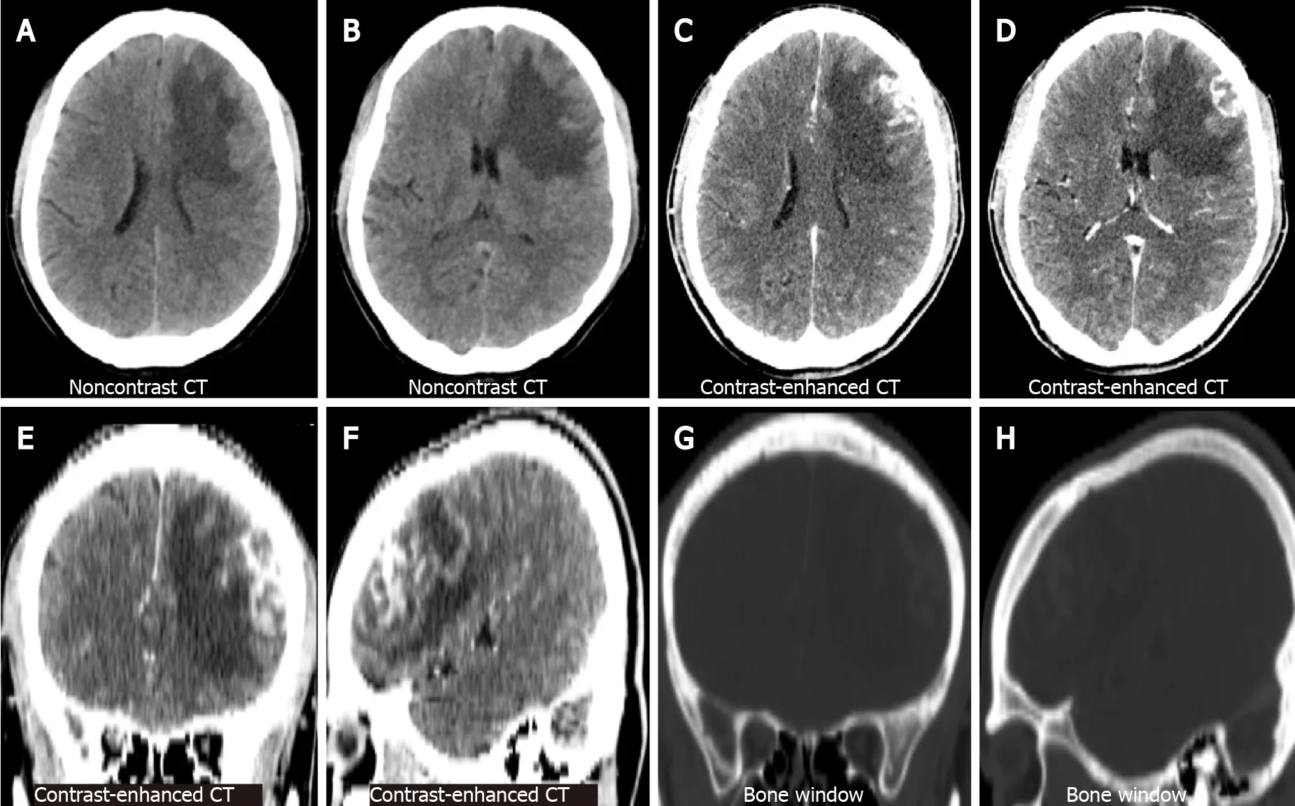
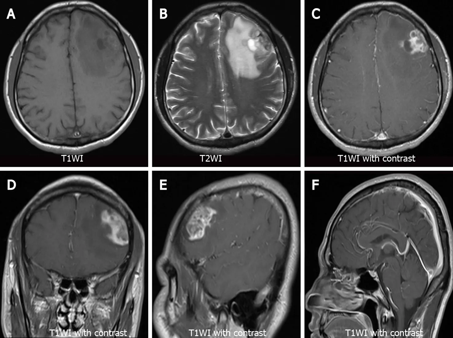
Non-contrast brain CT showed irregularly shaped nodular foci with isodensity at the left frontal corticomedullary junction.Large patches of hypodense edema were noted in the adjacent white matter (Figure 1A and B represent the lateral ventricular and basal ganglia levels,respectively).Contrast-enhanced brain CT showed significant heterogeneous enhancement in the left frontal foci (Figure 1C and D,the same level as the Figure 1A and B).Coronal and sagittal views of contrast-enhanced CT images showed irregular morphology of the lesion and poor demarcation with the adjacent skull (Figure 1E and F).Bone window CT showed no abnormalities in the adjacent skull (Figure 1G and H,the same level as the Figure 1E and F).Subsequently,the patient underwent a brain MRI.Axial T1-weighted images (T1WI) showed heterogeneous hypointense lesions in the left frontal lobe (Figure 2A).Axial T2-weighted images (T2WI) showed a heterogeneously mixed hyperintensity signals with hypointense areas in the left frontal lobe lesion (Figure 2B).After administration of gadolinium,the lesion showed heterogeneous enhancement on axial (Figure 2C),coronal (Figure 2D),and sagittal T1WI (Figure 2E).No abnormalities were found on sagittal T1WI of the sellar region (Figure 2F).
Physical examination
No rash or positive neurological signs were found on physical examination.
Laboratory examinations
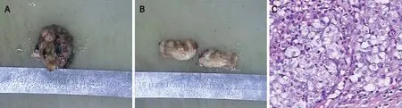
The patient underwent brain tumour resection.Gross examination showed that the specimen was a grey-brown solid tumour (Figure 5A) and the cut surface was greybrown and grey-white (Figure 5B).Histopathological examination revealed mononucleated and multinucleated histocytes with abundant cytoplasm and slight staining (haematoxylin and eosin,magnification,× 200;Figure 5C).On immunohistochemistry,the specimen stained positive for S100,CD207 (Langerin),CD4,and CD1a,and negative for CD3 and CD20.Ki67 (MIB-1) index was slightly>30%.
Imaging examinations
The patient had no known comorbidities or family history,but had a 15-year smoking history.
No recurrence was observed on brain MRI at the 12-mo follow-up (Supplementary Figure 3).
Further diagnostic work-up
Laboratory tests results showed increased in carcinoembryonic antigen (CEA) levels (7.03 ng/mL,reference:0-5 ng/mL),with no other abnormal findings.
FINAL DIAGNOSIS
The patient received chemotherapy (vindesine and prednisone acetate) after surgery.
TREATMENT
The final histological diagnosis was LCH.
OUTCOME AND FOLLOW-UP
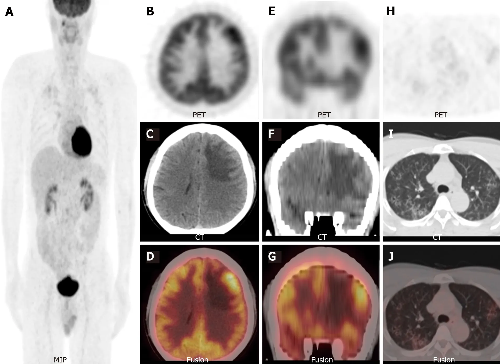
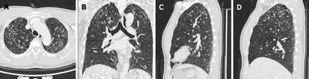
Considering the elevated CEA levels and CT and MRI manifestations,further investigation was required to rule out brain metastases.Therefore,F-FDG PET/CT was performed.Maximum-intensity-projection imaging showed a focal increase inFFDG uptake in the right maxillary sinus and multiple foci of increasedF-FDG uptake in the bilateral lung fields (Figure 3A).Axial (Figure 3B-D) and coronal (Figure 3E-G) views of the selected PET,non-enhanced CT (NE-CT),and fused PET/CT images showed moderately increasedF-FDG uptake in the left frontal nodule [the maximum standardized uptake value (SUVmax) of the lesions and surrounding tissues are shown in Supplementary Figure 1].No abnormalF-FDG uptake was observed in the sellar region (Supplementary Figure 2).Axial (Figure 3H-J) views of the selected PET,NE-CT,and fused PET/CT images showed multiple cysts with peripheral exudation in the upper lobes of bilateral lungs,with slightly increasedF-FDG uptake.HRCT was performed to further evaluate the pulmonary lesions.Axial (Figure 4A),coronal (Figure 4B),and sagittal (Figure 4C and D,left and right lungs,respectively) views of HRCT images showed multiple scattered small thick-walled irregular cysts and small nodules.Sinusitis was diagnosed in the right maxillary sinus.Bilateral lung manifestations should be differentiated from pulmonary LCH,but brain nodules are more difficult to diagnose and should be differentiated from gliomas.
DISCUSSION
The patient had no history of polyuria or polydipsia.No other illnesses were observed.
When I finally had my frozen yogurt and my friend was still complaining about the embarrassment47 I had caused her; I felt gratitude48 well up within me. At that very moment, I didn t care anymore what other people thought. I was going to do the right thing, even if it meant losing or embarrassing my friends. I smiled to myself because even though I had helped that lady in such a small way, she had helped me more by showing me how I could be different in the world and how good that could feel.
There are no previous reports ofF-FDG PET/CT for assessing the metabolic activity of brain parenchymal LCH.To our knowledge,our case report is the first with a PET/CT description.The SUVmax of the brain lesion was approximately 9.5,which was similar to the SUVmax of LCH lesions involving other regions reported in the literature[11].Additional bilateral lung lesions were found,and pulmonary manifestations were decisive for diagnosis[12].As 30% patients with LCH present with multiorgan system involvement,it is important to detect involvement of other tissues (such as the bone,soft tissue,the CNS,or the lungs)[3,13].Single or isolated brain lesions have previously been reported based on only brain CT or MRI,without whole-body scans[6,7].Without whole-body evaluation,reports of isolated brain lesions may be non-rigorous or biased.More recent studies that performed whole-body evaluations have identified a higher rate of focal LCH lesions than that previously reported[14,15].Therefore,PET/CT or PET/MRI seems to be more appropriate for evaluating this disease[16].This is especially true for combined bone and lung lesions as some case without obvious symptoms are incidentally detected;they may be missed by relying solely on radiography or CT[8].Several studies have confirmed the diagnostic value of systemic scans,such as PET/CT or PET/MRI for LCH[14,15,17,18].The diagnostic evaluation of LCH plays a crucial role in treatment planning.PET/CT or PET/MRI can be used to assess multiple foci throughout the body,guide biopsy sites,and assist with post-treatment strategies.
Now it happened one day, while they were sailing on the high seas, that Trusty John, sitting on the forepart of the ship, fiddling21 away to himself, observed three ravens22 in the air flying toward him
Based on prospective trials,the combination of vinblastine plus prednisolone is the most commomly used induction chemotherapy regimen and is administered over six weeks[19].
CONCLUSION
As a systemic disease,LCH has the potential to involve the brain parenchyma,and its diagnosis is extremely challenging.The use of multimodal imaging or whole-body imaging,combined with the manifestation of lesions at other sites,can be helpful in the diagnosis of this disease.Moreover,multimodality imaging is useful for assessing the systemic status of LCH,developing treatment plans,and evaluating post-treatment strategies.
 World Journal of Clinical Cases2022年4期
- World Journal of Clinical Cases的其它文章
- Surgical treatment of acute cholecystitis in patients with confirmed COVID-19:Ten case reports and review of literature
- Rituximab as a treatment for human immunodeficiency virusassociated nemaline myopathy:What does the literature have to tell us?
- Eustachian tube involvement in a patient with relapsing polychondritis detected by magnetic resonance imaging:A case report
- Endoscopic clipping for the secondary prophylaxis of bleeding gastric varices in a patient with cirrhosis:A case report
- Inflammatory myofibroblastic tumor after breast prosthesis:A case report and literature review
- Hoffa’s fracture in an adolescent treated with an innovative surgical procedure:A case report