Primary duodenal dedifferentiated liposarcoma:A case report and literature review
2022-03-18 02:16:54NahIhmKimJiShinLeeChanChoiJongHeeNamYooDukChoiHeeJoonKimSungSunKim
World Journal of Clinical Cases 2022年6期
lNTRODUCTlON
Liposarcoma is the most common form of malignant soft tissue tumor and tends to occur in the retroperitoneum,deep soft tissues of the trunk,and extremities[1].Primary liposarcoma of the gastrointestinal tract is extremely rare,with only a few cases reported in the literature[2-10].In this report,we describe a unique case of dedifferentiated liposarcoma(DDLPS)originating from the duodenum and review previously reported cases.Pathologists should keep liposarcoma in the differential diagnosis list.
So, by the help of his thread, he tried to mount upwards36, but he could go such a little way, and hurt himself dreadfully when he tumbled back to earth again
The Enchanter held his club close to the Princess s charming little nose, whereupon she woke up and shrieked85 with terror at finding herself in a strange place with the detested Grumedan
CASE PRESENTATlON
Chief complaints
A 64-year-old male presented with a 7-d duration of repeated abdominal pain.
History of present illness
The patient also complained of 8 kg weight loss in 3 mo.
It was 1997, in Chittagong, Bangladesh(), me and my family have just moved to a new apartment in a new area. So, after few weeks have passed, I started going back to school, since it was during Ramadan we moved. Well, I made some new friends in the neighborhood. This girl who was always hanging out with, her name was Ivy2.
History of past illness
The patient had been on antihypertensive medication for 20 years.
Personal and family history
He had no other significant personal or family medical history.
Physical examination
The tumor usually presents as a large painless mass in late adult life with an equal distribution between males and females.The retroperitoneum is the most common location and occurrence in the extremities and subcutaneous tissue is very rare[16].Since DDLPS primarily involving the intestine is extremely unusual,less than 10 cases have been reported to date[1-3,5-10,17].Of those tumors arising in the small bowel,four originated in the jejunum,five in the ileum,and two in the duodenum(Table 1).Clinical information on the precise locations of the other three small bowel tumors is not available.The literature reveals that occurrences of DDLPS in the small intestine can cause various symptoms,such as intussusception,bleeding,obstruction,and abdominal discomfort.Since Okabayashi[6]reported the first case in 2013,there has only been one additional report of DDLPS in the duodenum[10].
Laboratory examinations
Laboratory findings showed slight elevation of aspartate aminotransferase(55 U/L).Tumor markers including carcinoembryonic antigen(2.13 mg/mL),were within the normal range.
It had been a year since Susan, thirty-four, became blind. As the result of a medical accident she was sightless, suddenly thrown into a world of darkness, anger, frustration3 and self-pity. All she had to cling to was her husband Mark.
Imaging examinations
Abdominal computed tomography(CT)revealed a 3 cm-sized heterogeneously enhancing mass in the pancreaticoduodenal groove,causing the obstruction of the second portion of the duodenum.
FlNAL DlAGNOSlS
Kim NI,Choi YD,and Kim SS conceptualized the manuscript;reviewed the literature;and interpreted the H&E slides,immunohistochemistry slides,and fluorescencehybridization;Lee JS,Choi C,and Nam JH contributed to the manuscript draft;Kim HJ contributed to the operative performance;all authors read and approved the final manuscript.

TREATMENT
The patient underwent pylorus-preserving pancreaticoduodenectomy withright hemicolectomy,superior mesenteric vein segmental resection,and inferior vena cava wedge resection.Intraoperatively,the duodenal mass invaded the pancreas and hepatic flexure of the colon,resulting in gastric and duodenal distension.The tumor also appeared to invade adjacent large blood vessels.Although the duodenal mass was surgically removed,the entire tumor could not be completely removed as the tumor had already spread throughout the body.
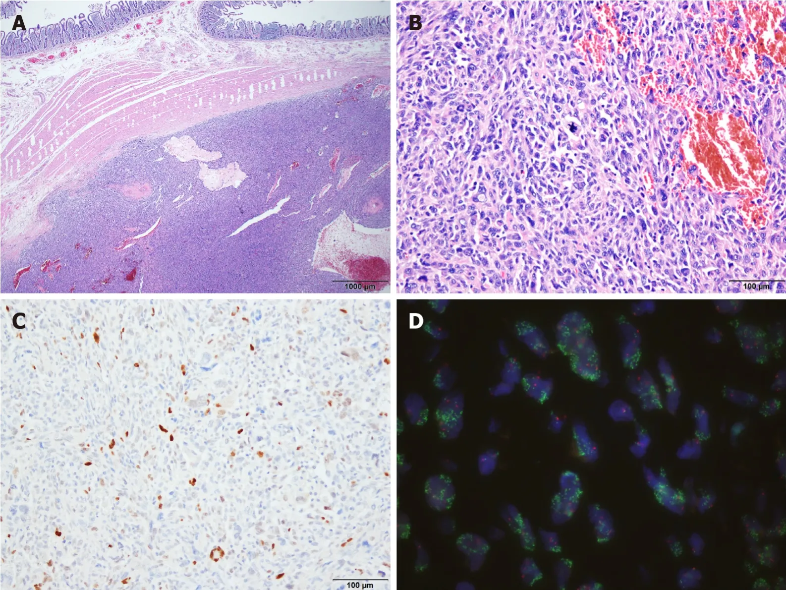
Upon macroscopic examination,the surgical resection specimen appeared to have originated from the submucosal layer of the duodenum,measuring 3x3 cm at its greatest dimension.The nodular mass showed a white-to-tan colored cut surface with a focal area of hemorrhage.Pathologic evaluation revealed a tumor arising from the duodenum and extending to the pancreas,colon,and omentum.Histology showed the epicenter of the tumor was the submucosal layer with normal-looking mucosa.The tumor was relatively well circumscribed and composed of high-grade pleomorphic cells.The tumor was arranged in a haphazard,fascicular growth pattern with telangiectatic-like feature.The majority of the tumor cells exhibited marked nuclear atypia with brisk mitotic activity.Undifferentiated tumor cells displayed large vesicular or hyperchromatic nuclei and prominent macronucleoli.Discohesive polygonal giant cells with abundant eosinophilic cytoplasm were also observed.Immunohistochemistry was positive for MDM2 and CDK4 but was negative for CK,Actin,Desmin,CD117,CD34,ERG,S100,TFE3,Melan A,and HMB45.The tumor was also found to harboramplificationfluorescencehybridization(FISH).
OUTCOME AND FOLLOW-UP
At last he spoke9 to the queen: Dear wife, this man has done me a great service, and has, besides, behaved like a gentleman in not allowing me to send back the money
Oh, dearest Princess! exclaimed the Caliph, say, when does he come, and where is the hall? The owl paused a moment and then said: Do not think me unkind, but I can only grant your request on one condition
However,due to the vessel invasion of the tumor,cancer seeding contributed to anastomotic dehiscence.Seven days after initial surgery,a 5 mm-sized leakage of the hepaticojejunal anastomosis occurred,leading to generalized peritonitis.The patient underwent a second operation.Large amounts of necrotic fluid had filled the abdominal cavity,and severe adhesions were observed.Hepaticojejunal anastomosis repair with superior mesenteric vein thrombectomy was performed.Fourteen days later,the patient developed sepsis due to the perforation of the gastrojejunal-anastomosis.A life saving emergency operation was performed.Intraoperatively,massive hematoma and bowel adhesion were observed in the upper abdomen.Overall,the tissue was very friable and edematous due to severe inflammation.An external stent was inserted and repaired using a T-tube in the gastrojejunal perforation site.Since the possibility of perforation was very high in the case of primary repair of the jejunal limb,an external stent was inserted using a hemovac drain.After massive irrigation,the operation was terminated.
49 The girl asked her if she knew the way to the Prince who lived with his stepmother in the castle which lay east of the sun and west of the moon, and who was to marry a princess with a nose which was three ells long
Unfortunately,the patient did not recover from disseminated intravascular coagulation and peritonitis,and he died 60 d after surgery.
DlSCUSSlON
Liposarcomas are subclassified as atypical lipomatous tumor/well-differentiated liposarcoma(ALT/WDLPS),DDLPS,myxoid liposarcoma,pleomorphic liposarcoma,or myxoid pleomorphic liposarcoma according to the World Health Organization classification[11].WDLPS is a typically indolent histologic subtype that presents as slowly growing masses but can be locally aggressive with minimal to no distant metastatic potential,while DDLPS is a higher grade histology with the potential for rapid growth and distant metastatic potential[12,13].
The term DDLPS was first introduced by Evans in 1979[14]and is defined as a combination of ALT/WDLPS and a high-grade non-lipogenic sarcoma-like component of variable histologic grade.DDLPS can occur(90%),with about 10% occurring from a pre-existing WDLPS[15].The histologic hallmark of DDLPS is the transition from ALT/WDLPS to non-lipogenic sarcoma,although a well-differentiated component may not be identifiable.
Physical examination revealed diffuse abdominal tenderness,but a palpable mass was not detected.

Microscopically,the tumor exhibits variable histologic features but mostly undifferentiated pleomorphic cells with striking nuclear atypia.Such tumors with unusual locations and histopathological features may pose a diagnostic challenge.Thus,sarcomatoid carcinoma,GIST,LMS,malignant melanoma and other high-grade sarcomas should be among the list of differential diagnoses.
Sarcomatoid carcinomas should be considered as the primary differential diagnosis.These tumors are predominantly composed of poorly differentiated spindle cells and/or undifferentiated bizarre anaplastic cells resembling fibrosarcoma or LMS.Sarcomatoid carcinomas can be diagnosed by immunohistochemistry using cytokeratin to demonstrate epithelial derivation.
GIST may occur anywhere in the gastrointestinal tract with 30% arising in the small bowel,including the duodenum.The tumor also exhibits a broad morphologic spectrum.While most instances of GIST are spindle or epithelioid cell tumors,progression to high-grade sarcomatous morphology can be seen rarely.The majority of GISTs show expression of CD117,DOG1 and CD34,which may be helpful for diagnosis.
We presented a unique case of DDLPS originating from the duodenum,one of the rarest locations for gastrointestinal sarcomas.DDLPS should be thoroughly distinguished from its morphological mimickers,as the tumor may be more highly aggressive and extensive than clinically and radiologically expected.While histopathologic features and immunohistochemistry offer evidence of DDLPS,FISH is essential to confirm the diagnosis.Pathologists should keep DDLPS among the initial histologic differential diagnoses for high-grade tumors of the gastrointestinal tract.
Most melanomas of the gastrointestinal tract are metastases from the skin,and primary small bowel melanoma of duodenal origin is extremely rare[24,25].Because malignant melanomas display various histologic appearance,strong clinical suspicion and precise evaluation are needed to diagnose primary duodenal melanoma.
Based on the morphology of tumor cells in DDLPS,other types of high-grade tumors such as undifferentiated pleomorphic sarcoma,malignant peripheral nerve sheath tumor,and angiosarcoma should be included among the differential diagnoses.The tumor in the present case showed positive immunoreactivity for MDM2 and CDK4.DDLPS was diagnosed based on the histological and immunohistological findings combined withamplificationFISH.
Dual staining with MDM2 and CDK4 has been shown to be both sensitive and specific to DDLPS[26].This is a result of the overexpression of the protein product from chromosomal amplification in the 12q13-15 region of theandoncogenes.Amplification of these genes can then be confirmed with FISH if diagnostic uncertainty remains[27,28].
However,MDM2 positivity by immunohistochemistry is not a specific indicator ofamplification because MDM2 positivity is observed in many other sarcomas,including WDLPS,DDLPS,intimal sarcoma,LMS,angiosarcoma,and myxofibro-sarcoma[29].Validation ofamplification by FISH,which is currently a gold standard,is mandatory to confirm the diagnosis of DDLPS.
But the hermit bade him call the man who had the fog in his sack, and the sack was opened and the fog flew out, and hung right round the king s ships, so that they could see nothing
While the biologic behavior of DDLPS appears to be unfavorable,the most effective treatment modality is surgical resection.There have been no published studies of adjuvant therapy due to the paucity of intestinal DDLPS.Because there are only a limited number of cases,we cannot predict the outcomes for DDLPS arising in the small bowel,but we expect similar prognoses compared with soft tissue DDLPS.
CONCLUSlON
Gastrointestinal LMS is also very rare,with fewer than 100 cases reported in the English-language literature since 2000[18-21].Typical LMS shows spindle cells with blunt-ended nuclei and eosinophilic fibrillary cytoplasm.The tumor cells are arranged in intersecting fascicles with varying degree of nuclear atypia,necrosis,and brisk mitotic activity.LMS can exhibit a poorly differentiated,pleomorphic appearance in addition to typical areas;this is known as pleomorphic LMS or dedifferentiated LMS[22,23].For this diagnosis to be established,morphological features characteristic of classic LMS must be present,and are usually positive for at least one myogenic marker,although staining is often weaker and more focal than in typical leiomyosarcomatous areas[22,23].The diagnosis of LMS should be made on the basis of immunohistochemical stains along with the appropriate morphological features.
ACKNOWLEDGEMENTS
We are grateful to the patient for allowing us to use his medical records in our case report.
FOOTNOTES
The preoperative differential diagnosis was duodenal adenocarcinoma,gastro-intestinal stromal tumor(GIST),or leiomyosarcoma(LMS).An accurate diagnosispreoperative upper endoscopy was not possible because only the mucosal surface was collected due to duodenal stenosis.The duodenal tumor was diagnosed as DDLPS by combining all clinical,radiologic,and intraoperative findings and histologic data(Figure 1 and Figure 2).
Grant from Chonnam National University Hospital Biomedical Research Institute,No.CRI17004-1.
Written informed consent was obtained from the patient for publication of this case report and any accompanying images.
Subsequently,chest CT and whole-body positron emission tomography/CT scans were performed to check for preexisting and unidentified intraabdominal liposarcoma outside the gastrointestinal tract and secondary duodenal involvement.No specific abnormalities or metastases were found.
The authors declare that they have no conflict of interest.
The authors have read the CARE Checklist(2016),and the manuscript was prepared and revised according to the CARE Checklist(2016).
This article is an open-access article that was selected by an in-house editor and fully peer-reviewed by external reviewers.It is distributed in accordance with the Creative Commons Attribution NonCommercial(CC BYNC 4.0)license,which permits others to distribute,remix,adapt,build upon this work non-commercially,and license their derivative works on different terms,provided the original work is properly cited and the use is noncommercial.See:https://creativecommons.org/Licenses/by-nc/4.0/
South Korea
On Christmas Eve, I placed the envelope on the tree, the note inside telling Mike what I had done and that this was his gift from me. His smile was the brightest thing about Christmas that year and in succeeding years.
Nah Ihm Kim 0000-0001-6215-8549;Ji Shin Lee 0000-0003-4634-2228;Chan Choi 0000-0002-9411-9568;Jong Hee Nam 0000-0001-5991-7102;Yoo Duk Choi 0000-0002-4385-1759;Hee Joon Kim 0000-0002-8636-5726;Sung Sun Kim 0000-0002-8569-2178.
Wang JL
It s kind of you, said Granddad, with his eyes cast down. Then, seeing the blanket at his feet, he stooped to pick it up. And will you look at this, he said. The fine blanket my son has given me to go away with.
A
Wang JL
 World Journal of Clinical Cases2022年6期
- World Journal of Clinical Cases的其它文章
- Vaginal enterocele after cystectomy:A case report
- Acute kidney injury due to intravenous detergent poisoning:A case report
- Bilateral pneumothorax and pneumomediastinum during colonoscopy in a patient with intestinal Behcet’s disease:A case report
- Successful embolization of an intrahepatic portosystemic shunt using balloon-occluded retrograde transvenous obliteration:A case report
- lmplant site development using titanium plate and platelet-rich fibrin for congenitally missed maxillary lateral incisors:A case report
- Median arcuate ligamentum syndrome:Four case reports