Volumetric assessment of hepatic grafts using a light detection and ranging system for 3D scanning: Preliminary data
2022-09-01 01:47:42GeorgiosKatsanosKonstantinaEleniKarakasiIonAnastasiosKarolosAthanasiosKofinasNikolaosAntoniadisVassiliosTsioukasGeorgiosTsoulfas
World Journal of Hepatology 2022年7期
Georgios Katsanos, Konstantina-Eleni Karakasi, Ion-Anastasios Karolos, Athanasios Kofinas, Nikolaos Antoniadis, Vassilios Tsioukas, Georgios Tsoulfas
Georgios Katsanos, Konstantina-Eleni Karakasi, Athanasios Kofinas, Nikolaos Antoniadis, Georgios Tsoulfas, Department of Transplantation, Medical School, Aristotle University of Thessaloniki, Hippokration General Hospital, Thessaloniki 54642, Greece
lon-Anastasios Karolos, Vassilios Tsioukas, Department of Rural and Surveying Engineering, Aristotle University of Thessaloniki, Thessaloniki 54642, Greece
Abstract BACKGROUND Liver transplantation has evolved into a safe life-saving operation and remains the golden standard in the treatment of end stage liver disease. The main limiting factor in the application of liver transplantation is graft shortage. Many strategies have been developed in order to alleviate graft shortage, such as living donor partial liver transplantation and split liver transplantation for adult and pediatric patients. In these strategies, liver volume assessment is of paramount importance, as size mismatch can have severe consequences in the success of liver transplantation.AIM To evaluate the safety, feasibility, and accuracy of light detection and ranging (LIDAR) 3D photography in the prediction of whole liver graft volume and mass.METHODS Seven liver grafts procured for orthotopic liver transplantation from brain deceased donors were prospectively measured with an LIDAR handheld camera and their mass was calculated and compared to their actual weight.RESULTS The mean error of all measurements was 17.03 g (range 3.56-59.33 g). Statistical analysis of the data yielded a Pearson correlation coefficient index of 0.9968, indicating a strong correlation between the values and a Student’s t-test P value of 0.26. Mean accuracy of the measurements was calculated at 97.88%.CONCLUSION Our preliminary data indicate that LIDAR scanning of liver grafts is a safe, cost-effective, and feasible method of ex vivo determination of whole liver volume and mass. More data are needed to determine the precision and accuracy of this method.
Key Words: Light detection and ranging; Graft volume; 3dscan; Ex vivo volumetry; Liver grafts
lNTRODUCTlON
Liver transplantation (LT) has evolved into a safe life-saving operation and remains the golden standard in the treatment of end stage liver disease[1]. The main limiting factor in the application of LT in the vast range of diseases that progress to end stage liver failure, as well as in the developing transplant oncology, is graft shortage, affecting thousands of adult and pediatric patients[2].
Over the years, many strategies have been developed in order to alleviate graft shortage, such as living donor liver transplantation[3] and split liver transplantation[4]. In these strategies, liver volume assessment is of paramount importance, as size mismatch can have severe consequences in the success of LT[5].
Although several techniques have been developed in order to assess liver graft volumes, few data and methods can accurately calculate partial split graft volumes in split liver transplantation[6], especially in the scenario of donors that have not been subjected to abdominal imaging studies.
Reality capture, on the other hand, is the use of various technical means to capture a digital 3D model representation of a subject from the real world. Recent technological advancements have made reality capture hardware such as light detection and ranging (LIDAR) 3D technology available to the public at reasonable prices. This technology has a multitude of applications and its value has not been extensively explored in liver surgery and liver transplantation[7,8]. We conducted a preliminary proof-of-concept study in order to evaluate the feasibility, safety, and accuracy of 3D LIDAR scanning photography of whole liver grafts and the prediction of liver volume and mass.
MATERlALS AND METHODS
Seven liver grafts procured for orthotopic liver transplantation from brain deceased donors were prospectively measured in this single blind, ongoing study. During the standard back table procedure, grafts were weighed and their mass in grams was recorded using a DSW200D weight scale (DELMAC Group, Athens, Greece). Before graft storage in the traditional nylon bags, the graft was placed on a flat sterile surface and photographed using an Original Structure 3D Scanning Sensor from the Occipital company (Occipital inc., Boulder, United States) (Figure 1). This particular sensor can be adapted to any device with the iOS and iPadOS operating system (Figure 2A), using a special bracket suitable for each corresponding model of iPhone or iPad of the end user. For the purposes of this study, an iPad (6th generation; Apple Inc., California, United States) was used (Figure 2B). The structure sensor communicates with the iPadviaa USB to a lightning cable, while the 3D scanning process is done using a suitable iPadOS compatible application provided by Occipital. This application provides the user with the ability to convert the point cloud resulting from the scanning process into a Mesh 3D digital .obj format. The Occipital structure sensor is a mobile based structure light system (SLS). This SLS consists of a laser-emitting diode, an infrared radiation range projector, and an infrared sensor and the iPad’s RGB sensor that provide measuring data to an included system on a chip (SOC) for processing. The output stream from the structure sensor alone consists of a point dataset, with a VGA resolution (640 × 480 pixels), where every pixel records the distance from sensor to the target. The infrared sensor records the reflectance intensity of the infrared (IR) light pattern projected by the IR projector onto the target while its SOC triangulates the 3D scene using specific algorithmic patterns. The main advantage of the above procedure is that the extraction of the 3D model does not require any kind of contact with the physical object (in our case the liver transplant). All measurements were conducted in fully sterile conditions with no contact with the grafts. All measurements lasted less than 3 min.
After completing the 3D reconstruction of the liver graft, the final .obj model is imported into the 3D Mesh and Point Cloud management and editing software, the 3D Slicer, a free, open source and multiplatform software package used for medical, biomedical, and related imaging research. A detailed view of an exported model participating in this study is shown in Figure 2C.
To extract the final volume of the liver model, the part of the surface on which the implant is placed (blue background) is removed from the model. The side of the graft that is in contact with the table is considered as a completely flat surface (Figure 2D). The complete flowchart of the procedure is presented on Figure 3.
In this study, mass and volume calculations were conducted by two separate teams that were blinded as to the other team’s results and measurements.
LIDAR calculated volume was converted into mass using a fixed value of liver density defined by convention at 1.07 gr/mL[9,10] .
Calculated liver mass was compared to the actual weighted liver mass of each graft.
Statistical analysis
R studio for windows (R studio, Boston MA, United States) version 4.1.1 was used to perform all the statistical analyses employing packages “rstatix” and “tidyverse”.
RESULTS
From June 2021 until January 2022, seven liver grafts from deceased donors were included in the study. The average donor age was 52.4 years, and the men-to-women ratio was 3:4. Apart from gender and age, we recorded weight, height, body mass index, and body surface area (BSA). Liver core biopsy was performed for all liver grafts as a standard practice in our department. Donor demographics are presented in Table 1. Graft weight was measured in grams (g). LIDAR imaging analysis provided the calculated graft volumes expressed in millilitres (mL). Considering the mean human liver density at 1.07 g/mL, calculated LIDAR volumes were converted to mass in grams by multiplying the volumes by 1.07. The theoretical volume of the grafts was also recorded using the Vauthey-Abdalla formula[11] [total liver volume = -794.41 + 1267.28 × body surface area). Table 2 depicts the results.
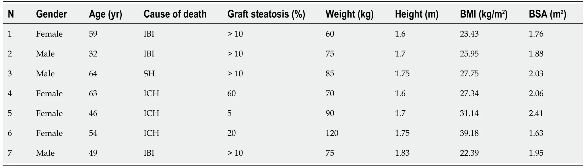
Table 1 Donor demographics.
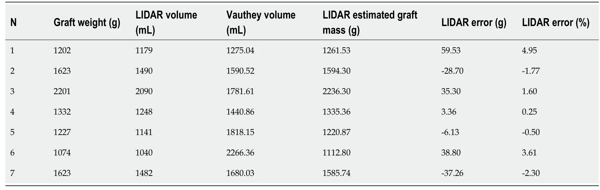
Table 2 Results
The mean duration of the measurement was 123 (74-171) s. No incidence was recorded during the procedure, which was conducted during the usual graft preparation by the surgical team. One graft was discarded due to severe steatosis. In the other six grafts, no cases of graft dysfunction or non-function were recorded in the subsequent transplantation.
LIDAR assisted graft volume and mass calculation results were compared with the actual weighed mass of the grafts. The mean error of all measurements was 17.03 g (range 3.56-59.33 g). Initially, data fluctuation analysis was performed for one factor (ANOVA). Average values, fluctuation, and degrees of freedom were calculated, and the null hypothesis (F < Fcit) was confirmed (Table 3). Statistical analysis of the data yielded a Pearson correlation coefficient index of 0.9968, indicating a strong correlation between the values and a Student’st-testPvalue of 0.26. Mean accuracy of the measurements was calculated at 97.88%. Results are depicted in Figure 4.
DlSCUSSlON
Liver graft mass and volume and their relations to recipient somatometric characteristics are essential factors for the outcome of LT. Although in standard whole liver adult to adult orthotopic LT, size is usually not an issue and the already existing methods of graft volume evaluation might be sufficient, accurate prediction of partial liver volumes in living donor[12] and split liver transplantation presents a more complex challenge[13]. Up to date, the main methods for partial liver volume calculation rely on preoperative imaging studies[14,15], which present their own set of challenges[16]. In the present work, we conducted a preliminary proof-of-concept study for the evaluation of the available handheld LIDAR technology for the evaluation of hepatic graft volume, as the first step in the development of a method that could eventually accurately estimate partial split liver volumes of grafts evaluated for split liver transplantation. The use of whole grafts aimed at calibrating the method and detecting eventual technical issues, as well as overcoming the technical issues associated with the split liver surgical technique and the fact that split liver transplantation is not currently performed in Greece. Our preliminary data tend to validate the concept of the study; however, it does not have a valuable clinical applicationper se, as whole liver mass and volume can be easily calculated by simply weighing the graft or by the water displacement method. However, due to the asymmetric structure of the liver, the calculation of partial liver volumes is more complex, and the existing mathematic formulas cannot accurately predict the segmental hepatic volumes that can vary considerably between patients[17], leaving the preoperative imaging studies of the graft in the form of either a computed tomography (CT) or magnetic resonance imaging (MRI) scan as the most used and valuable option. LIDAR assisted liver volumetry could add a useful tool forex vivopartial liver volume calculation mainly in cases of split liver transplantation for donors that for various reasons did not have a pre-procurement CT or MRI study. Compared to traditional methods for liver volumetry such as CT and MRI, LIDAR volumetric assessment is more cost-effective, less time-consuming, and less operator-dependent. Triple phase liver CT scans or MRI scans can be difficult to obtain even in tertiary hospitals, let alone in the setting of a small rural donor hospital. Moreover, the multi-organ donor is not burdened with intravenous contrast media administration, which may affect kidney function. Liver 3D model capture using the LIDAR camera is performedex vivo, just after backtable liver preparation, in less than 3 min and under sterile conditions. Actual volume measurement is done utilizing an open, free software package without the need of an expert radiologist. One obvious drawback in comparison to preoperative donor imaging is that the internal anatomy of the liver cannot be assessed and surgical plane planning is not possible. Another issue is that liver volume is measured during a state of non-perfusion, so liver mass and volume may differ if compared to a perfused organin vivo[10]. LIDAR assisted volumetry showed a better accuracy than the theoretical volume calculation using the VAUTHEY formula. This is probably mainly due to the lack of precise donor data (mainly donor weight), as many rural hospitals do not have the ability to weigh bedridden patients and the donor weight data derive from crude estimation or medical records. Finally, the main flaw of the present study is the inability to scan the inferior surface of the liver and segment I, which lie against a flat surface, and by convention this surface is considered
completely flat in our calculations. The subsequent steps in this ongoing study will be the refinement of the measuring technique, and the evaluation of the method in cadaveric livers with simulation of theex situsplitting procedure and measurement of partial liver volumes (mainly left lateral section volumes), before moving in the actual setting of real world split liver transplantation.
CONCLUSlON
Our preliminary data indicate that LIDAR scanning of liver grafts is a safe, cost-effective, and feasible method ofex vivodetermination of whole liver volume and mass. More data are needed to determine the precision and accuracy of this method.
ARTlCLE HlGHLlGHTS
Research background
Split liver transplantation is a viable option of increasing the number of available grafts, as one liver graft can yield two partial grafts for two donors. In this procedure, partial liver volume estimation,particularly left lateral segment volume estimation, is critical to the outcome of the procedure.
Research motivation
To assess the application of light detection and ranging technology in the ex vivo estimation of whole liver grafts.
Research objectives
To evaluate the feasibility, safety, and accuracy of 3D light detection and ranging (LIDAR) scanning photography of whole liver grafts and the prediction of liver volume and mass.
Research methods
Seven liver grafts procured for orthotopic liver transplantation from brain deceased donors were prospectively measured in this single blind, ongoing study. All measurements were conducted in fully sterile conditions with no contact with the grafts. LIDAR calculated volume was converted into mass using a fixed value of liver density defined by convention at 1.07 gr/mL. Calculated liver mass was compared to the actual weighted liver mass of each graft.
Research results
From June 2021 until January 2022, seven liver grafts from deceased donors were included in the study.Graft weight was measured in grams (g). LIDAR imaging analysis provided the calculated graft volumes expressed in millilitres (mL). Considering the mean human liver density at 1.07 g/mL,calculated LIDAR volumes were converted to mass in grams by multiplying the volumes by 1.07.Statistical analysis of the data yielded a Pearson correlation coefficient index of 0.9968, indicating a strong correlation between the values, and a Student’s t-test P value of 0.26. Mean accuracy of the measurements was calculated at 97.88%.
Research conclusions
Our preliminary data indicate that LIDAR scanning of liver grafts is a safe, cost-effective, and feasible method of ex vivo determination of whole liver volume and mass. More data are needed to determine the precision and accuracy of this method.
Research perspectives
LIDAR assisted liver volumetry could add a useful tool for ex vivo partial liver volume calculation mainly in cases of split liver transplantation for donors that for various reasons did not have a preprocurement computed tomography (CT) or magnetic resonance imaging (MRI) study. Compared to traditional methods for liver volumetry such as CT and MRI, LIDAR volumetric assessment is more cost-effective, less time-consuming, and less operator-dependent.
FOOTNOTES
Author contributions:Katsanos G and Karakasi KE contributed equally to this work; Katsanos G, Karakasi KE, and Tsoulfas G designed the research study; Katsanos G, Karakasi KE, Karolos IA, and Kofinas A performed the research; Antoniadis N and Karakasi KE conducted the data analysis and statistical analysis; Katsanos G, Tsoulfas G, and Tsioukas V analyzed the data and wrote the manuscript; Katsanos G, Kofinas A, and Tsoulfas G revised the manuscript; all authors have read and approved the final manuscript.
Supported bythe European Union and Greek national funds through the Operational Program Competitiveness, Entrepreneurship and Innovation, No. T1EDK-03599.
lnstitutional review board statement:The study was reviewed and approved by the Aristotle University of Thessaloniki Institutional Review Board (Approval No. 3.479).
lnformed consent statement:All study participants, or their legal guardian, provided written consent prior to study enrollment.
Conflict-of-interest statement:All authors of this manuscript having no conflicts of interest to disclose.
Data sharing statement:No additional data are available.
CONSORT 2010 statement:The authors have read the STROBE Statement-checklist of items, and the manuscript was prepared and revised according to the STROBE Statement-checklist of items.
Open-Access:This article is an open-access article that was selected by an in-house editor and fully peer-reviewed by external reviewers. It is distributed in accordance with the Creative Commons Attribution NonCommercial (CC BYNC 4.0) license, which permits others to distribute, remix, adapt, build upon this work non-commercially, and license their derivative works on different terms, provided the original work is properly cited and the use is noncommercial. See: https://creativecommons.org/Licenses/by-nc/4.0/
Country/Territory of origin:Greece
ORClD number:Georgios Katsanos 0000-0002-5845-8175; Konstantina-Eleni Karakasi 0000-0003-2448-556X; Ion-Anastasios Karolos 0000-0003-0491-1293; Athanasios Kofinas 0000-0002-3180-1930; Nikolaos Antoniadis 0000-0002-3988-4515; Vassilios Tsioukas 0000-0002-2104-4315; Georgios Tsoulfas 0000-0001-5043-7962.
S-Editor:Wang LL
L-Editor:Wang TQ
P-Editor:Wang LL
 World Journal of Hepatology2022年7期
- World Journal of Hepatology的其它文章
- Retraction Note: Screening and identification of bioactive compounds from citrus against non-structural protein 3 protease of hepatitis C virus genotype 3a by fluorescence resonance energy transfer assay and mass spectrometry
- Challenge of managing hepatitis B virus and hepatitis C virus infections in resource-limited settings
- Gut microbiota contribution to hepatocellular carcinoma manifestation in non-alcoholic steatohepatitis
- “Starry liver” - Von Meyenburg complex clinical case presentation and differential diagnosis discussion: A case report
- Hepatitis B virus markers in hepatitis B surface antigen negative patients with pancreatic cancer: Two case reports
- Hepatitis C virus burden: Treating and educating people without prejudice