STAT3基因沉默對胰腺癌細胞株的增殖及對吉西他濱敏感性的影響
2014-08-04 02:43:20潘雪ThiruvengadamArumugamVijayaRamachandranCraigLogsdon
中華胰腺病雜志 2014年2期
潘雪 Thiruvengadam Arumugam Vijaya Ramachandran Craig D Logsdon
吉西他濱目前仍代表著胰腺癌化療的標準傳統(tǒng)方案。然而由于其內(nèi)源耐藥性導致其臨床效用一般,因此一系列體外實驗嘗試檢測吉西他濱耐藥的分子指標。越來越多的證據(jù)顯示,信號轉(zhuǎn)導與轉(zhuǎn)錄激活因子3(signal transducer and activator of transcription 3, STAT3)在惡性腫瘤的轉(zhuǎn)化中起著重要作用[1-3]。STAT蛋白是細胞內(nèi)信號轉(zhuǎn)導因子家庭的一員,轉(zhuǎn)導細胞外信號,激活酪氨酸蛋白激酶(JAKs)和酪氨酸激酶受體,而它們又反過來進一步激活STATs。激活的STATs移位至細胞核,從而調(diào)節(jié)基因轉(zhuǎn)錄[4]。文獻報道[5],STAT3在白血病、淋巴瘤、乳腺癌和前列腺癌中表達,抑制STAT3活性可下調(diào)它的靶基因Bcl-XL、c-myc和cyclin D1等的表達,誘導腫瘤細胞凋亡。本研究應(yīng)用RNA干擾技術(shù)沉默STAT3基因表達,探討STAT3基因在胰腺癌細胞株對吉西他濱耐藥機制中的作用。
材料和方法
一、材料
人胰腺癌細胞株BxPC3、L3.6pl、CFPAC-1、MPanc-96、PANC1和MiaPaCa-2均購自美國ATCC(American Type Culture Collection),人胰腺癌上皮細胞(HPDE)由加拿大多倫多大學Tsao博士友情饋贈。除BxPC3在RPMI液中培養(yǎng)外,其他胰腺癌細胞株均在含10%胎牛血清的DMEM液中培養(yǎng)。HPDE細胞在含角質(zhì)形成細胞的無血清培養(yǎng)液中培養(yǎng)。靶向STAT3的siRNA(siSTAT3)購自Dharmacon Res公司,陰性對照siRNA(siNC)購自Ambion公司。Hiperfect 轉(zhuǎn)染試劑購自Qiagen公司。細胞增殖試劑盒購自Promega公司。
二、方法
1.STAT3活性檢測:將癌細胞接種于96孔板,應(yīng)用前期構(gòu)建的STAT3熒光素慢病毒、對照的海腎螢光素酶慢病毒感染細胞[6],獲得穩(wěn)定轉(zhuǎn)染的帶熒光素報告基因的胰腺癌細胞株。將感染細胞接種于96孔板,待細胞生長至70%融合后,收集細胞裂解液,應(yīng)用雙螢光報告基因試劑盒(Promega公司)定量檢測STAT3及磷酸化STAT3(pSTAT3)的活性。
2.STAT3 基因表達沉默實驗:取對數(shù)生長期細胞接種于6孔板或96孔板,待細胞長至50%融合時,應(yīng)用Hiperfect轉(zhuǎn)染試劑將siSTAT3或siNC分別轉(zhuǎn)染癌細胞,在不含抗生素的無血清培養(yǎng)液中孵育48 h,采用蛋白質(zhì)印跡法確認基因沉默效果。
3.細胞增殖實驗:取轉(zhuǎn)染的細胞孵育24 h,加入10 μmol/L的吉西他濱(耐藥株)或1 μmol/L的吉西他濱(敏感株)繼續(xù)培養(yǎng)48 h,應(yīng)用MTS試劑(Promega公司)檢測細胞增殖,以未用吉西他濱處理的細胞數(shù)為100%,計算生長抑制率。
4.細胞凋亡檢測:取轉(zhuǎn)染siSTAT3或siNC的培養(yǎng)24 h的對數(shù)生長期細胞,加入上述濃度吉西他濱后繼續(xù)孵育72 h,收集細胞,用冷PBS洗滌,應(yīng)用低張液(0.1% sodium citrate, 0.1% Triton X-100,20 mg/ml RNase,50 mg/ml propidium iodide)孵育30 min,上流式細胞儀EPICS-XO(Beckman Coulter)檢測細胞凋亡。
5.蛋白質(zhì)印跡法:收集轉(zhuǎn)染的胰腺癌細胞,提取蛋白,常規(guī)行蛋白質(zhì)印跡法檢測細胞STAT3和pSTAT3(Tyr705)的表達,以β-actin為內(nèi)參。兔抗人一抗購自Cell Signaling Technology Inc,工作濃度1∶250,熒光標記的羊抗兔二抗IRDye 680購自Santa Cruz Biotech公司,工作濃度1∶100。最后通過Odyssey紅外熒光成像儀(Li-Cor Biosciences, Lincoln, Nebraska)檢測。
三、統(tǒng)計學處理

結(jié) 果
一、胰腺癌細胞株STAT3表達水平及其與吉西他濱耐藥性的相關(guān)性
根據(jù)本研究組以前的研究報道[6],PANC1、MPanc96、MiaPaCa細胞株為吉西他濱耐藥菌株,BxPC3、CFPAC-1、L3.6pl細胞株為吉西他濱敏感細胞株。6株胰腺癌細胞株STAT3及pSTAT3蛋白表達量均顯著高于HPDE細胞,但胰腺癌細胞的表達量與其對吉西他濱耐藥性無明顯相關(guān)(圖1)。
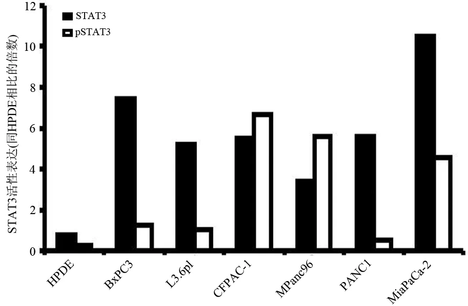
圖1 人胰腺癌細胞株和HPDE細胞STAT3、pSTAT3蛋白表達
二、siRNA轉(zhuǎn)染后胰腺癌細胞的沉默效果
轉(zhuǎn)染siSTAT3的BxPC3、CFPAC、L3.6pl、PANC1、MPanc96、MiaPaCa細胞STAT3蛋白的表達均較轉(zhuǎn)染siNC的相應(yīng)細胞顯著下調(diào)(圖2,表1)。

圖2 轉(zhuǎn)染細胞的STAT3 蛋白表達
三、STAT3基因沉默對胰腺癌細胞增殖、凋亡及與吉西他濱耐藥的影響
STAT3基因表達沉默后各胰腺癌細胞的增殖、凋亡均未受影響。但STAT3基因沉默可以增加所有6株細胞對吉西他濱的敏感性,吉西他濱的殺傷效應(yīng)增加了10%~15%(圖3)。

表1 轉(zhuǎn)染細胞的STAT3蛋白表達量
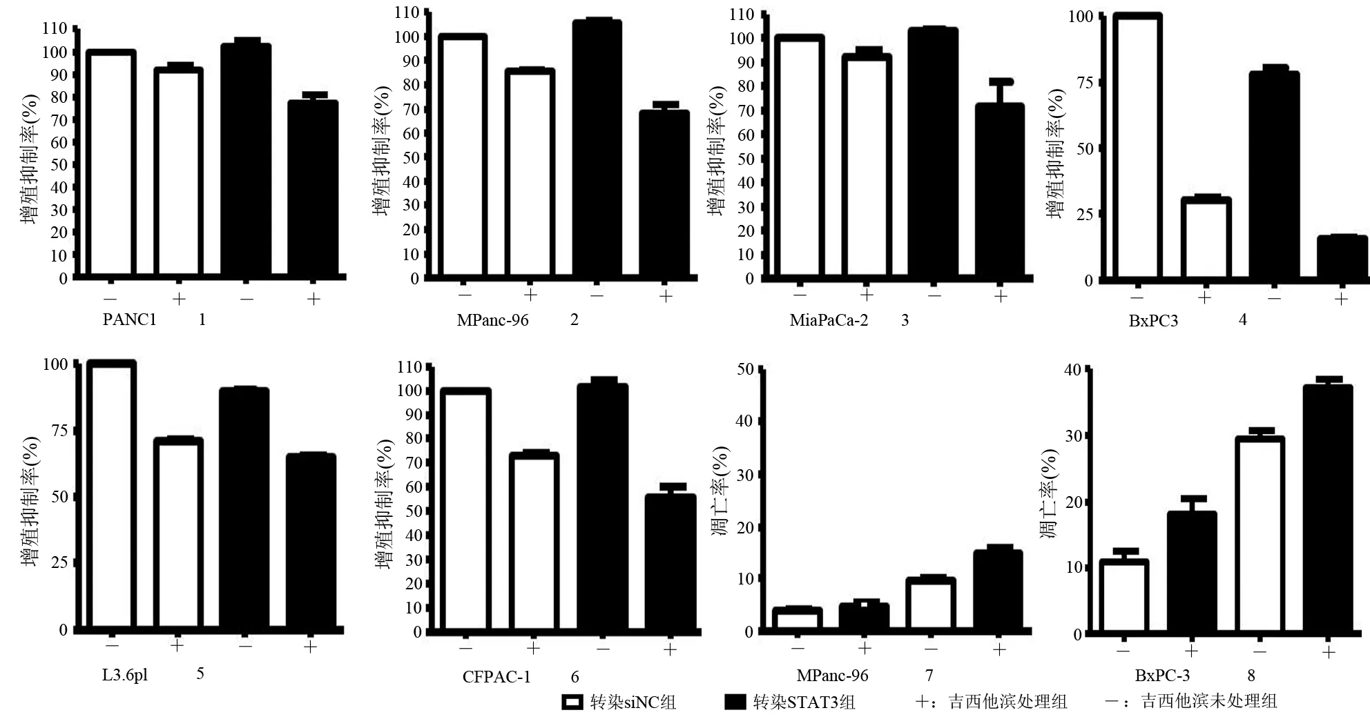
圖3 吉西他濱對轉(zhuǎn)染siSTAT3、siNC細胞的增殖(1~6)及凋亡(7~8)的影響
討 論
腫瘤化療中一個關(guān)鍵問題是腫瘤細胞對化療藥物的耐藥性。目前明確的是化療主要是通過上調(diào)凋亡、衰老或有絲分裂來誘導細胞周期阻滯或死亡。因為STAT3能夠誘導存活蛋白(Survival protein)表達、防止細胞周期阻滯或衰老的發(fā)生,因此,推測這種信號途徑不僅參與細胞轉(zhuǎn)化過程,而且也可能參與其內(nèi)源性耐藥機制。有研究認為[7-9],STAT3也許同內(nèi)源性耐藥性有關(guān)。
很多胰腺癌患者對聯(lián)合化療效果較差的原因主要是胰腺癌細胞存在內(nèi)源性的化療藥物耐藥性。在腫瘤細胞中STAT3被認為是通過抑制凋亡來產(chǎn)生化療藥物耐藥性的。一些研究證明,應(yīng)用Jak2激酶抑制劑AG490抑制STAT3活性均可導致細胞凋亡發(fā)生[10-11]。本研究結(jié)果顯示,STAT3基因沉默的胰腺癌細胞株,不管是對吉西他濱敏感株還是對耐藥菌株,其增殖并未受到影響,但其對吉西他濱的敏感性均有不同程度的增加,提示阻滯STAT3信號途徑可能會逆轉(zhuǎn)內(nèi)源性的藥物耐藥性。
參 考 文 獻
[1] Scholz A, Heinze S, Detjen KM, et al. Activated signal transducer and activator of transcription 3 (STAT3) supports the malignant phenotype of human pancreatic cancer[J]. Gastroenterology, 2003,125(3): 891-905.
[2] Wei D, Le X, Zheng L, et al. Stat3 activation regulates the expression of vascular endothelial growth factor and human pancreatic cancer angiogenesis and metastasis[J]. Oncogene,2003, 22(3): 319-329.
[3] Bromberg JF, Wrzeszczynska MH, Devgan G, et al. Stat3 as an oncogene[J]. Cell, 1999, 98: 295-303.
[4] Darnell JE, Kerr IM, Stark GR. Jak-STAT pathways and transcriptional activation in response to IFNs and other extracellular signaling proteins[J]. Science, 1994, 264(5164): 1415-1421.
[5] Gao Z, Huang C, Qiu ZJ,et al. Effect of RNAi-mediated STAT3 gene inhibition on metastasis of human pancreatic cancer cells[J]. Zhonghua Wai Ke Za Zhi, 2008,46(13):1010-1013.
[6] Pan X, Arumugam T, Yamamoto T, et al. Nuclear factor-kappaB p65/relA silencing induces apoptosis and increases gemcitabine effectiveness in a subset of pancreatic cancer cells[J]. Clin Cancer Res, 2008,14(24):8143-8151.
[7] Kato K, Nomoto M, Izumi H, et al. Structure and functional analysis of the human STAT3 gene promoter: alteration of chromatin structure as a possible mechanism for the upregulation in cisplatin-resistant cells[J]. Biochim Biophys Acta, 2000, 1493(1-2):91-100.
[8] Rebbaa A, Chou PM, Mirkin BL. Factors secreted by human neuroblastoma mediated doxorubicin resistance by activating STAT3 and inhibiting apoptosis[J]. Mol Med, 2001,7(6):393-400.
[9] Real PJ, Sierra A, De Juan A, et al. Resistance to chemotherapy via Stat3-dependent overexpression of Bcl-2 in metastatic breast cancer cells[J]. Oncogene, 2002,21(50):7611-7618.
[10] Bowman T, Broome MA, Sinibaldi D, et al. Stat3-mediated Myc expression is required for Src transformation and PDGF-induced mitogenesis[J]. Proc Natl Acad Sci U S A, 2001, 98(13):7319-7324.
[11] Burke WM, Jin X, Lin HJ, et al. Inhibition of constitutively active Stat3 suppresses growth of human ovarian and breast cancer cells[J]. Oncogene, 2001, 20(55):7925-7934.
猜你喜歡
保健醫(yī)苑(2022年5期)2022-06-10 07:46:38
現(xiàn)代臨床醫(yī)學(2022年3期)2022-06-06 07:59:40
昆明醫(yī)科大學學報(2022年1期)2022-02-28 07:43:40
中學生數(shù)理化·七年級數(shù)學人教版(2021年6期)2021-11-22 07:50:58
中學生數(shù)理化·七年級數(shù)學人教版(2021年6期)2021-11-22 07:50:58
中學生數(shù)理化·七年級數(shù)學人教版(2021年6期)2021-11-22 07:50:58
中學生數(shù)理化·七年級數(shù)學人教版(2020年12期)2021-01-18 06:57:46
中學生數(shù)理化·七年級數(shù)學人教版(2020年12期)2021-01-18 06:57:46
科學大眾(2020年12期)2020-08-13 03:22:22
海峽科技與產(chǎn)業(yè)(2016年3期)2016-05-17 04:32:12