磷酸化PKC-δ對地塞米松誘導大鼠成骨細胞凋亡的影響
2016-01-20 13:55:08付文舉張德志王國喜劉玉林泰州市第二人民醫院江蘇泰州5500遼寧醫學院附屬第一醫院
山東醫藥 2015年31期
付文舉,張德志,王國喜,劉玉林( 泰州市第二人民醫院,江蘇泰州5500;遼寧醫學院附屬第一醫院)
磷酸化PKC-δ對地塞米松誘導大鼠成骨細胞凋亡的影響
付文舉1,張德志2,王國喜1,劉玉林1
( 1泰州市第二人民醫院,江蘇泰州225500;2遼寧醫學院附屬第一醫院)
摘要:目的觀察磷酸化PKC-δ對地塞米松( Dex)誘導的大鼠成骨細胞凋亡的影響。方法取新生SD大鼠顱骨第3代成骨細胞,分為對照組、Dex組( 1×10⁻⁷mol/L Dex)、PMA預處理組( 1×10⁻⁷mol/L Dex + 100 nmol/L PMA)、Rottlerin預處理組( 1×10⁻⁷mol/L Dex +2 μmol/L Rottlerin),培養24 h后采用MTT法檢測細胞增殖活性,采用Annexin-V-FITC/PI染色流式細胞術和吖啶橙/溴乙錠( AO/EB)熒光染色法檢測細胞凋亡,采用Western blotting法檢測凋亡相關蛋白Bcl-2、Bax、Caspase-3及磷酸化PKC-δ。結果與對照組相比,Dex組細胞增殖活性低,細胞凋亡多,磷酸化PKC-δ、Caspase-3、Bax表達高,Bcl-2表達低( P均<0.05)。與Dex組相比,PMA預處理組細胞增殖活性低,細胞凋亡多,磷酸化PKC-δ、Caspase-3、Bax表達高,Bcl-2表達低( P均<0.05)。與Dex組相比,Rottlerin預處理組細胞增殖活性高,細胞凋亡少,磷酸化PKC-δ、Caspase-3、Bax表達低,Bcl-2表達高( P均<0.05)。結論磷酸化PKC-δ可促進Dex誘導的大鼠成骨細胞凋亡。
關鍵詞:蛋白激酶C-δ;半胱氨酸蛋白3; B細胞淋巴瘤相關蛋白x; B細胞淋巴瘤2;成骨細胞;細胞調亡
Effect of phosphorylated PKC-δ on Dexamethasone-induced osteoblast apoptosis of rats
FU Wen-jyu1,ZHANG De-zhi,WANG Guo-xi,LIU Yu-lin
( Second People's Hospital of Taizhou,Taizhou 225500,China)
Abstract:Objective To observe the effect of phosphorylated protein kinase C-δ( PKC-δ) on Dexamethasone ( Dex) -induced osteoblast apoptosis of rats.Methods The osteoblasts of the third generation from newly born SD rats' calvaria were divided into four groups: the control group,Dex group ( 1×10⁻⁷mol/L Dex),PMA preconditioning group ( 1× 10⁻⁷mol/L Dex +100 nmol/L PMA) and Rottlerin preconditioning group ( 1×10⁻⁷mol/L Dex + 2 μmol/L Rottlerin).The cells of each group were incubated for 24 h.Then,the cell proliferation activity was measured by MTT assay,the apoptosis was detected by AnnexinⅤ-FITC/propidium iodide ( PI) -flow cytometry ( FCM) and acridine oringe/ethidium bromide ( AO/EB) staining,and the expression of the apoptosis-related proteins ( Bcl-2,Bax and Caspase-3) and phosphorylated PKC-δ was detected by Western blotting.Results Compared with the control group,the proliferation activity was reduced,the apoptosis was increased,the expression of phosphorylated PKC-δ,Caspase-3 and Bax was increased,while the Bcl-2 expression was decreased in the Dex group,and the difference was statistically significant ( all P<0.05).Compared with the Dex group,the cell proliferation activity was reduced,the apoptosis was increased,the expression of phosphorylated PKC-δ,Caspase-3 and Bax was increased,while the Bcl-2 expression was decreased in the PMA preconditioning group,and the difference was statistically significant ( all P<0.05).Compared with the Dex group,the cell proliferation activity was obviously higher,the apoptosis was decreased,the expression of phosphorylated PKC-δ,Caspase-3 and Bax was decreased,while the Bcl-2 expression was increased in the Rottlerin preconditioning group,and the difference was statistically significant ( all P<0.05).Conclusion The phosphorylated PKC-δ can promote the Dex-induced osteoblast apoptosis of rats.
Key words:protein kinase C-δ; caspase-3; B-cell lymphoma related protein x; B-cell lymphoma 2; osteoblasts; apoptosis
糖皮質激素誘導的股骨頭壞死是股骨頭壞死的主要原因之一,其病理生理機制中涉及蛋白激酶C ( PKC)信號途徑的激活[1]。PKC-δ是PKC家族的重要成員,可參與多種細胞凋亡因子刺激誘導的細胞凋亡[2~4],但在成骨細胞凋亡方面的研究較少。在細胞凋亡的調控因素中,凋亡相關蛋白Caspase-3、Bcl-2、Bax一直備受關注。因此,本研究觀察了磷酸化PKC-δ對Dex誘導的大鼠成骨細胞凋亡的影響,為明確Dex誘導成骨細胞凋亡的分子機制提供依據。
1 材料與方法
1.1實驗動物與試劑出生24 h內SD大鼠10只,購自遼寧醫學院動物培養中心。胎牛血清、DMEM/F12培養基、胰蛋白酶購自Heclone公司,吖啶橙、溴乙錠、Ⅰ型膠原酶、MTT購自Sigma公司,茜素紅為國產,PKC-δ選擇性抑制劑Rottlerin、PKC激活劑佛波酯( PMA)購自美國Calbiochem公司,抗磷酸化PKC-δ一抗購自北京博奧森試劑有限公司,Annexin V-FITC流式檢測試劑盒購自北京四正柏生物科技有限公司。
1.2方法
1.2.1成骨細胞的分離培養與鑒定取所有大鼠顱骨,采用酶消化法分離成骨細胞,按1×10⁵/mL濃度接種于25 mL細胞培養瓶中,用含10% FBS的DMEM/F12培養,置于5%CO2、37℃培養箱中,24 h后首次換液,以后每2 d換液1次,待細胞融合至80%左右時,進行傳代,取第3代成骨細胞用于實驗。采用倒置相差顯微鏡觀察細胞形態和茜素紅染色鈣結節的方法鑒定。24 h內細胞貼壁,形態上呈多形性,主要為長梭形,胞質有突起,向外伸展; 1周后,細胞呈“鋪路石”樣排列,多數呈鱗片狀,體較大,多偽足,胞質豐富,胞核較大;繼續培養,可見局部多層生長,并有黑色鈣結節生成。
1.2.2成骨細胞凋亡模型建立取70%~80%融合的第3代成骨細胞,更換新鮮培養液,分為對照組(未做任何處理)、Dex組(調節培養液中Dex濃度為1×10⁻⁷mol/L)、PMA預處理組(調節培養液中Dex濃度為1×10-7mol/L、PMA濃度為100 nmol/L)、Rottlerin預處理組(調節培養液中Dex濃度為1× 10-7mol/L、Rottlerin濃度為2 μmol/L),置于培養箱中繼續培養24 h。
1.2.3成骨細胞增殖活性檢測采用MTT法。取上述四組細胞,以1×10⁵/mL接種于96孔板,每組8復孔,置于培養箱中繼續培養24 h后吸棄上清,加入培養液稀釋的MTT( 1 mg/mL),每孔10 μL,置培養箱中4 h;棄上清,加入等體積二甲亞砜,振蕩10 min,結晶溶解,呈紫色溶液; BIO-RAD680型酶標儀490 nm波長測吸光度值。實驗重復3次,取平均值。
1.2.4成骨細胞凋亡檢測采用Annexin-V-FITC/PI染色流式細胞術和吖啶橙/溴乙錠( AO/EB)熒光染色法檢測。參照Annexin V-FITC流式檢測試劑盒說明收集上述四組細胞,流式細胞儀檢測細胞凋亡。實驗重復3次,取平均值。取上述四組細胞,PBS沖洗2次,0.1 g/L的AO/EB混合液染色,凋亡細胞核呈現黃綠色熒光或黃綠色碎片顆粒,正常細胞呈綠色熒光,在熒光顯微鏡下觀察200倍視野下,隨機計數100個細胞中的凋亡細胞所占的比例。細胞凋亡率( %) = (凋亡細胞/計數細胞總個數)× 100%。
1.2.5各組成骨細胞Bcl-2、Bax、Caspase-3、磷酸化PKC-δ檢測采用Western blotting法。取上述四組細胞,以添加磷酸酶抑制劑的細胞裂解液裂解細胞,獲取胞質蛋白。等量蛋白進行10% SDS-PAGE電泳,然后轉至硝酸纖維素膜。以5%脫脂奶粉室溫封閉2 h,與抗Bcl-2抗體、抗Bax抗體、抗Caspase-3抗體及抗磷酸化PKC抗體( 1∶400稀釋) 4℃孵育過夜,與HRP標記羊抗兔IgG室溫孵育1 h,用ECL顯影在柯達膠片上。同一膜用洗脫液洗脫,檢測β-actin表達( Abcam一抗1∶3 000稀釋)作為內對照。實驗重復3次,取平均值。
1.3統計學方法采用SPSS17.0統計軟件。計量資料以珋x±s表示,組間比較用方差分析。P<0.05為差異有統計學意義。
2 結果
2.1各組細胞增殖活性比較對照組、Dex組、PMA預處理組、Rottlerin預處理組細胞吸光度值分別為0.437±0.013、0.379±0.012、0.369±0.013、0.410± 0.010,Dex組與其他三組比較,P均<0.05。
2.2各組細胞凋亡率比較流式細胞儀檢測結果顯示,對照組、Dex組、PMA預處理組、Rottlerin預處理組細胞凋亡率分別為3.5%±0.46%,19.0%± 0.54%、26.7%±0.69%、10.5%±0.35%,Dex組與其他三組比較,P均<0.05。
AO/EB雙熒光檢測結果顯示,對照組、Dex組、PMA預處理組、Rottlerin預處理組細胞凋亡率分別為2.9%±0.45%、20.6%±0.50%、33.4%± 0.55%、14.0%±0.30%,Dex組與其他三組比較,P均<0.05。
2.3各組細胞Bcl-2、Bax、Caspase-3、磷酸化PKC-δ表達比較結果見表1。
表1 各組成骨細胞中Caspase-3、Bax、Bcl-2、磷酸化PKC-δ表達比較(±s)
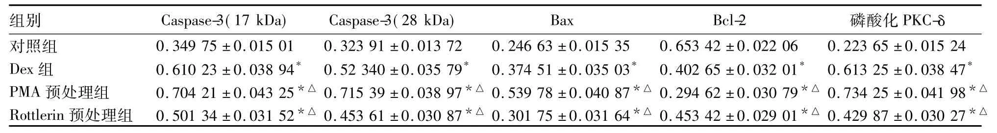
表1 各組成骨細胞中Caspase-3、Bax、Bcl-2、磷酸化PKC-δ表達比較(±s)
注:與對照組比較,*P<0.05;與Dex組比較,△P<0.05。
組別 Caspase-3( 17 kDa) Caspase-3( 28 kDa) Bax Bcl-2 磷酸化PKC-δ對照組 0.349 75±0.015 01 0.323 91±0.013 72 0.246 63±0.015 35 0.653 42±0.022 06 0.223 65±0.015 24 Dex組 0.610 23±0.038 94* 0.52 340±0.035 79* 0.374 51±0.035 03* 0.402 65±0.032 01* 0.613 25±0.038 47*PMA預處理組 0.704 21±0.043 25*△0.715 39±0.038 97*△0.539 78±0.040 87*△0.294 62±0.030 79*△0.734 25±0.041 98*△Rottlerin預處理組 0.501 34±0.031 52*△0.453 61±0.030 87*△0.301 75±0.031 64*△0.453 42±0.029 01*△0.429 87±0.030 27*△
3 討論
Weinstein等[5]最早提出糖皮質激素能夠誘導成骨細胞和骨細胞凋亡,進而導致骨細胞數量減少。隨后的臨床研究及動物實驗研究[6,7]發現,激素性骨壞死所致股骨頭中可見大量凋亡的骨細胞。本實驗中我們用Dex成功誘導建立成骨細胞凋亡模型。另外,我們的前期實驗研究[1]證明糖皮質激素誘導的成骨細胞凋亡與PKC通路相關。
PKC是一類富含絲/蘇氨基酸的激酶,在機體組織細胞內廣泛存在,目前已發現12種亞型,作為細胞信號的重要成分對機體多種細胞的增殖、分化及凋亡等方面起重要作用。在不同細胞中PKC各亞型作用各異,且受刺激因子不同和條件的影響[8,9]。PKC-δ可通過多種信號通路及相關蛋白誘導細胞凋亡[10,11]。Rottlerin為PKC-δ的特異性抑制劑,PMA為PKC激活劑。本實驗中MTT法檢測結果顯示,Dex組細胞增殖活性低于對照組,Rottlerin預處理組細胞增殖活性高于Dex組,PMA預處理組細胞增殖活性低于Dex組。流式細胞儀檢測及AO/EB雙熒光染色結果均表明,與對照組比較,Dex組細胞凋亡明顯增加,且PMA預處理組較Dex組細胞凋亡增加,Rottlerin預處理組較Dex組細胞凋亡減少。可見,Dex誘導的成骨細胞凋亡與PKC-δ的磷酸化有關。Bcl-2的同系物Mcl-1與PKC-δ參與的HaCaT細胞凋亡相關[12]。本實驗檢測結果顯示磷酸化PKC-δ表達的變化與促凋亡蛋白Caspase-3 和Bax一致,與凋亡保護性蛋白Bcl-2相反。由此,進一步確認PKC-δ的高表達與細胞的凋亡相關,推測PKC-δ可能是通過促使Caspase-3、Bax的活化及抑制Bcl-2的表達水平,從而影響Dex誘導的成骨細胞細胞凋亡。但其具體的作用途徑尚待進一步研究。
PKC-δ作用底物種類眾多,該信號傳導通路復雜,目前仍不斷有新的PKC-δ作用途徑提出,其最終的作用機制以及調解因子目前仍然沒有得到確定。本實驗在離體細胞水平上證實了PKC-δ是Dex誘導的成骨細胞凋亡中PKC途徑的重要一環,Rottlerin可通過抑制PKC-δ的表達減少細胞的凋亡,但PKC-δ究竟是通過介導何種信號通路來參與Dex誘導的成骨細胞凋亡還有待進一步研究。
參考文獻:
[1]仲興,張德志,韓鴻賓,等.地塞米松誘導成骨細胞凋亡的蛋白激酶C途徑[J].中國組織工程研究,2013,17( 41) : 7205-7212.
[2]Khwaja A,Tatton L.Caspase-mediated proteolysis and activation of protein kinase Cdelta plays a central role in neutrophil apoptosis [J].Blood,1999,94( 1) : 291-301.
[3]Patel R,Apostolatos A,Carter G,et al.Protein kinase C δ( PKC-δ) splice variants modulate apoptosis pathway in 3T3L1 cells during adipogenesis: identification of PKC-δⅡinhibitor[J].J Biol Chem,2013,288( 37) : 26834-26846.
[4]Belot A,Kasher PR,Trotter EW,et al.Protein kinase cδ deficiency causes mendelian systemic lupus erythematosus with B celldefective apoptosis and hyperproliferation[J].Arthritis Rheum,2013,65( 8) : 2161-2171.
[5]Weinstein RS,Jilka RL,Parfitt AM,et al.Inhibition of osteoblast ogenesis and promotion of apoptosis of osteoblasts and osteocytes by glucocorticoids.Potential mechanisms of their deleterious effects on bone[J].J Clin Invest,1998,102( 2) : 274-282.
[6]Kothapalli R,Aya-ay JP,Bian H,et al.Ischaemic injury to femoral head induces apoptotic and oncotic cell death[J].Pathology.2007,39( 2) : 241-246.
[7]Wang FS,Chuang PC,Lin CL,et al.MicroRNA-29a protects against glucocorticoid-induced bone loss and fragility in rats by orchestrating bone acquisition and resorption[J].Arthritis Rheum,2013,65( 6) : 1530-1540.
[8]Shu L,Zhang W,Su G,et al.Modulation of HERG K Channels by Chronic Exposure to Activators and Inhibitors of PKA and PKC: Actions Independent of PKA and PKC Phosphorylation[J].Cell Physiol Biochem,2013,32( 6) : 1830-1844.
[9]Qiu S,Jiang Z,Huang Z,et al.Migration of retinal pigment epithelium cells is regulated by protein kinase Cα in vitro[J].Invest Ophthalmol Vis Sci,2013,54( 10) : 7082-7090.
[10]Patel R,Apostolatos A,Carter G,et al.Protein kinase C δ( PKC-δ) splice variants modulate apoptosis pathway in 3T3L1 cells during adipogenesis: identification of PKC-δII inhibitor[J].J Biol Chem,2013,288( 37) : 26834-26846.
[11]Belot A,Kasher PR,Trotter EW,et al.Protein kinase cδ deficiency causes mendelian systemic lupus erythematosus with B celldefective apoptosis and hyperproliferation[J].Arthritis Rheum,2013,65( 8) : 2161-2171.
[12]Opferman JT,Iwasaki H,Ong CC,et al.Obligate role of anti-apoptotic MCL-1 in the survival of hematopoietic stem cells[J].Science,2005,307( 5712) : 1101-1104.
·基礎研究·
收稿日期:( 2015-05-02)
作者簡介:第一付文舉( 1986-),男,住院醫師,主要研究方向為骨壞死的病因與機制。E-mail: fuwenju2011@163.com
基金項目:國家自然科學基金資助項目( 81041063)。
文章編號:1002-266X( 2015) 31-0020-03
文獻標志碼:A
中圖分類號:R681
doi:10.3969/j.issn.1002-266X.2015.31.007
