Exploring the effect of acupuncture plus mild hypothermia on miRNA-204 and its target gene expressions in CIRI rat brain tissues based on MAPK signal pathway
2021-10-27 08:27:32TangYulan唐雨蘭LiuMailan劉邁蘭LuoJian羅堅LiNan李南ZhangGuoshan張國山YuJie郁潔YangQianyun楊茜蕓
Tang Yu-lan (唐雨蘭), Liu Mai-lan (劉邁蘭), Luo Jian (羅堅), Li Nan (李南), Zhang Guo-shan (張國山), Yu Jie (郁潔),Yang Qian-yun (楊茜蕓)
Hunan University of Chinese Medicine, Changsha 410208, China
Abstract
Keywords: Acupuncture Therapy; Reperfusion Injury; Hypothermia, Induced; Brain Ischemia; MicroRNAs; Gene Expression;Rats
Cerebral ischemia-reperfusion injury (CIRI) refers to the worsened ischemic injury after the blood supply is restored and recanalized by various treatments in the ischemic brain tissues[1], which is the main mechanism of damage in cerebral ischemic disease. According to the statistics, there are 1.5-2.0 million new cases of ischemic stroke in China every year. Ischemic stroke will not only affect the physical and mental health of patients, but also increase the economic burden of the family and society[2].
Acupuncture has been proved to effectively treat CIRI,and has attracted extensive attention from the medical community at home and abroad. The research team of professor Li Tie-lang revealed that acupuncture at Dazhui(GV 14), Baihui (GV 20) and Shuigou (GV 26) alleviated the symptoms of neurologic impairment in CIRI model rats and reduced the cerebral infarction area[3-4].Meanwhile, it was reported that acupuncture affected microRNA (miRNA) expression in the brain tissues of CIRI rats, such as miRNA-106, -204, -136, -181, -29c, etc.[5]Because the mitogen-activated protein kinase (MAPK)pathway is closely related to cell apoptosis, we speculated that the miRNAs mentioned above belonged to certain pathways and may be most closely related to the MAPK pathway.
Mild hypothermia is an important clinical treatment method for CIRI. Therefore, based on previous research,miRNA-204 and its target gene expressions, cell apoptosis and damage repair were observed in CIRI rats after acupuncture plus mild hypothermia therapy in this study. These findings provide experimental basis for acupuncture treatment of CIRI.
1 Materials
1.1 Experimental animal
Sixty specific pathogen free (SPF), male or female, 8 weeks old, and 250-280 g Sprague-Dawley rats were provided by the Animal Experiment Center of Hunan University of Chinese Medicine. The animal license number: SCXK (Xiang) 2016-0002.
1.2 Main reagents
Agarose (Shanghai Yuanye Biotechnology Co., Ltd.,China); loading buffer (6X, Shanghai Beyotime Biotechnology Co., Ltd., China); miRNA transcription kit,trizol, UltraSYBR Mixure, and 100 bp DNA marker (Beijing ComWin Biotech Co., Ltd., China); EDTA, tris and DEPO[Sigma-Aldrich (Shanghai) Trading Co., Ltd., China];primer [Sangon Biotechnology (Shanghai) Co., Ltd.,China]; SYBRGREENI nucleic acid dye (Beijing Probe Biotechnology Co., Ltd., China).
1.3 Main instruments
Vortex mixer (Qilin Bell Instrument Manufacturing Co.,Ltd., China); Benchtop refrigerated centrifuge(Eppendorf Corporate, Germany); fluorescence quantitative polymerase chain reaction (PCR) instrument and plate [Roche Diagnostics Products (Shanghai) Co.,Ltd., China]; homogenizer (Beijing Solarbio Science &Technology Co., Ltd., China).
2 Methods
2.1 Animal groups
SPF rats were free to have food and water, and adaptively fed for 1 week before experiments. One week later, 60 rats were numbered from 1 to 60 with the random number table method, and then divided into 6 groups, including a blank control group, a sham operation group, a model group, a mild hypothermia group, an acupuncture group and an acupuncture plus mild hypothermia group, with 10 rats in each group. Rat CIRI models were prepared in the model group, the mild hypothermia group, the acupuncture group and the acupuncture plus mild hypothermia group.
2.2 Animal modeling and modeling standards
2.2.1 Modeling method
Except for the blank control group and the sham operation group, rats in the other 4 groups all received CIRI modeling referring to the literatures with modified method[6]. The specific procedures included: rats were fasted for 12 h before surgery, anesthetized intraperitoneally with 10% chloral hydrate[3 mL/(kg·bw)] after weighing, fixed on the rat board after anesthesia, and received median neck incision after shaving of hair around the neck to identify the three arteries on the right by blunt dissection, the common carotid artery (CCA), the external carotid artery (ECA)and the internal carotid artery (ICA). For the right middle cerebral artery (MCA) embolization, the ECA and CCA were ligated, and the ICA was temporarily clamped; an oblique incision was made in the CCA to insert the No.3 fishing line through the incision for about 19 mm along the ICA by slowly loosening the arterial clamp on the CCA. Finally, the skin was sutured. About 8 mm fishing line for embolization was drawn out after embolization for 2 h, indicating completed CIRI modeling. Rats in the blank control group only received weighing and anesthesia, and in the sham operation group only received inserting about 8 mm fishing line without embolization. All rats were intramuscularly injected with penicillin after surgery to prevent infection, kept in dry cages and paid close attention to the reaction. The 5-level 4-point method reported in the literature was used as the modeling standard[6]. Rats scored 1-3 points indicated that the modeling was successful and were included in this study.
2.2.2 Modeling exclusion criteria
Rats, with neurological impairment score <1 point,being complicated with subarachnoid hemorrhage or dead, without ischemic focus after 2,3,5-triphenyltetrazolium chloride (TTC) staining, were excluded, and were replenished with the same numbers of rats after modeling again.
2.3 Acupoint selection and acupuncture methods
According to the methods provided by the
Experimental Acupuncture Science[7]and the bone measurement method analogous to the human body acupoints, Dazhui (GV 14), Baihui (GV 20) and Shuigou(GV 26) were positioned.
Baihui (GV 20): At the center of the parietal bone receiving horizontal insertion of needle for 2 mm.
Shuigou (GV 26): At the center of the cleft lip and 1 mm below the apex nasi receiving perpendicular needling for 2 mm.
Dazhui (GV 14): Located between the 7th cervical vertebra and the 1st thoracic vertebra in the middle of the back receiving perpendicular needling for 3 mm.
2.4 Mild hypothermia method
Rats, in the mild hypothermia and the acupuncture plus mild hypothermia groups, were placed in metabolic cages equipped with ice packs and crushed ice for cooling, until the rectal temperature was reduced to 32-34 ℃ and the tympanic membrane temperature was reduced to 32-34 ℃. Body temperature was checked once an hour. If the body temperature did not meet the requirement, the ice bags and crushed ice in the metabolic cages were adjusted at any time to maintain the body temperature mentioned above for 72 h. Body temperature of rats was rewarmed after 72 h.
2.5 Experimental protocols
Rats were included in the experiment after successful modeling (the successful modeling rate was 100%without any death) and treated following each step accordingly.
The first step was intervention according to the groups.Rats in the blank control group were bred routinely for 72 h without any interventions. Rats in the sham operation group and in the model group were bred routinely for 72 h, and only received binding without other intervention after surgery. Rats in the acupuncture group were bred routinely for 72 h, and received acupuncture at Dazhui (GV 14), Baihui (GV 20) and Shuigou (GV 26) after binding with needle retaining for 30 min and needling manipulation for 1 min every 15 min.Acupuncture was performed once every 12 h for 6 times in total within 72 h. Rats in the mild hypothermia group were bred routinely for 72 h, and received mild hypothermia therapy for 72 h after binding[8-9]. Rats in the acupuncture plus mild hypothermia group were bred routinely for 72 h, followed by acupuncture as in the acupuncture group and mild hypothermia intervention as in the mild hypothermia group after binding.
The second step was to determine the neurological impairment score 72 h after intervention.
The third step was to separate the brain after cervical dislocation, and collect the ischemic brain tissues on ice.
The fourth step was TTC staining to determine the cerebral infarction area ratio; real-time quantitative polymerase chain reaction (RT-qPCR) method was used to detect the expressions of miRNA-204 and its target genes.
2.6 Neurological impairment score
The 5-level 4-point method reported in the literature was used to score the neurological impairment before and after intervention[6].
2.7 Infarction area ratio
The ischemic tissues were white and the normal tissues were red after TTC staining. The infarction areas of the rat brain slices in each group were first observed and compared on gross examination. A slice with the largest ischemic section was selected. Then the Image-Pro Plus image analysis software was used to scan and calculate the total area of healthy side and the noninfarcted area of the affected side, following the method of Swanson RA,et al[8]to calculate the percentage of infarcted area (IA%). IA% = (S1- S2) ÷ S1× 100% (S1= total area of the healthy side; S2= non-infarcted area of the affected side).
2.8 Expressions of miRNA-204 and its target genes(Map3k8, Ppp3r1 and Ntrk2) in brain tissues
Total RNA from tissues was extracted with trizol and subjected to agarose gel electrophoresis. Reverse transcription of miRNA and mRNA was achieved by a two-step method. All primers were synthesized by Sangon Biotech (Shanghai) Co., Ltd., China. RT-qPCR method was used to determine the expressions of miRNA-204 and its target genes.
2.9 Statistical processing
All data were statistically analyzed using SPSS 20.0 software. Quantitative data were first checked by normality test, and data conforming to the normal distribution were statistically described as mean ±standard deviation (±s). To compare within the group,the least significant difference (LSD)-ttest was used for data with uniform variance, Tamhane's T2 method was used for data with uneven variance; one-way analysis of variance was used for comparison between groups.Median (M) and interquartile range (IQR) statistical description [M (IQR)] were used for data not conforming to the normal distribution analyzed by non-parametric test.P<0.05 indicated a statistically significant difference.
3 Results and Analysis
3.1 Neurological impairment score
Before intervention, compared with the blank control group and sham operation group, the neurological impairment scores of the modeled rats were statistically significantly increased (allP<0.01), indicating that the model was successful. Before intervention, there was no significant difference in the neurological impairment score of modelled rats among groups (P>0.05), indicating that the model preparation baseline was consistent.After 72 h of intervention, compared with the model group, the neurological impairment scores of the three intervention groups were statistically significantly reduced (allP<0.01), indicating that the three intervention options all improved the neurological impairment score (Table 1).
3.2 Cerebral infarction area ratio
Compared with the blank control group and the sham operation group, the infarction area ratio of each modeled rat was statistically significantly increased (allP<0.01), indicating that the model was successful. After 72 h of intervention, compared with the model group,infarction area ratio of rats in the 3 intervention groups decreased significantly (allP<0.01); compared with the acupuncture group and the mild hypothermia group, the infarction area ratio in the acupuncture plus mild hypothermia group was statistically significantly reduced(bothP<0.01), (Table 2 and Figure 1).
GroupnBefore intervention After intervention
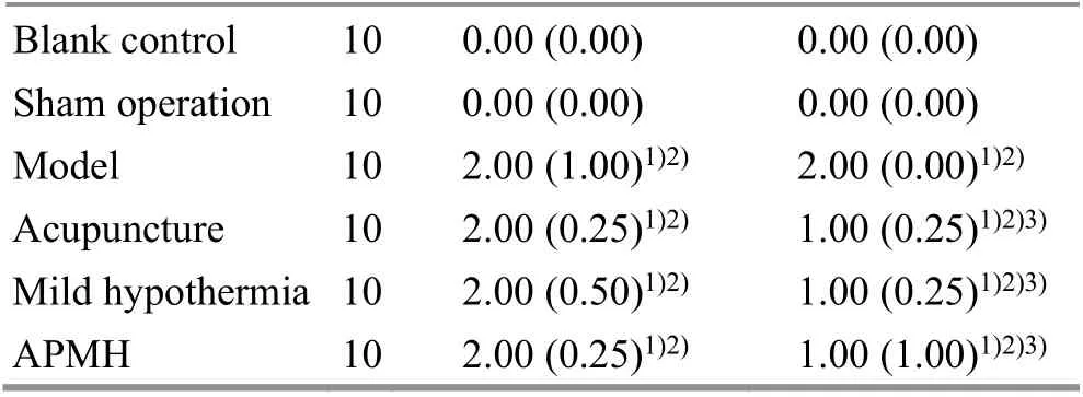
Table 1. Comparison of neurological impairment score of rats among groups [M (IQR), point]
Table 2. Comparing infarction area ratio of brain tissues among groups ( ±s, %)

Table 2. Comparing infarction area ratio of brain tissues among groups ( ±s, %)
Note: APMH=Acupuncture plus mild hypothermia group;compared with the blank control group, 1) P<0.01; compared with the sham operation group, 2) P<0.01; compared with the model group, 3) P<0.01; compared with the acupuncture group, 4) P<0.01;compared with the mild hypothermia group, 5) P<0.01
Group n Cerebral infarction area ratio Blank control 5 0.00±0.00 Sham operation 5 0.00±0.00 Model 5 36.96±0.611)2)Acupuncture 5 20.69±1.011)2)3)Mild hypothermia 5 22.14±0.611)2)3)APMH 5 10.27±1.621)2)3)4)5)
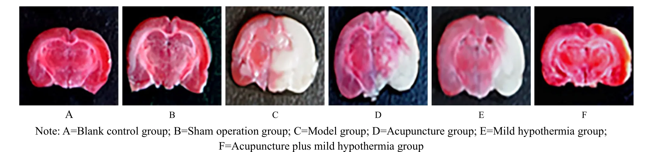
Figure 1. Cerebral infarction area in each group
3.3 Expression of miRNA-204 in rat brain tissues
Compared with the model group, the miRNA-204 expressions in brain tissues of the three intervention groups were significantly decreased (allP<0.01).Compared with the acupuncture group and mild hypothermia group, the acupuncture plus mild hypothermia group showed no obvious advantage in down-regulating the miRNA-204 expression (bothP>0.05), (Table 3).
3.4 Expression of Map3k8 in rat brain tissues
Compared with the model group, the Map3k8 expressions in brain tissues of the three intervention groups were significantly increased (allP<0.01).Compared with the acupuncture group and mild hypothermia group, the acupuncture plus mild hypothermia group showed a more statistically significant effect in up-regulating the Map3k8 expression(bothP<0.05), (Table 4).
Table 3. Comparing miRNA-204 expression in rat brain tissues among groups ( ±s)

Table 3. Comparing miRNA-204 expression in rat brain tissues among groups ( ±s)
Note: APMH=Acupuncture plus mild hypothermia group;compared with the blank control group, 1) P<0.01; compared with the sham operation group, 2) P<0.01; compared with the model group, 3) P<0.01
Group n miRNA-204 expression Blank control 3 0.91±0.23 Sham operation 3 1.10±0.27 Model 3 3.12±0.251)2)Acupuncture 3 1.22±0.323)Mild hypothermia 3 1.18±0.403)APMH 3 0.99±0.413)
Table 4. Comparing Map3k8 expression in rat brain tissues among groups ( ±s)
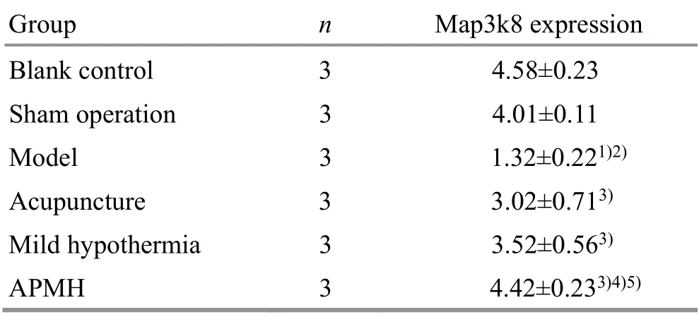
Table 4. Comparing Map3k8 expression in rat brain tissues among groups ( ±s)
Note: APMH=Acupuncture plus mild hypothermia group;compared with the blank control group, 1) P<0.01; compared with the sham operation group, 2) P<0.01; compared with the model group, 3) P<0.01; compared with the acupuncture group, 4) P<0.05;compared with the mild hypothermia group, 5) P<0.05
Group n Map3k8 expression Blank control 3 4.58±0.23 Sham operation 3 4.01±0.11 Model 3 1.32±0.221)2)Acupuncture 3 3.02±0.713)Mild hypothermia 3 3.52±0.563)APMH 3 4.42±0.233)4)5)
3.5 Expression of Ppp3r1 in rat brain tissues
Compared with the model group, the Ppp3r1 expressions in brain tissues of the three intervention groups were decreased (allP<0.01). Compared with the mild hypothermia group and the acupuncture group, the acupuncture plus mild hypothermia group showed no statistically significant advantage in regulating the Ppp3r1 expression of rat brain tissues (bothP>0.05),(Table 5).
3.6 Expression of Ntrk2 in rat brain tissues
Compared with the model group, the Ntrk2 expressions of brain tissues in the three intervention groups were statistically significantly increased (allP<0.01). Compared with the acupuncture group and mild hypothermia group, the acupuncture plus mild hypothermia group showed no statistically significant advantage in regulating the brain tissue Ntrk2 expression(allP>0.05), (Table 6).
GroupnPpp3r1 expression
Table 5. Comparing Ppp3r1 expression in rat brain tissues among groups ( ±s)
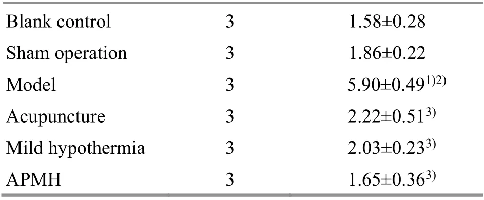
Table 5. Comparing Ppp3r1 expression in rat brain tissues among groups ( ±s)
Note: APMH=Acupuncture plus mild hypothermia group;compared with the blank control group, 1) P<0.01; compared with the sham operation group, 2) P<0.01; compared with the model group, 3) P<0.01
Blank control 3 1.58±0.28 Sham operation 3 1.86±0.22 Model 3 5.90±0.491)2)Acupuncture 3 2.22±0.513)Mild hypothermia 3 2.03±0.233)APMH 3 1.65±0.363)
Table 6. Comparing Ntrk2 expression in rat brain tissues among groups ( ±s)
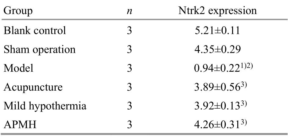
Table 6. Comparing Ntrk2 expression in rat brain tissues among groups ( ±s)
Note: APMH=Acupuncture plus mild hypothermia group;compared with the blank control group, 1) P<0.01; compared with the sham operation group, 2) P<0.01; compared with the model group, 3) P<0.01
Group n Ntrk2 expression Blank control 3 5.21±0.11 Sham operation 3 4.35±0.29 Model 3 0.94±0.221)2)Acupuncture 3 3.89±0.563)Mild hypothermia 3 3.92±0.133)APMH 3 4.26±0.313)
4 Discussion
Acupuncture therapy has a long history of treating ischemic cerebrovascular disease and has been widely recognized by the medical community at home and abroad. Clinical and experimental research results proved that mild hypothermia had convinced brain protection effect. Mild hypothermia therapy reduced brain tissue oxygen consumption, inhibited nerve cell apoptosis, reduced the production of active metabolites such as oxygen free radicals, inhibited synthesis and release of excitatory amino acids, and promoted activity of superoxide dismutase with a neuroprotective effect[9-10]. The experimental results of Pan J,et al[11]showed that the effective time window of reperfusion therapy in CIRI rats after mild hypothermia intervention was increased to 4 h, indicating that local mild hypothermia application during reperfusion period prolonged the time window, and the mechanism may be to reduce brain cell apoptosis, protect blood-brain barrier, and inhibit inflammation.
The latest research has found that the MAPK signaling pathway is intricate with multiple pathways in parallel.One of them is the highly conservative tripeptide motif‘TGY’ (P38 MAPK) structure in cells[12], which is a classic cellular pathway, not only participating in cell inflammation and apoptosis, but also being an important pathway conducting pain information[13]. When stimulated by the external environment, such as physical and chemical factors or inflammatory factors, the protein kinase cascade stimulates the phosphorylation of this pathway, which in turn causes a series of cellular responses[14].
In recent years, mild hypothermia therapy has gradually become well-known. After CIRI, mild hypothermia therapy can reduce brain tissue oxygen consumption, inhibit synthesis and release of excitatory amino acids, up-regulate Bcl-2 and down-regulate Bax expressions, and inhibit Caspase-3 expression[15-17]. At the same time, some scholars discovered that after CIRI,the MAPK/ERK pathway was activated to regulate cell differentiation and apoptosis. Apoptosis co-exists during normal development and aging process, which is a protective response and achieved by different apoptosis signaling pathways[18-19]. Lin YP,et al[20]identified that one of the mechanisms of acupuncture, mild hypothermia, and acupuncture plus mild hypothermia in CIRI treatment may be associated with inhibiting the phosphorylation levels of MEK-2 and ERK1/2, Bax expression and cell apoptosis, while increasing Bcl-2 expression.
Based on the experimental results of Lin YP,et al[21],we speculated that one of the mechanisms of acupuncture, mild hypothermia, and acupuncture plus mild hypothermia therapy for CIRI may be downregulating the phosphorylation levels of Raf-1, MEK-2 and ERK1/2 proteins, and reducing cell apoptosis.
The results of this study indicated that acupuncture or mild hypothermia alone was effective in CIRI treatment,which also briefly explained their possible effect on brain tissue protection. Our previous findings showed that the therapeutic effect of acupuncture plus mild hypothermia was significantly better than that of either acupuncture or mild hypothermia alone. The corresponding gene expression was measured and their relationship with CIRI was speculated based on the MAPK pathway. The effect of acupuncture plus mild hypothermia was better than that of acupuncture or mild hypothermia alone,which may be caused by the superimposed or synergistic effect of acupuncture and mild hypothermia, and further decreased phosphorylation of Raf-1, MEK-2 and ERK1/2 proteins.
The results of this study also showed that acupuncture plus mild hypothermia therapy significantly reduced the neurological impairment score, the area of cerebral infarction, and the miRNA-204 expression in brain tissues,which further confirmed that acupuncture plus mild hypothermia therapy reduced neurological impairment and promoted neurological recovery. However, the sample size of the current study was relatively small;therefore, more in-depth experiments for further verification are needed.
Conflict of Interest
The authors declare that there is no potential conflict of interest in this article.
Acknowledgments
This work was supported by Opening Fund Project of Domestic First-class Discipline Construction of Traditional Chinese Medicine of Hunan University of Chinese Medicine(湖南中醫藥大學中醫學一流學科開放基金項目, No.2018ZYX33).
Statement of Human and Animal Rights
The treatment of animals conformed to the ethical criteria in this experiment.
Received: 10 April 2020/Accepted: 19 December 2020
 Journal of Acupuncture and Tuina Science2021年5期
- Journal of Acupuncture and Tuina Science的其它文章
- Study on the regulatory effect of herbal cakepartitioned moxibustion on colonic CD206, AMPK and TSC2 in rats with Crohn disease
- Acupuncture combined with medication for postherpetic neuralgia affecting the head and face:a randomized controlled trial
- Tendon-regulating and bone-setting manipulation plus endurance resistance exercises for female with chronic neck pain
- Efficacy and effect on related brain-gut peptides of acupoint sticking therapy for functional dyspepsia
- Effects of acupuncture plus language training on language function and cerebral blood flow in patients with motor aphasia after ischemic stroke
- Clinical observation on moxibustion therapy plus tuina in treating children with recurrent respiratory tract infections due to qi deficiency of spleen and lung