Giant retroperitoneal lipoma presenting with abdominal distention:A case report and review of the literature
2022-03-15 11:59:52ZhiYanChenXianLongChenQiYuQingBoFan
World Journal of Clinical Cases 2022年5期
lNTRODUCTlON
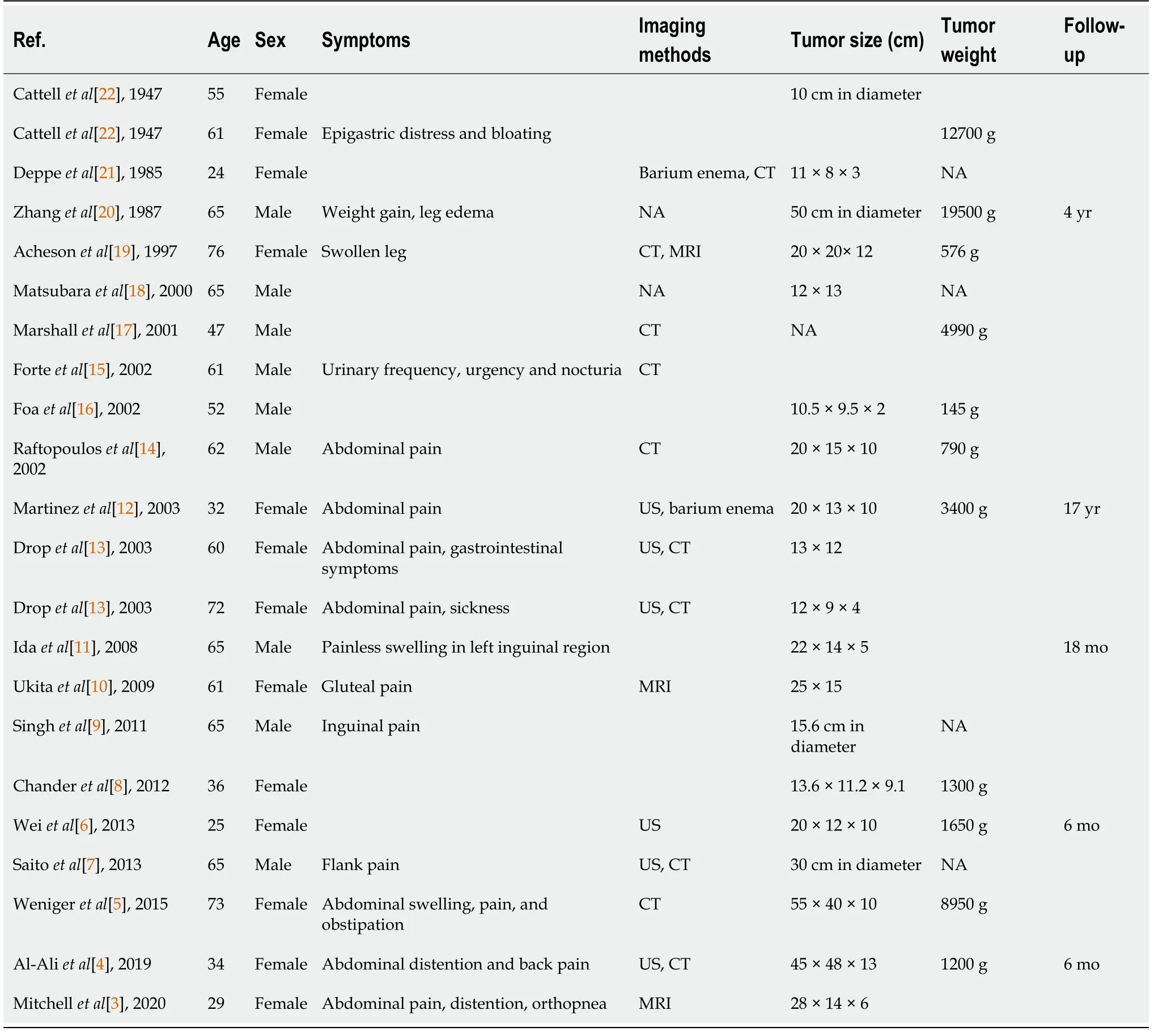
Lipomas are a category of benign tumors originating from well-differentiated adipocytes.Their predilection sites are subdermal tissues of the trunk and extremities[1,2].Retroperitoneal lipomas are a rare condition,with just 22 case reports describing the tumour in adults in the previous literature of PubMed since 1970[3-22].They account for 2.9% of primary retroperitoneal tumors,approximately 80% of which are known as malignant[6,23,24].Unlike subcutaneous lipomas,which are related to obesity,hyperlipidaemia,and injuries,retroperitoneal lipomas have an unknown aetiology[5,25-27].On account of the rarity and limited knowledge of these tumors,further reports and investigations are necessary.In this report,we describe the case of a 53-year-old postmenopausal woman who presented with a massive fatty retroperitoneal mass measuring 28.6 cm× 16.6 cm and weighing 7.126 kg.
I would like to say that this first date was ideal4. However, we had an argument in the book store while we were reading our zodiac signs. Not a very good sign for a first date. I will have to admit that by the time we left the book store and made it to the end of the block we were both laughing and holding hands like we were best friends forever.
CASE PRESENTATlON
Chief complaints
A 53-year-old postmenopausal woman who complained about progressive abdominal distention and aggravating satiety was referred to the department of gynaecology in our center.
History of present illness
The patient started to feel intensifying abdominal distention and satiety for the last 2 mo.She also found a significant increase in abdominal circumference and thinning of the limbs.The patient denied other discomforts,including fever,abdominal pain,nausea,and vomiting.The patient had been postmenopausal for 5 years and did not report abnormal vaginal bleeding.
History of past illness
The patient had a 10-year history of hypertension and took Loxone once per day,with stable control of blood pressure.The patient denied any history of diabetes,coronary heart disease,or malignancy.She also reported no drug allergy or other physical impairment.Additionally,the patient did not receive regular physical examinations,and the last medical examination had occurred more than 10 years prior.
Personal and family history
Due to the large size,the measurement of retroperitoneal tumors by preoperative imaging examinations can be inaccurate.Despite their typical presentations on CT and MRI,both imaging modalities may not rule out the possibility of WDLPS[29].Approximately 80% of retroperitoneal tumors appear to be malignant,most of which are softtissue sarcomas,a category of very uncommon neoplasms,with an overall incidence of 0.3% to 0.4% per 100000 people[10].Liposarcomas account for 41% of sarcomas,and the majority of the cases are malignant from the start.A few outliers arise from benign lipomas in the early stages[30].The final diagnosis of lipomas depends on histopathology.Tissue for pathology can be acquired by fine-needle aspiration or coreneedle biopsy,but it is nearly impossible to distinguish lipoma-like WDLPS and lipomas due to the limited tissue sample obtained by these methods for detecting atypia and hyperchromatic cells.Postoperative histopathology remains the gold standard for diagnosis.Histologic characteristics for WDLPS include mature adipocytes punctuated with big atypical hyperchromatic cells.However,WDLPS are likely to be misdiagnosed,because atypia may be localized,especially in deep lesions with tiny samples.Murine double minute(,located at 12q14-15)and cyclindependent kinase 4 gene are regularly amplified in WDLPS,which cannot be observed in benign lipomas.Hence,fluorescencehybridization has emerged as a promising method for differential diagnosis[11,16,18].
The patient reported no relevant clinical symptoms after the operation.During a series of follow-ups for 18 mo,the laboratory tests and imaging examinations were normal and indicated no signs of relapse.
Physical examination
Ultrasound is generally used for the initial diagnosis and screening of abdominal masses.Radiography,especially CT and magnetic resonance imaging(MRI),is a crucial diagnostic tool for further evaluation of retroperitoneal tumors.The characteristics of adipose tissues are consistent on CT and MRI,but they differ on ultrasonography depending on the physical properties and histologic types.The fatty content is the fundamental feature to identify fat-containing retroperitoneal tumors during imaging examinations.Typical lipomas appear as extensive hyperechoic lesions on ultrasound,while they appear as homogeneous fat-containing masses with thin septa on CT and MRI.Retroperitoneal lipomas are difficult to identify preoperatively since they mimic liposarcomas,which account for the majority of fat-containing retroperitoneal tumors.Liposarcomas present heterogeneous signal intensity and variable appearances on MRI and CT due to the varying subtypes,which included well-differentiated liposarcoma(WDLPS),dedifferentiated liposarcoma,myxoid/round cell liposarcoma,pleomorphic liposarcoma,and mixed liposarcoma.The increased vascularity in liposarcomas that present as low-intensity signals on T1-weighted images can be used for differentiation.However,both lipomas and WDLPS are accompanied by a large amount of fat and minimal soft tissue and have identical appearances on CT and MRI,making it hard to distinguish lipomas from well-differentiated liposarcomas preoperatively.
Laboratory examinations
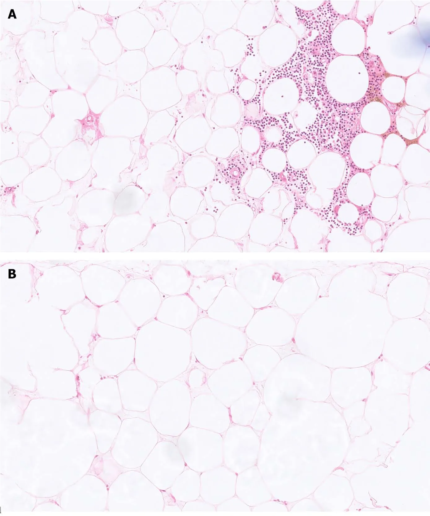
After 1 wk of uneventful hospitalization,the patient was discharged from the hospital with full recovery from her clinical symptoms.The final paraffin pathology showed that the tumor was composed of mature adipose tissues and hematopoietic cells,without cytologic atypia,and confirmed the diagnosis of multiple lipomas and multiple myelolipomas(Figure 4).
Imaging examinations
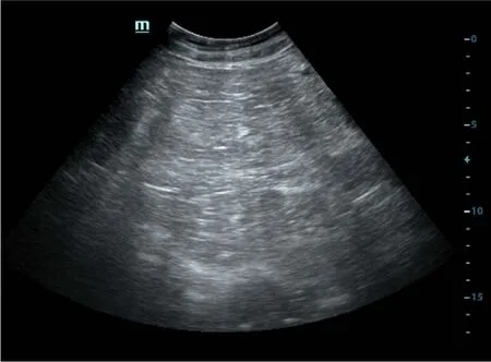
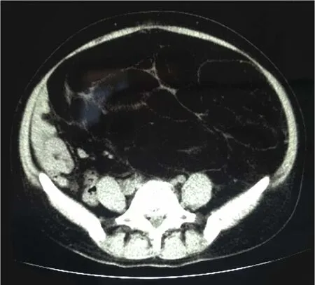
The patient had received a computed tomography(CT)plain scan at another hospital,and was re-evaluated by ultrasonography at our hospital.A massive hyperechoic mass,approximately 30 cm × 17 cm in size,was visualizedultrasound.The mass was clearly defined and had internal echogenicity,filled with stripe-like structures.Minimal blood signals were detected by colour Doppler ultrasound(Figure 1).A CT plain scan demonstrated a giant homogeneous mass mainly consisting of fatty tissue and thin septa.It measured 16.6 cm × 28.6 cm in volume and pushed the peritoneal contents,such as the bowel loops and uterus,to the right part of the abdomen(Figure 2).
FlNAL DlAGNOSlS
Based on the clinical manifestations,normal laboratory examinations,and imaging examinations indicating its adipose origin,the clinicians considered the mass to be a giant retroperitoneal lipoma.However,the possibility of malignancy cannot be overlooked due to its large size.
TREATMENT
We herein report a massive retroperitoneal lipoma,which consisted of multiple conventional lipomas and multiple myelolipomas.Retroperitoneal lipomas are rare mesenchymal-originated tumors.It was first reported in 1947[22],and since then,a total of 22 cases have been reported in adults sporadically(Table 1).The peak incidence of adult retroperitoneal lipomas occurs between the ages of 40 and 60,with no discernible gender predisposition.According to the morphologic characteristics,lipomas can be subdivided into conventional lipoma,fibrolipoma,angiolipoma,spindle cell lipoma,pleomorphic lipoma,and myelolipoma[7],of which,almost all myelolipomas have been identified inside the adrenal gland,with just about 50 cases of myelolipomas being identified in extra-adrenal locations,such as the retroperinoneum[28].The exact underlying aetiology of retroperitoneal lipomas is not well understood.Seeding after fibroid excision,exogenous hormone treatment,or chronic abnormalities in glucose homeostasis have all been blamed for these benign tumors.And genetic factors are thought to have an important role in adipocyte proliferation[5].
After choosing a vast quantity, which she divided between her sisters for she had made a heap of the wonderful dresses for each of them she opened the last chest, which was full of gold
After hospitalization,the patient received a series of laboratory examinations for testing liver and kidney function,faecal occult blood,blood coagulation factors,electrolyte panel,and tumor biomarkers.The laboratory findings fell within the normal range.
OUTCOME AND FOLLOW-UP
On the fourth morning, when my own odor had become indistinct from the smell of the horses, I woke to a beautiful sight. There was Sprite, standing19 on her own spindly legs, nudging me gently with her nose to get up.
DlSCUSSlON
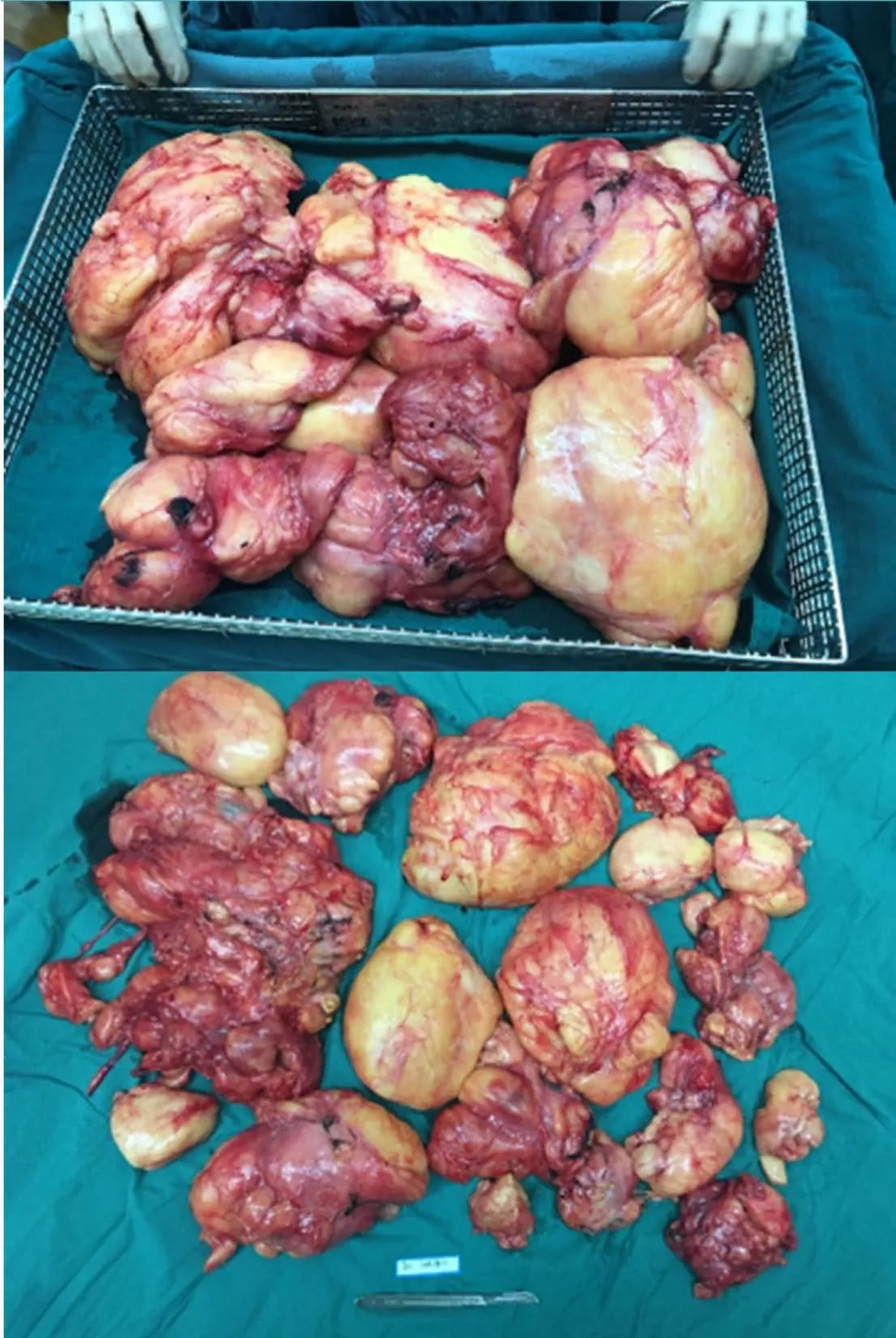
After completing the examinations and preoperative assessments,the patient underwent an exploratory laparotomy with the suspicion of malignancy,most likely retroperitoneal liposarcoma.During the operation,a bulky yellowish tumor originating from perirenal fatty tissues in the left retroperitoneal region was found to occupy the retroperitoneum.The uterus and adnexa were displaced by the mass.The mass adhered to the left psoas major muscle and wrapped around the left ureter,making it unfeasible to performresection.After carefully separating the left ureter,we performed tumor debulking and resection of the left adnexa,which was also tightly adhered to the tumor.The total weight of the mass was 7.126 kg(Figure 3).The frozen pathological results suggested that the mass mainly consisted of adipose tissues,and a retroperitoneal lipomatous tumor was considered.
Retroperitoneal tumors are often asymptomatic for a long period of time throughout their early clinical course,owing to the vast potential spaces in the retroperitoneum.Local compression of surrounding organs and tissues,which can manifest obstructive urinary/bowel symptoms such as stomach pain,fullness,early satiety,or lower extremity oedema,may occur once the tumors have grown to gigantic sizes.Theclinical presentations tend to be variable and nonspecific[4].Hence,imaging examinations play an essential role in the diagnosis of these lesions.
The patient’s height was 161 cm,and her weight was 60 kg(body mass index:23.3,within the normal range).The physical examination revealed a palpable giant abdominal mass reaching the xiphisternum with a rubbery consistency.Other clinical symptoms,including tenderness,rebound tenderness,and mobile turbid sounds,were found.
No noteworthy personal or family history was reported by the patient.
It is of great importance to discern tumor characteristics intraoperatively and make decisions about the resection extent subsequently.In cases of the pathological diagnosis of liposarcoma,resection with negative margins(R0)is crucial.If infiltrative growth is detected by frozen pathology,a broad excision should be performed.Surgeons should also tailor personalized surgical strategies for patients with important involved adjacent structures who are unsuitable for an entire resection.Commonly,removal of the involved structures is required[3,5].The prognosis and recurrence risk for patients with benign retroperitoneal lipomas are unclear due to the limited number of case reports.Patients are often recommended to receive regular clinical and radiologic follow-ups.
If you can, I will give you a robe of honour; but if you fail, I will order you to receive twelve strokes on your cheeks, and five-and-twenty on the soles of your feet, because you have been falsely called Selim the learned
Back home at the dinner table that evening, Amy was quiet. Her mother knew that things were not going well at school. That’s why Patti Hagadorn was happy to have some exciting news to share with her daughter.
CONCLUSlON
Retroperitoneal lipomas are rare benign tumors originating from adipose tissues and they tend to have large sizes.Imaging examinations,especially CT and MRI,are fundamental diagnostic tools for these tumors.Surgical resection is the main treatment method.resection is commonly required.Postoperative histopathology determines the final diagnosis,and immunohistochemical analysis could be useful in the differentiation of liposarcomas.Regular follow-ups are also required for the patients.
ACKNOWLEDGEMENTS
The authors are deeply grateful to Dr.Zhao H and Dr.Shi J for their help in reviewing the postoperative pathology.
 World Journal of Clinical Cases2022年5期
- World Journal of Clinical Cases的其它文章
- Subclavian artery stenting via ilateral radial artery access:Four case reports
- Neurothekeoma located in the hallux and axilla:Two case reports
- Diffuse invasive signet ring cell carcinoma in total colorectum caused by ulcerative colitis:A case report and review of literature
- Tacrolimus treatment for relapsing-remitting chronic inflammatory demyelinating polyradiculoneuropathy:Two case reports
- Aseptic abscess in the abdominal wall accompanied by monoclonal gammopathy simulating the local recurrence of rectal cancer:A case report
- Unusual magnetic resonance imaging findings of brain and leptomeningeal metastasis in lung adenocarcinoma:A case report