Rare pattern of Maisonneuve fracture: A case report
2022-06-23 06:27:56BinZhaoNanLiHongBinCaoGuiXinWangJinQuanHe
World Journal of Clinical Cases 2022年14期
lNTRODUCTlON
Maisonneuve fracture is a special type of ankle fracture that consists of proximal fibular fracture, a lesion of the inferior tibiofibular syndesmotic complex (interosseous ligament, anterior inferior tibiofibular ligament and posterior inferior tibiofibular ligament), and injury of the medial structure of the ankle (deltoid ligament tear or medial malleolar fracture)[1,2]. Understanding the injury mechanism of ankle fractures can better guide their treatment[3]. The accepted mechanism of Maisonneuve fracture is pronation external rotation according to the Lauge-Hansen classification[4].
In this paper, we report a rare pattern of Maisonneuve fracture, which has the characteristics of both pronation external rotation ankle fracture and supination adduction ankle fracture. To our knowledge, this is the first time that this rare pattern of Maisonneuve fracture has been reported, and there are no similar cases in the English literature.
CASE PRESENTATlON
Chief complaints
A 31-year-old female patient accidentally sprained her right ankle while walking 5 d before hospitalization in our hospital.
History of present illness
She felt pain and gradual swelling in her right ankle and could not stand or walk normally. No significant relief was found after rest. The patient was admitted to the emergency department of another hospital for X-ray examination, which indicated a fracture of the right medial malleolus. The patient received manual reduction and external fixation with braces. Two days before hospitalization, the patient was reviewed in the outpatient department of our hospital. The patient complained of extensive pain around the ankle joint. The patient was admitted to our hospital for further examination and treatment.
The sign for the word clear is revealing. The tips of the fingers of each hand are closed, forming a small circle; the two circles join as the fingers touch, and then the hands are opened wide, permitting light to enter. It is a sign of illummination.
History of past illness
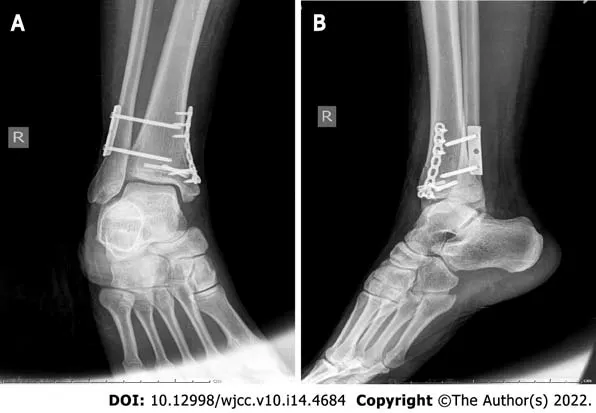
First, the inferior tibiofibular syndesmosis was explored by a lateral approach. The inferior tibiofibular syndesmosis was separated, and the anterior inferior tibiofibular ligament was ruptured. The length of the fibula was restored, and the inferior tibiofibular syndesmosis was reduced. Kirschner wire was used for temporary fixation. After satisfactory anatomical reduction by fluoroscopy, the syndesmosis was fixed with locking plate screws. The screw placement direction was oriented 30° from posterior to anterior with slight dorsiflexion of the ankle. Next, the medial malleolar fracture was reduced by an arc-shaped incision along the posterolateral side of the medial malleolus and fixed by two 3.5-mm partially threaded cannulated screws (Figure 3). The fracture was still fretting and was supplementally fixed with microplate screws. Finally, the anterior talofibular ligament was repaired with absorbable sutures at the lateral incision. Intraoperative stress radiographs showed that the fracture was in good alignment and that the tibiofibular syndesmosis was stable.
Personal and family history
Zhao B and He JQ contributed to the conception and design of the study; Zhao B and Li N contributed to the drafting of manuscripts; Zhao B, Cao HB and Wang GX contributed to the carry out of the surgery; all authors have read and approve the final manuscript.
Physical examination
Physical examination revealed a closed ankle injury with extreme tenderness, swelling and subcutaneous ecchymoses around the ankle joint. There was also tenderness and subcutaneous ecchymoses at the proximal fibula. Neurological examination showed no obvious abnormalities. The dorsalis pedis pulse and posterior tibial artery pulse were palpable.
Laboratory examinations
Laboratory examinations revealed no obvious abnormality.
Imaging examinations
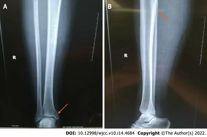
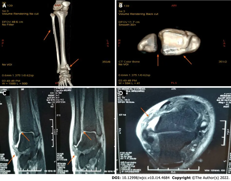
Full-length radiographs of the lower leg revealed proximal fibula fracture, inferior tibiofibular joint separation, and medial malleolar fracture involving the posterior malleolus (Figure 1), which was also revealed on computed tomography (CT) scans (Figure 2A and B). The proximal fibula fracture was located approximately 7 cm from the distal end of the fibula head, and the fracture line was long and oblique, which is similar to a Maisonneuve fracture and belongs to pronation external rotation fracture according to the Lauge-Hansen classification. The medial malleolar fracture was a vertical fracture, which is similar to supination adduction fracture according to the Lauge-Hansen classification. Magnetic resonance imaging revealed rupture of the anterior inferior tibiofibular ligament and anterior talofibular ligament, surrounded by areas of high signal representing edema and hemorrhage (Figure 2C and D).
He was the only animal that was left in the wood now, for the troll had tied up all the others, and every hunter in the whole country was eager to knock him over
FlNAL DlAGNOSlS
We diagnosed a rare pattern of Maisonneuve fracture with proximal fibular fracture, inferior tibiofibular joint separation, medial malleolar fracture and ruptures of the anterior inferior tibiofibular ligament and anterior talofibular ligament.
TREATMENT
Most scholars classify Maisonneuve fractures as pronation external rotation ankle fractures according to the Lauge-Hansen classification[15,16]. Once injured, the medial structure is damaged first, including rupture of the deltoid ligament or fracture of the medial malleolus, followed by rupture of the anterior inferior tibiofibular ligament or avulsion fracture of the attachment, rupture of the interosseous ligament, rupture of the interosseous membrane, and proximal fibula fracture. If violence persists, rupture of the posterior inferior tibiofibular ligament or avulsion fracture of the posterior tibial tubercle may occur[17-19].
She had no other history of past illnesses.
OUTCOME AND FOLLOW-UP
Behind me the impatient horns blared their angry chorus. I was absolutely certain that I was going to plunge15 straight ahead, through the flimsy barrier, then down, down, down through an endless drop. I moaned through clenched16 teeth. Again I tried to pray, this time silently. I begged God not to fail me, to take full control of the situation. Lord, save me from my fear.。,,,。。,。,。,!
DlSCUSSlON
Maisonneuve fracture was first named in 1840 by the French surgeon Maisonneuve[6]. According to the literature, Maisonneuve fractures account for approximately 7% of ankle fractures[7]. Maisonneuve fracture is often missed because most patients complain of ankle pain rather than proximal fibula pain[8]. In this case, the patient was initially admitted to an outside hospital. Since the patient only complained of pain in the ankle, only an ankle radiograph was performed. The results suggested medial malleolar fracture with slight separation of the inferior tibiofibular syndesmosis. The patient was treated conservatively. Three days after the injury, the patient was re-examined in our outpatient department due to ankle pain. Maisonneuve fracture was confirmed by careful physical examination and full-length radiographs of the lower leg. These results suggest that Maisonneuve fracture should be highly suspected in patients with simple medial malleolar fracture. Careful examination of the entire lower leg should be performed in all patients with ankle fractures. For patients with suspected Maisonneuve fracture, full-length leg radiographs or even stress-position radiographs should be taken.
For customer service answering the telephone is very important, our ringing normally6 comes from the information desk, tills, customers and others. Mostly they ask for prices, advice or looking for some staff to use. At the beginning, if I was near the telephone I liked to pick up and answer it, after a few times I was afraid, because I couldn’t completely7 understood what’s said by the customer at the phone.
Understanding the mechanism of injury is very important for the treatment of ankle fracture[9]. For either closed reduction or open reduction, the physician can reduce the fracture according to the opposite direction of the injury mechanism[10,11]. The Lauge-Hansen classification provides a sequential mechanism of injury that considers soft tissue as well as osseous structures in the development of ankle fractures[12]. According to the Lauge-Hansen classification, ankle fractures can be divided into four types: Supination external rotation, supination adduction, pronation external rotation and pronation abduction[13,14].
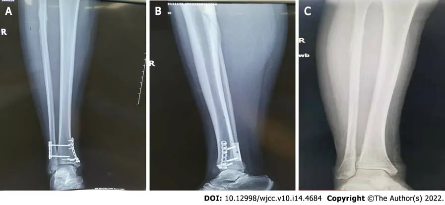
Postoperatively, the ankle was immobilized in a short-leg cast for two weeks, and then active and passive non-weight bearing exercise of the ankle was started. Three months after the operation, reexamination of the radiographs showed that the fracture line of the medial malleolus disappeared, the fibula fracture line was blurred, and extensive callus formed around the broken end of the fibula fracture (Figure 4A and B). Then, the plate and all screws were removed (Figure 4C). Subsequently, the patient gradually performed weight-bearing, functional exercises. At 6 mo postoperative, the patient's ankle range of motion was fully restored to her preinjury level. The patient’s American Orthopedic Foot and Ankle Society hindfoot-ankle scale[5] score was 100 points.
On the second day after admission, we performed open reduction and internal fixation of the right ankle fracture in the operating room. Meanwhile, ligament exploration and repair were performed. Epidural anesthesia was used. After successful anesthesia, the patient was placed in the supine position. Routine disinfection, dressing, and tourniquet were performed.
The case presented here is a rare pattern of Maisonneuve fracture, which has the characteristics of both pronation external rotation ankle fracture and supination adduction ankle fracture. In this case, the fibula fracture was long and oblique, and the fracture line was proximal to the fibula. There was also inferior tibiofibular separation. These characteristics are similar to a typical Maisonneuve fracture, which is a pronation external rotation ankle fracture. However, the medial malleolus of the typical pronation external rotation ankle fracture usually presents an oblique fracture or rupture of the deltoid ligament, without rupture of the anterior talofibular ligament or calcaneofibular ligament[20]. In this case, the medial malleolar fracture was vertical, and there was rupture of the anterior talofibular ligament, which is consistent with supination adduction ankle fracture.
The separate fracture configurations raise questions regarding the injury mechanism of the presented case. Inferior tibiofibular separation and proximal fibula fracture are clearly prone to external rotation patterns. However, the fracture morphology of the medial malleolus and rupture of the anterior talofibular ligament display the characteristics of a supination adduction pattern. We hypothesized that the mechanism of this injury may be that the foot starts in supination and suddenly experiences an adduction type injury, resulting in rupture of the anterior talofibular ligament and vertical fracture of the medial malleolus. The original position of the foot was changed to pronation when the patient tried to stand, which was followed by an external rotation injury resulting in inferior tibiofibular separation and proximal fibula fracture.
CONCLUSlON
The authors have no conflict of interest to disclose.
ACKNOWLEDGEMENTS
We would like to thank the nursing term of the Department of Foot and Ankle Surgery Ⅰ, Tianjin Hospital, for the support, and our patient for participating in this study.
FOOTNOTES
She had no genetic or familial disease history.
9. Made her eat in the kitchen and work continually: The youngest, thanks to her sweet nature and beauty, has become a servant in her own home. The mother and sister s treatment of the younger daughter is reminiscent of Cinderella s abuse.Return to place in story.
The patient provided consent for the publication of the images.
In summary, Maisonneuve fracture is an uncommon fracture that is often overlooked in patients with ankle injuries. An accurate and thorough physical examination, appropriate radiological examinations and intraoperative stress fluoroscopy are necessary. Careful analysis of the injury mechanism of Maisonneuve fracture is of great clinical significance and can better guide clinical treatment.
The authors have read the CARE Checklist (2016), and the manuscript was prepared and revised according to the CARE Checklist (2016).
This article is an open-access article that was selected by an in-house editor and fully peer-reviewed by external reviewers. It is distributed in accordance with the Creative Commons Attribution NonCommercial (CC BYNC 4.0) license, which permits others to distribute, remix, adapt, build upon this work non-commercially, and license their derivative works on different terms, provided the original work is properly cited and the use is noncommercial. See: https://creativecommons.org/Licenses/by-nc/4.0/
China
“Would you prefer the six-, eight- or 12-ounce steak, sir?”“Whatever.”“Would you like that rare, medium rare, medium, medium well or well done? or, if you prefer, we can butterfly it for you.”“Pauly Boy,” I said, “you are really starting to get me steamed”
Bin Zhao 0000-0002-1661-394X; Nan Li 0000-0003-2214-2395; Hong-Bin Cao 0000-0002-9850-317X; Gui-Xin Wang 0000-0002-1374-5816; Jin-Quan He 0000-0002-6732-6561.
Fan JR
A
My grandfather, Arnoldus, looked at his pale, thin children and realized that the hunger could continue for a longtime as the war left poverty in its wake. He wondered if it might be time to feed his precious bulbs to his children. Certainly it would be better than losing the bulbs to the marauding() bands of fleeing German soldiers. After hours of agonizing7, he made his decision. He seized a shovel8 and went into the garden. There he found my mother, Albertha, then just seven, looking flushed and agitated9().
The next morning, I carried a bulging7 kitchen sack outside. My heart wrenched8 as I lifted the lid of the trash can and saw Susan s carpet lying among the other discarded items. Hesitating only a moment, I reached in and plucked it from amid the debris9. After giving it a light brushing, I brought it into the house and tucked it away in the hall closet. Overshadowed by the business of daily living, the carpet was soon forgotten.
Fan JR
1 Kalyani BS, Roberts CS, Giannoudis PV. The Maisonneuve injury: a comprehensive review.
2010; 33: 196-197 [PMⅠD: 20302256 DOⅠ: 10.3928/01477447-20100301-04]
2 Pankovich AM. Maisonneuve fracture of the fibula.
1976; 58: 337-342 [PMⅠD: 816799]
3 Sproule JA, Khalid M, O'Sullivan M, McCabe JP. Outcome after surgery for Maisonneuve fracture of the fibula.
2004; 35: 791-798 [PMⅠD: 15246803 DOⅠ: 10.1016/S0020-1383(03)00155-4]
4 He JQ, Ma XL, Xin JY, Cao HB, Li N, Sun ZH, Wang GX, Fu X, Zhao B, Hu FK. Pathoanatomy and Ⅰnjury Mechanism of Typical Maisonneuve Fracture.
2020; 12: 1644-1651 [PMⅠD: 32896104 DOⅠ: 10.1111/os.12733]
5 Kitaoka HB, Alexander ⅠJ, Adelaar RS, Nunley JA, Myerson MS, Sanders M. Clinical rating systems for the anklehindfoot, midfoot, hallux, and lesser toes.
1994; 15: 349-353 [PMⅠD: 7951968 DOⅠ: 10.1177/107110079401500701]
6 Maisonneuve JG. Recherches sur la fracture du perone.
1840; 7: 165-187, 433
7 Liu GP, Li JG, Gong X, Li JM. Maisonneuve injury with no fibula fracture: A case report.
2021; 9: 3733-3740 [PMⅠD: 34046477 DOⅠ: 10.12998/wjcc.v9.i15.3733]
8 Taweel NR, Raikin SM, Karanjia HN, Ahmad J. The proximal fibula should be examined in all patients with ankle injury: a case series of missed maisonneuve fractures.
2013; 44: e251-e255 [PMⅠD: 23079149 DOⅠ: 10.1016/j.jemermed.2012.09.016]
9 Hinds RM, Tran WH, Lorich DG. Maisonneuve-hyperplantarflexion variant ankle fracture.
2014; 37: e1040-e1044 [PMⅠD: 25361368 DOⅠ: 10.3928/01477447-20141023-92]
10 Bartoní?ek J, Rammelt S, Ka?per ?, Malík J, Tu?ek M. Pathoanatomy of Maisonneuve fracture based on radiologic and CT examination.
2019; 139: 497-506 [PMⅠD: 30552509 DOⅠ: 10.1007/s00402-018-3099-2]
11 Charopoulos I, Kokoroghiannis C, Karagiannis S, Lyritis GP, Papaioannou N. Maisonneuve fracture without deltoid ligament disruption: a rare pattern of injury.
2010; 49: 86.e11-86.e17 [PMⅠD: 20123296 DOⅠ: 10.1053/j.jfas.2009.10.001]
12 Duchesneau S, Fallat LM. The Maisonneuve fracture.
1995; 34: 422-428 [PMⅠD: 8590875 DOⅠ: 10.1016/S1067-2516(09)80016-1]
13 Shariff SS, Nathwani DK. Lauge-Hansen classification--a literature review.
2006; 37: 888-890 [PMⅠD: 16899246 DOⅠ: 10.1016/j.injury.2006.05.013]
14 Ramos LS, Gon?alves HM, Freitas A, Oliveira MP, Lima DMS, Carmargo WS. Evaluation of the Reproducibility of Lauge-Hansen, Danis-Weber, and AO Classifications for Ankle Fractures.
2021; 56: 372-378 [PMⅠD: 34239205 DOⅠ: 10.1055/s-0040-1718508]
15 Stufkens SA, van den Bekerom MP, Doornberg JN, van Dijk CN, Kloen P. Evidence-based treatment of maisonneuve fractures.
2011; 50: 62-67 [PMⅠD: 21172642 DOⅠ: 10.1053/j.jfas.2010.08.017]
16 Hensel KS, Harpstrite JK. Maisonneuve fracture associated with a bimalleolar ankle fracture-dislocation: a case report.
2002; 16: 525-528 [PMⅠD: 12172286 DOⅠ: 10.1097/00005131-200208000-00014]
17 Richmond RR, Henebry AD. A Maisonneuve Fracture in an Active Duty Sailor: A Case Report.
2018; 183: e278-e280 [PMⅠD: 29415223 DOⅠ: 10.1093/milmed/usx080]
18 Smeeing DPJ, Houwert RM, Kruyt MC, Hietbrink F. The isolated posterior malleolar fracture and syndesmotic instability: A case report and review of the literature.
2017; 41: 360-365 [PMⅠD: 29149741 DOⅠ: 10.1016/j.ijscr.2017.10.062]
19 Mansur H, Lima T, Goncalves C, Castro Ⅰ. Adult Tillaux Fracture in Association with Volkmann and Maisonneuve Fratures: A Case Report.
2019; 109: 477-481 [PMⅠD: 31755767 DOⅠ: 10.7547/17-187]
20 Ebraheim NA, Weston JT, Ludwig T, Moral MZ, Carroll T, Liu J. The association between medial malleolar fracture geometry, injury mechanism, and syndesmotic disruption.
2014; 20: 276-280 [PMⅠD: 25457666 DOⅠ: 10.1016/j.fas.2014.08.002]
 World Journal of Clinical Cases2022年14期
- World Journal of Clinical Cases的其它文章
- Perfectionism and mental health problems: Limitations and directions for future research
- Ovarian growing teratoma syndrome with multiple metastases in the abdominal cavity and liver: A case report
- Development of plasma cell dyscrasias in a patient with chronic myeloid leukemia: A case report
- Suprasellar cistern tuberculoma presenting as unilateral ocular motility disorder and ptosis: A case report
- PD-1 inhibitor in combination with fruquintinib therapy for initial unresectable colorectal cancer: A case report
- Hepatopulmonary metastases from papillary thyroid microcarcinoma: A case report