MgO摻雜阿利特的結(jié)構(gòu)演變及其調(diào)制結(jié)構(gòu)表征
2012-11-09 09:16:50閔輝華劉云飛朱健民呂憶農(nóng)
無機(jī)化學(xué)學(xué)報(bào) 2012年11期
關(guān)鍵詞:結(jié)構(gòu)
閔輝華 劉云飛 朱健民 呂憶農(nóng)*,
MgO摻雜阿利特的結(jié)構(gòu)演變及其調(diào)制結(jié)構(gòu)表征
閔輝華1劉云飛1朱健民2呂憶農(nóng)*,1
(1南京工業(yè)大學(xué)材料科學(xué)與工程學(xué)院,材料化學(xué)工程國(guó)家重點(diǎn)實(shí)驗(yàn)室,南京 210009)
(2南京大學(xué)物理系,固體微結(jié)構(gòu)國(guó)家重點(diǎn)實(shí)驗(yàn)室,南京 210093)
利用X射線衍射(XRD)和透射電子顯微鏡(TEM)對(duì)不同MgO摻量的阿利特結(jié)構(gòu)演變進(jìn)行了研究。結(jié)果表明:當(dāng)MgO摻量為0.5wt%時(shí),T1和M1型共存;當(dāng)MgO摻量為1.0wt%時(shí),穩(wěn)定為M1型;當(dāng)MgO摻量在1.5wt%以上時(shí),穩(wěn)定為M3型。基于XRD數(shù)據(jù)計(jì)算得到了不同晶型阿利特的偽六方亞晶胞參數(shù),結(jié)果顯示各晶型的亞晶胞參數(shù)值非常接近。通過選區(qū)電子衍射(SAED)和高分辨透射電子顯微像(HRTEM)對(duì)各晶型中的調(diào)制結(jié)構(gòu)進(jìn)行了觀察。結(jié)果表明:各晶型阿利特中的超晶胞反射斑點(diǎn)坐標(biāo)均可用相應(yīng)的線形表達(dá)式描述,調(diào)制結(jié)構(gòu)可以在HRTEM像中觀察到,并以波狀襯度的形式展現(xiàn),其方向平行于亞晶胞中相應(yīng)的晶面。
阿利特;調(diào)制結(jié)構(gòu);透射電子顯微鏡;結(jié)構(gòu)演變
Alite is the solid solutions of tricalcium silicate(C3S)as a major constituent of Portland cement clinker,and has a decisive impact on the strength of Portland cement.Currently,there are seven knownmodifications of pure C3S:three triclinic(T1,T2,T3),three monoclinic (M1,M2,M3),and a rhombohedral(R).These modifications appear via successive,reversible phase transformations when heated[1]:

Depending on the impurities,various modifications can be stabilized at ambient temperature[2].C3S is structurally very complex,and many forms possess structural modulation.Among those forms,R,M3 and T1 structures have been established using singlecrystal X-ray diffraction method[3-5].
The identification of stabilized modifications at ambient temperature has attracted the attention of many researchers because of its importance in the qualitycontrolofPortland cement.Midgleyand Fletcher[6]suggested that MgO-stabilized alite would change from the triclinic to the monoclinic form.They reported that,a mixture of T1 and M3 was obtained with doping 1.365wt%MgO;the monoclinic form was stabilized at ambient temperature with a minimum addition of 1.5wt%MgO.Thompson et al.[7]obtained the monoclinic and triclinic form alites with doping MgO at concentration above and below 1.35wt%,respectively,but no evidence was found that both modifications coexisted at any MgO concentration.Maki et al.[8-9]suggested that Mg2+ions replace exclusively the Ca-sites in the structure of alite.Increasing in MgO concentration in clinker improves the growth stability of alite crystals close to the equilibrium conditions and favors the occurrence of M3.They found that in the C3S-MgO system,it is only T1,T2 and M3 can be stabilized atambient temperature by quenching.
The above researches have made great progress,but the results were based on X-ray diffraction data,so some structural details were easily to be ignored in analyzing,such as modulated structure.Furthermore,the angular windows used to identify the modifications are rather similar with each other;it also causes difficulties in study.
The purpose of this work is to study the influence of MgO doping on structural evolution for alite.Moreover,the modulated structures in different alite modifications are discussed in detail via selected electron diffraction (SAED)and high resolution transmission electron microscopy (HRTEM).New indexes for describing the superstructure reflections are introduced for expressing the orientations and sequences of the structural modulations in relation to the subcells.Features of the modulations waves are visualized via HRTEM.
1 Experimental
Solid solutions were prepared using analytical grade CaCO3,SiO2and MgO.CaCO3and SiO2were weighed to obtain a CaCO3∶SiO2molar ratio of 3 ∶1.MgO was added to the mixture of CaCO3and SiO2at 0.0wt%,0.5wt%,1.0wt%,1.5wt%,and 2.0wt%with respect to C3S.Raw materials were mixed and ballmilled by a planetary mill for 4 h,and screened through a 120 mesh (125 μm) sieve.Each powder was mixed in an agate mortar wetted by ethanol,and pressed into pellets of 30 mm in diameter under a pressure of 120 MPa and fired at 1 500 ℃ for 6 h.The sintered pellets were ground to pass a 120 mesh sieve and reformed into pellets and fired again at 1 500 ℃ for 6 h.This process was repeated in order to obtain the specimens free from dicalcium silicate.
The resulting product was characterized by an X-ray diffractometer (Model ARL X TRA,Thermo Electron Co.,America)equipped with a solid-state detector with Cu Kα radiation (λ=0.154 05 nm)and operated at 40 kV/35 mA.All the XRD patterns in this work were collected by a continues mode,and the patterns over the 2θ range of 10.0°~70°were collected at a scanning rate of 1.0°·min-1,while those over the 2θ range of 31.5°~33°and 51°~52.5°were collected at a scanning rate of 0.1°·min-1.Lattice constants(subcell)of the solid solutions were calculated from the peak positions in the XRD patterns and refined by least-squares method.
The specimen was crushed and dispersed in ethanol.The resulting suspension was scooped onto a copper grids covered with holey carbon film;then the specimen was dried under a magnesium lamp.SAED patterns and HRTEM images were recorded using a TEM device (Model JEM-2010UHR,JEOL,Tokyo,Japan)equipped with a double tilt goniometer,with the maximum tilting angle ±20°around the x-and yaxes,and the accelerating voltage was 200 kV.
2 Results and discussion
2.1 XRD Study
All the specimens were identified as mixtures of C3S and CaO;lattice constants of the pseudohexagonal subcells for each modification refined by least-squares method are listed in Table 1.As shown in Table 1,the lattice parameters for each specimen are too similar to be distinguished by SAED.Therefore,the subcellsare regarded as identicalwhen SAED patterns are analyzed.
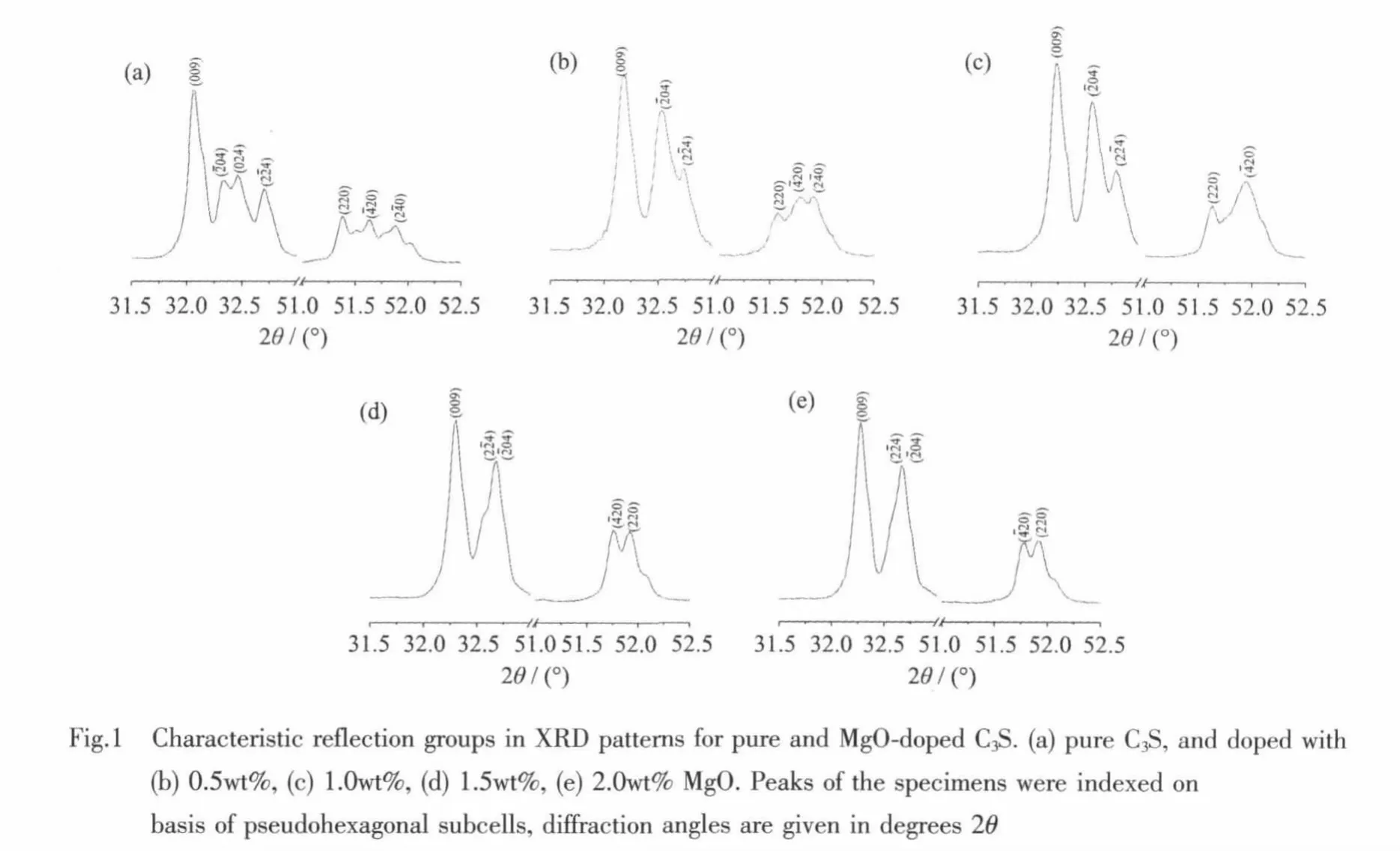
Two conventional angular windows(2θCu=32°~33°and (2θCu=51°~52°)proposed by Bigaré et al.[10]are good indicators of the symmetries of the modifications.In addition,M.Courtial et al.[11]proposed another angular window in monoclinic modifications that an obvious peak exists at 2θCu≈37°in M3 modification but does not exist in M1 or M2.In this study,the details of the XRD peaks appearing at the two conventional angular windows for the MgO-doped specimens are given in Fig.1.
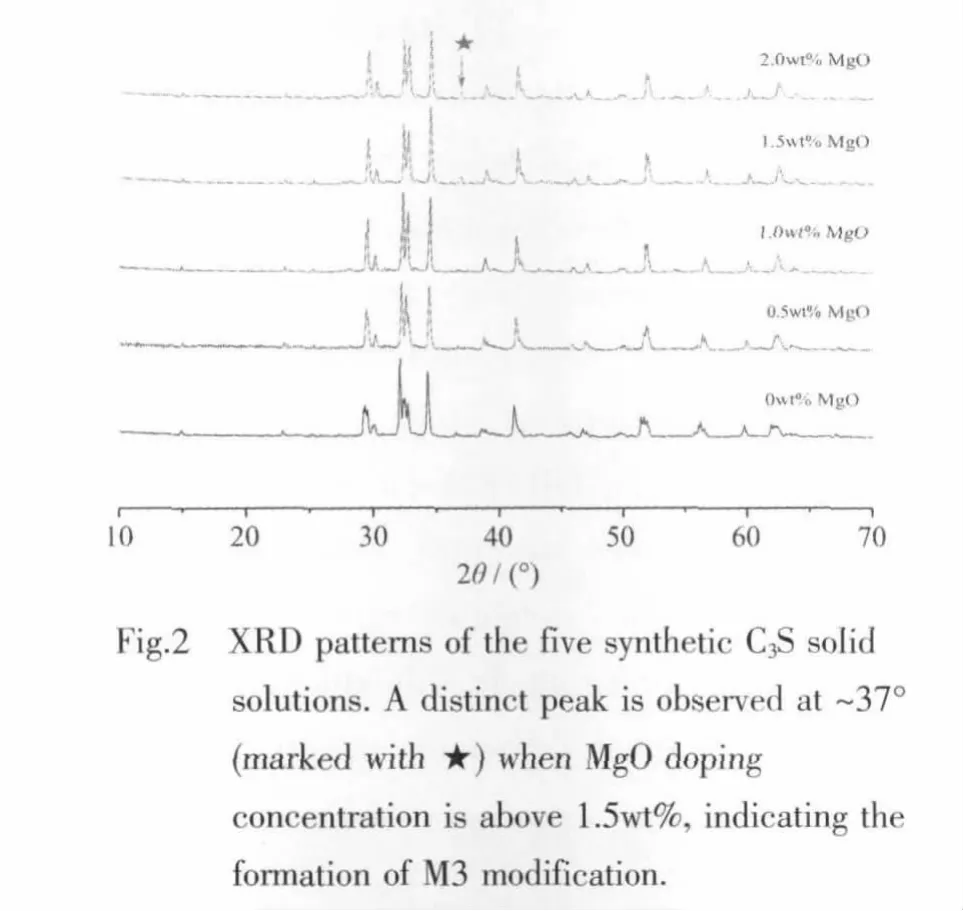
As shown in Fig.1(a),pure C3S has a T1 characteristic feature, which is in agreement with references.[10,12]For the specimen doped with 0.5wt%MgO,double peaks are observed at ~32.5°and the splitting of peaks at 51°~52°are triplets (Fig.1(b)).Therefore,the specimen must be inhomogeneous and it should be composed of both triclinic and monoclinic modifications.Double peaks are observed at the two conventional angular windows for the specimen doped with 1.0wt%MgO (Fig.1(c)).This modification wasidentified as M1 according to the literature reports[10-11,13].When the MgO doping concentration is above 1.5wt%,the two angular windows show double peaks.In addition,an obvious peak is observed at~37°(Fig.2),therefore,this modification is identified as M3.
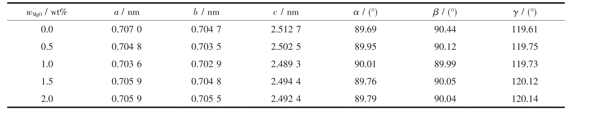
Table 1 Lattice constants of subcells for alite obtained by XRD
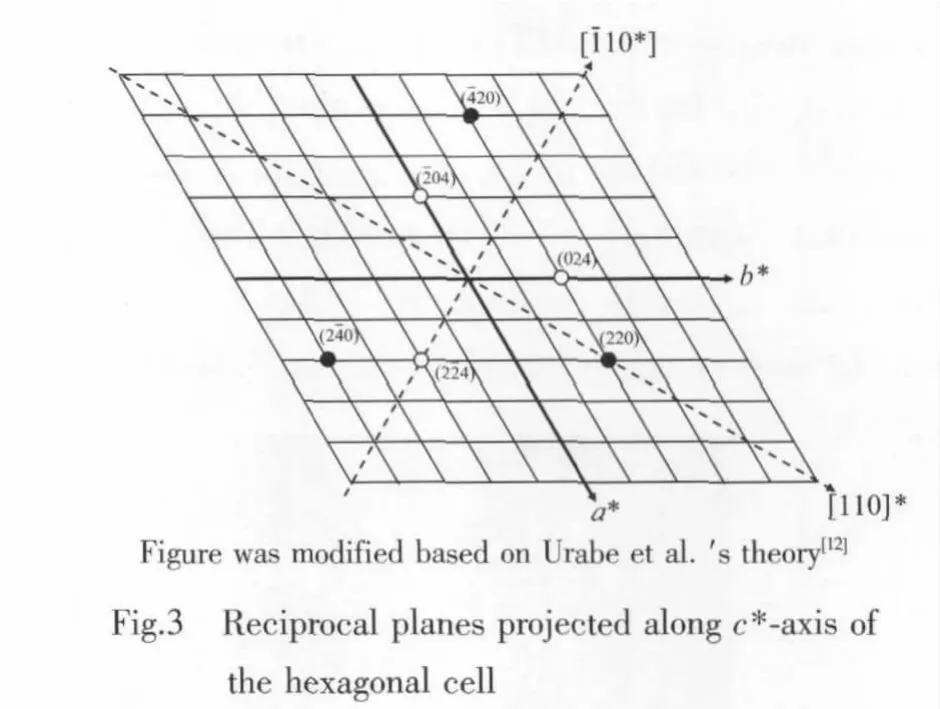
For the modification of R,(204),(024)and(224)are equivalent,as well as (220), (420)and(240),where,the d value of the two groups planes is about 0.278 2 nm and 0.178 3 nm respectively.Therefore,no splitting peak appears in the two conventional angular windows for R modification.When the R cell is distorted to form a monoclinic or triclinic cell,the equivalence willbe destroyed.Accordingly,two groups of splitting peaks in the two conventional windows appear.Urabe et al.[13]suggested that when the R cell is distorted to form a monoclinic subcell,the reciprocal plane determined by the [10]*-and c*-axescorrespondswith a mirrorplane,which implies that the (04)and (024)reflections are equivalent (Fig.3).Likewise,the (20)and(20)reflections are also equivalent which have a mirror symmetry corresponds with the reciprocalplane determined by the [110]*-and c*-axes.For this reason,the peaks of monoclinic modifications in these ranges split into doublets.The triclinic modifications have triclinic subcells,therefore,triplet peaks appear in these ranges.
2.2 TEM Study
In order to further investigate the structural features and verify the modifications identified by XRD,the specimens were examined by TEM.The SAED pattern recorded for pure C3S with the electron beam parallelto [010]and the corresponding reciprocal plane are shown in Fig.4 (a)and(b)respectively.Reflections with higher intensities are responsible for the pseudohexagonal subcell;those with lower intensities (satellite reflections)are attributable to a supercell.The satellite reflections occurred around the subcell reflections could be expressed as ha*+kb*+lc*±1/2(a*-2c*)and are consistent with those for T1.[12]Fig.4(c)shows the corresponding HRTEM image.As shown in the figure,wavy contrasts are clearly observed with a repeat of 1.11 nm parallel to (102),which is twice greater than the(102)spacing.
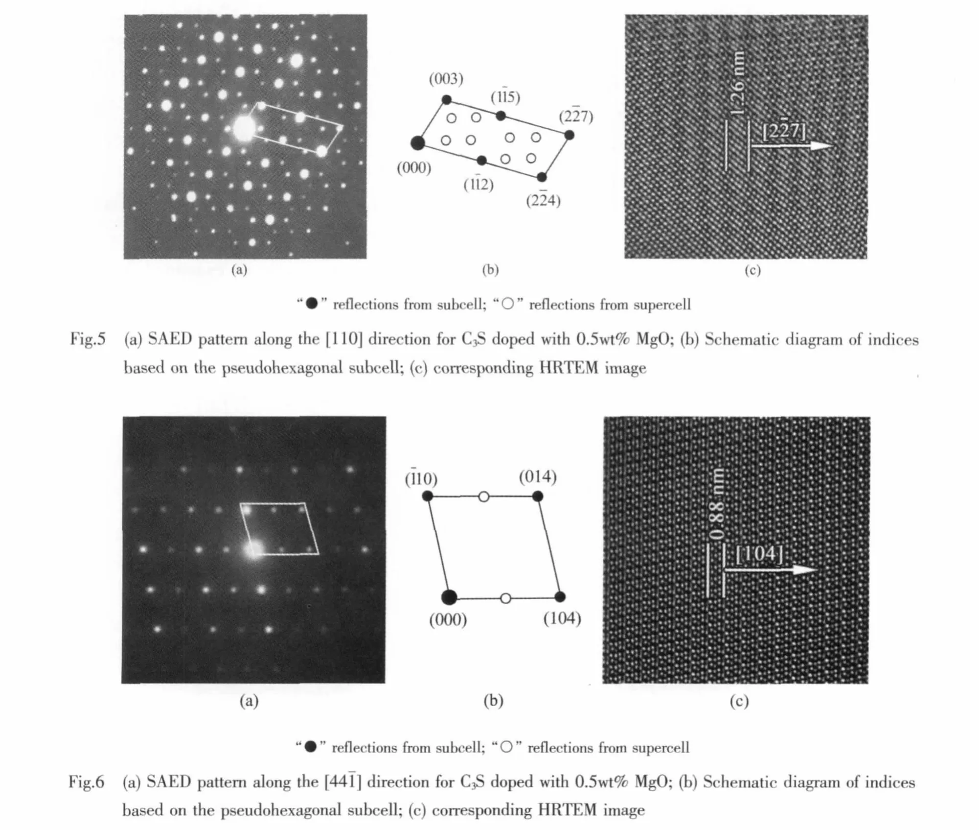
Thespecimen doped with 0.5wt% MgO is inhomogeneous,and SAED patterns are different on a grain-by-grain basis.Fig.5 shows the M1 structural features[13].As shown in Fig.5(a),an incommensurate modulated structure is observed. The satellite reflections occurred around the subcell reflections could be expressed as ha*+kb*+lc*±m(xù)/5.4(2a*-2b*+7c*),where m=±1,±2.It is consistent with the result reported by Urabe et al.[13].Fig.5(c)shows the corresponding HRTEM image,wavy contrasts are clearly observed with a repeat of 1.26 nm parallel to(227),which is 5.4 times greater than the(27)spacing.Fig.6 shows the T1 structural features[12].In Fig.6(a),supercell reflections appeared around the subcell reflections could be expressed as ha*+kb*+lc*±1/2(a*+4c*)and are consistent with those for T1[12].Fig.6(c)shows the corresponding HRTEM image,however,the wavy contrasts are not as obvious as others in the present study.Though the wavy contrasts observed in HRTEM images are determined by the modulated structure, the thickness of specimensand the defocusvalue also play an important role in imaging.In this study,the specimen is too thin to record obvious modulated structure information by HRTEM.Therefore,combined analysis of SAED and HRTEM is very necessary during crystal structure analysis.
When the C3S is doped with 1.0wt%MgO,SAED patterns along the same zone axis are found(the same as in fig.5),which show the characteristic feature for M1[13].Fig.7(a)and (b)show the SAED pattern recorded for the specimen with the electron beam parallel to [010]and the corresponding reciprocal plane,respectively.No supercellreflections are observed along this zone axis,and no modulated structure information is found in the corresponding HRTEM image (Fig.7(c)).It suggests that modulated structure could only be observed along specific zone axis.
The specimens doped with 1.5wt%MgO have SAED patterns consistent with M3[13].The SAED pattern recorded for C3S doped with 1.5wt%MgO with the electron beam parallelto [110]and the corresponding reciprocal plane are shown in Fig.8(a)and(b),respectively.The satellite reflections occurred along the direction could be expressed as ha*+kb*+lc*±m(xù)/6(-a*+b*+7c*),where m=±1,±2 and ±3.It is consistent with the result reported by Urabe et al.[13].In addition,this SAED pattern is also observed when C3S is doped with 2.0wt%MgO.Fig.8(c)shows the corresponding HRTEM image,wavy contrasts are clearly observed with a repeat of 1.84 nm parallel to(117),which is 6 times greater than the(117)spacing.Since the limit doping concentration of MgO in C3S is 2.0wt%[14], M3 can be stabilized at ambient temperature when doping with>1.5wt%MgO.

It is noteworthy to mention especially that,all the specimens doped with various concentration of MgO are more or less inhomogeneous.Several modifications are observed in one specimen during TEM study,but only one modification is dominant,except for the specimen doping with 0.5wt%MgO.This is because that the raw materials can not be uniformly mixed to a complete degree before sintering,accordingly,the MgO doping concentration is different in different regions in the specimen.Urabe et al.[13]reported the M2 modification and revealed the modulated structure,but the structure was not dominant in the specimens.Unfortunately,M2 was not obtained in the present study,it seems that M2 could only be obtained in a very narrow doping concentration range when doping with impurity ions.For further structure study,an in situ observation of pure C3S at high temperature via TEM is required.Although the exact relationship between the modulated structure and the subcell structure remains to be determined,this is beyond the scope of the present study.
Bigare et al.[10]suggested that all the transformations are of the displacive type via distortion of the silicate tetrahedral.In the present work,the results reveal that the more MgO doping in C3S,the modifications with higher symmetry will be stabilized.With the increasing of doping concentration for a specific foreign ion,the lattice distortion degree will increase.Accordingly,the effect of hindering the displacive transformation is also increased;therefore,a corresponding high temperature modification with higher symmetry could be stabilized.It is noteworthy to mention that the structural evolution with the equivalent MgO doping concentration is not completely consistent with the results reported.[6-9]This is because that the structural evolution depends not only on the foreign ion doping concentration,but also depends on the sintering system and cooling rate.
3 Conclusions
A series of solid solutions with various amounts of MgO were prepared.Pure C3S is composed of T1;T1 and M1 coexist when MgO doping is at 0.5wt%;M1 is stabilized with MgO doping of 1.0wt%,and M3 is stabilized with MgO doping above 1.5wt%.In addition,M2 is not stabilized with MgO doping.
All the modifications obtained in this study have pseudohexagonal subcells,which are modulated to form supercells.The pesudohexagonal subcells lattice constants are calculated based on XRD data,and all of them are similar with each other.
An incommensurate modulated structure is found in M1,while commensurate modulated structure is detected in other modifications.The coordinates of the supercell reflections in various modifications could be described as relative linear expressions,and the modulated structures could be observed in HRTEM images as wavy contrast streakings parallel to the corresponding subcell planes with relative long intervals.
[1]Taylor H F W.Cement Chemistry.2nd Ed.London:Thomas Telford,1997.
[2]Dunstertter F,de Noirfontaine M N,Courtial M.Cem.Concr.Res.,2004,36(1):39-53
[3]Nish F,Takèuchi Y.Zeit.Krist.,1984,168:197-212
[4]Nish F,Takèuchi Y,Maki I.Zeit.Krist.,1985,172:297-314
[5]Golovastikov N I,Matveeva R G,Belov N V.Sov.Phys.Crystallogr.,1976,20(4):441-445
[6]Midgley H G,Fletcher K E.Trans.Brit.Ceram.Soc.,1963,62(11):917-937
[7]Thompson R A,Killoh D C,Forrester J A.J.Am.Ceram.Soc.,1975,58(1/2):54-57
[8]Maki I,Fukuda K,Yoshida H,et al.J.Am.Ceram.Soc.,1992,75(11):3163-3165
[9]Maki I,Kato K.Cem.Concr.Res.,1982,12(1):93-100
[10]Bigaré M,Guinier A,Mazières C,et al.J.Am.Ceram.Soc.,1967,50(11):609-619
[11]Courtial M,de Noirfontaine M N,Dunstertter F,et al.Powder diffr.,2003,18(10):7-15
[12]Urabe K,Shirakami T,Iwashima M.J.Am.Ceram.Soc.,2000,83(5):1253-1258
[13]Urabe K,Nakano H,Morita H.J.Am.Ceram.Soc.,2002,85(2):423-429
[14]Woermann E,Hahn T,Eysel W.Zem.Kalk Gips,1963,16(9):370-375
Structural Evolution and Characterization of Modulated Structure for Alite Doped with MgO
MIN Hui-Hua1LIU Yun-Fei1LU Hong-Jiang1ZHU Jian-Min2LU Yi-Nong*,1
(1State Key Laboratory of Materials-Oriented Chemical Engineering,College of Materials Science and Engineering,Nanjing University of Technology,Nanjing 210009,China)
(2State Key Laboratory of Solid State Microstructures,Department of Physics,Nanjing University,Nanjing 210093,China)
Structural evolution for alite with various doping amounts of MgO was investigated via X-ray diffraction(XRD)and transmission electron microscopy (TEM).T1 and M1 coexist in alite with MgO doping of 0.5wt%,M1 was stabilized with MgO doping of 1.0wt%,and M3 was stabilized with MgO doping above 1.5wt%.The pesudohexagonal subcells lattice constants of all the modifications were calculated based on XRD data,and they were nearly identical.Modulated structures in each modification were observed via selected area electron diffraction (SAED)and high resolution transmission electron microscopy (HRTEM).The coordinates of the supercell reflections in various modifications were described as relative linear expressions and the modulated structures were observed in HRTEM images as wavy contrast streakings parallel to the corresponding subcell planes.
alite;modulated structure;transmission electron microscopy;structural evolution
TQ172
A
1001-4861(2012)11-2444-07
2012-03-14。收修改稿日期:2012-04-28。
國(guó)家重點(diǎn)基礎(chǔ)研究發(fā)展計(jì)劃(No.2009CB623101),江蘇省普通高校研究生科研創(chuàng)新計(jì)劃(No.CXZZ11_0322),江蘇高校優(yōu)勢(shì)學(xué)科建設(shè)工程資助項(xiàng)目。
*通訊聯(lián)系人。 E-mail:yinonglu@njut.edu.cn
猜你喜歡
小獼猴智力畫刊(2023年4期)2023-04-23 08:49:58
哲學(xué)評(píng)論(2021年2期)2021-08-22 01:53:34
中華詩詞(2019年7期)2019-11-25 01:43:04
模具制造(2019年3期)2019-06-06 02:10:54
中學(xué)生數(shù)理化·高一版(2018年1期)2018-02-10 05:20:03
影視與戲劇評(píng)論(2016年0期)2016-11-23 05:26:01
七彩語文·寫字與書法(2016年7期)2016-07-28 21:40:22
七彩語文·寫字與書法(2016年6期)2016-07-15 19:36:34
人間(2015年21期)2015-03-11 15:23:21
現(xiàn)代企業(yè)(2015年9期)2015-02-28 18:56:50