烏司他丁對熱打擊血管內皮細胞增殖及炎癥因子釋放的影響
2015-06-28 14:37:09潘志國邵玉耿焱陳鏡合蘇磊
解放軍醫學雜志 2015年5期
關鍵詞:劑量
潘志國,邵玉,耿焱,陳鏡合,蘇磊
烏司他丁對熱打擊血管內皮細胞增殖及炎癥因子釋放的影響
潘志國,邵玉,耿焱,陳鏡合,蘇磊
目的 探討烏司他丁對熱打擊血管內皮細胞增殖及炎癥因子釋放的影響。方法 將人臍靜脈內皮細胞株(HUVEC)細胞分為對照組及不同劑量(1000、2000、3000、4000、5000、6000、7000U/ml)烏司他丁組,采用CCK-8法檢測不同劑量烏司他丁對熱打擊下HUEVC細胞增殖的影響。另將HUVEC分為對照組、43℃熱打擊組及43℃熱打擊+烏司他丁組,采用ELISA法測定烏司他丁對熱打擊條件下HUVEC細胞炎癥因子白介素6(IL-6)和腫瘤壞死因子α(TNF-α)釋放的影響。結果 不同劑量烏司他丁對HUVEC細胞增殖均無明顯抑制作用(P>0.05)。ELISA檢測結果顯示,培養后即刻(0h),43℃熱打擊+烏司他丁組IL-6、TNF-α的釋放較對照組及43℃熱打擊組明顯降低(P<0.05);6h時43℃熱打擊+烏司他丁組IL-6、TNF-α的釋放較43℃熱打擊組仍明顯降低(P<0.05)。結論 烏司他丁對熱打擊下的HUEVC細胞增殖無抑制作用,但可抑制細胞IL-6和TNF-α的釋放。
烏司他丁;中暑;內皮細胞;細胞增殖
中暑是一種嚴重威脅人民群眾生命的疾病,是我國南方地區夏季的一種常見病,尤其在軍事訓練中時有發生[1]。國外流調顯示中暑的病死率為10%~15%,一旦發展為重癥中暑合并多器官功能衰竭(MODS),則病死率可達40%以上,即使存活也有30%以上遺留長期的神經系統等各類后遺癥[2-4]。多項研究表明,在重癥中暑的病理生理過程中,血管內皮細胞既是參與炎癥應答的主要細胞之一,亦是炎癥損害的靶細胞[5]。我們前期的臨床研究通過檢測重癥中暑患者外周血循環血管內皮細胞數量、血清血管假性血友病因子(vWF)以及血栓調節蛋白(TM)溶度,發現烏司他丁使重癥中暑患者培養上清脫落血管內皮細胞數量明顯減少,vWF和TM濃度下降[6]。筆者前期實驗亦證實熱打擊對血管內皮細胞具有細胞毒效應,可抑制其增殖,并促進炎癥因子白介素6(IL-6)和腫瘤壞死因子α(TNF-α)的釋放[7]。本實驗旨在初步探討烏司他丁對熱打擊下體外培養人血管內皮細胞增殖及炎癥因子釋放的影響,以進一步研究血管內皮細胞在重癥中暑發生及發展中的作用,尋找可能有效的治療藥物。
1 材料與方法
1.1 實驗材料及試劑 人臍靜脈內皮細胞株(HUEVC)為廣州總醫院醫學實驗科創建并保存。人IL-6和TNF-α ELISA檢測試劑盒分別購自中國CUSABIO和ExCell公司。CCK-8試劑盒購自日本同仁公司,烏司他丁購自天普公司。
1.2 方法
1.2.1 HUVEC細胞培養 HUVEC細胞復蘇后,置于離心管中1000r/min離心10min,棄上清,用高糖DMEM培養液(含10%胎牛血清,10萬U/L青霉素、100mg/L鏈霉素)混勻細胞,轉入25ml培養瓶中,在37℃、5%CO2濃度及飽和濕度條件下培養,次日換液,以后根據細胞生長情況每48~72h更換培養液一次,每周傳代1~2次。
1.2.2 CCK-8法檢測烏司他丁對熱打擊下HUVEC細胞增殖的影響 取對數生長期HUVEC細胞,按5×10³個/孔密度鋪入可拆卸96孔板,培養24h后,更換無血清高糖DMEM(100μl/孔)培養過夜,細胞分為對照組和1000、2000、3000、4000、5000、6000、7000U/ml組,除對照組外,其余各組加入相應濃度烏司他丁,并設置4個復孔,過夜培養。于每組細胞上清中加入10μl CCK-8,重新置于標準37℃、5%CO2濃度培養箱中孵育2h,用酶聯免疫檢測儀在450nm處讀取吸光度(A)值,計算細胞增殖率。繼續培養24、48、72h后,再次計算細胞增殖率。每個獨立實驗重復3次[8]。
1.2.3 雙抗體夾心ELISA法測定烏司他丁對HUVEC熱打擊下IL-6、TNF-α釋放的影響 取對數生長期細胞,按5×10⁵個/皿密度鋪入60mm×15mm細胞培養皿,培養至單層融合后,更換無血清高糖DMEM(1.5ml/皿)培養過夜。實驗分為對照組(細胞于37℃培養箱中培養1h)、43℃熱打擊組(細胞于43℃培養箱中培養1h)和43℃熱打擊+烏司他丁組(細胞培養液中加入3000U/ml烏司他丁并于43℃培養箱中培養1h)。分別于培養后即刻(0h)以及6h(置于37℃)后取上清待檢。按CUSABIO和ExCell公司試劑盒說明,采用ELISA法檢測培養上清中的IL-6 和TNF–α濃度。
1.3 統計學處理 采用SPSS 13.0軟件進行統計分析。計量資料以±s表示,在方差齊性基礎上多組間比較采用方差分析,進一步兩兩比較采用Bonferroni檢驗。P<0.05為差異有統計學意義。
2 結 果
2.1 烏司他丁對HUVEC細胞增殖的影響 CCK-8檢測結果顯示,與對照組(0h)比較,各種劑量組細胞24、48、72h增殖率均無明顯下降(P>0.05)。且相對于未加烏司他丁的對照組,其他各劑量烏司他丁組對HUVEC細胞的增殖均無明顯抑制作用(P>0.05,圖1)。
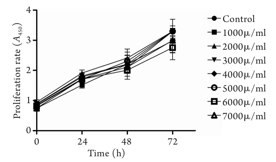
圖1 不同劑量烏司他丁對HUVEC細胞活力及增殖的影響Fig.1 Influence of ulinastatin on activity and proliferation of HUVEC cells
2.2 烏司他丁對熱打擊下HUVEC細胞炎癥因子釋放的影響 與對照組比較,43℃熱打擊+烏司他丁組在0h時的IL-6、TNF-α釋放均較對照組及43℃熱打擊組明顯降低(P<0.05)。43℃熱打擊+烏司他丁組HUVEC細胞在6h時的IL-6、TNF-α釋放均較43℃熱打擊組明顯降低(P<0.05,圖2)。
3 討 論
目前認為重癥中暑是一種繼發于熱損傷之后的全身炎癥反應綜合征(SIRS),進而發展為類膿毒癥,引發多臟器功能衰竭的過程[9]。血管內皮細胞與炎癥細胞的功能異常被視為重癥中暑發生發展的病理基礎[3,10]。大量的動物實驗表明,重癥中暑的死因包括:①全身炎癥細胞的活化,如單核細胞的遷移和跨血管壁運動;②廣泛出血和血栓形成(彌散性血管內凝血即DIC);③廣泛血管床內皮細胞損傷[11-12],以上病理變化最后均作用于血管內皮細胞。有研究發現,在重癥中暑動物模型中多臟器血管內皮結構和功能均受到損害,損傷血管內皮可促進白細胞和血小板黏附,為中暑炎癥損傷和血栓形成提供了結構基礎[13]。目前重癥醫學將血管內皮看作一個特殊的器官系統,有研究發現,血管內皮細胞損傷是影響重癥中暑病理生理序貫進展的關鍵因素[5],血管內皮細胞不僅僅是炎癥反應中的被動靶細胞,也是一種效應細胞,影響重癥中暑的發生發展。
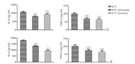
圖2 烏司他丁對熱打擊條件下HUVEC細胞炎癥因子釋放的影響Fig.2 Influence of ulinastatin on IL-6 and TNF-α release of HUVEC under heatshock
烏司他丁是從人尿液中提取的一種彈性蛋白酶抑制物,因其具有抗炎、減輕細胞與組織損傷、改善微循環與內皮細胞通透性等作用,廣泛應用于胰腺炎、急性肺損傷和膿毒癥等的治療[14-17]。有研究稱烏司他丁能維護血管正常舒縮功能及內皮細胞完整性,使內皮細胞表達的細胞間黏附分子-1和CD11b明顯減少,白細胞與血管內皮細胞的黏附減少,明顯減輕血管內皮細胞損傷[14]。因此,烏司他丁對于抑制全身過度的炎癥反應,保護機體免受炎癥傷害具有重要意義。有文獻指出,烏司他丁對患者機體無明顯毒副作用,并建議增大治療劑量[18-19]。本研究CCK-8實驗結果顯示,與對照組(0h)比較,各實驗組細胞24、48、72h增殖率均無明顯下降(P>0.05),且各劑量組烏司他丁對HUVEC細胞增殖均無明顯抑制作用(P>0.05)。
在本研究中,相對于37℃對照組,在給予3000U/ml烏司他丁與HUVEC細胞共孵育1h后,檢測0h及6h組細胞上清炎癥因子IL-6、TNF-α的濃度,結果顯示,在加入烏司他丁共孵育且經歷43℃熱打擊1h后,不但在0h組HUVEC細胞IL-6、TNF-α的釋放受到抑制(P<0.05),在6h后,加入烏司他丁共孵育的HUVEC細胞上清中的IL-6、TNF-α均較43℃有所下降(P<0.05)。提示烏司他丁可抑制熱打擊后IL-6、TNF-α的分泌,且可持續6h。
總之,針對烏司他丁在熱打擊中對HUVEC細胞作用機制的進一步研究,將有助于進一步了解重癥中暑發生、發展的機制以及尋找新的有效治療藥物。
[1]Su L, Guo ZH, Qian HJ. Epidemiological study and key-point analysis of severe heatstroke patients[J]. Med J Chin PLA, 2006, 31(9): 909-911. [蘇磊, 郭振輝, 錢洪津. 重癥中暑住院病人流行病學調查與分析[J]. 解放軍醫學雜志, 2006, 31(9): 909-911.]
[2]Glazer JL. Management of heatstroke and heat exhaustion[J]. Am Fam Physician, 2005, 71(11): 2133-2140.
[3]Bouchama A, Knochel JP. Heat stroke [J]. N Engl J Med, 2002, 346(25): 1978-1988.
[4]Hammami MM, Bouchama A, Al-Sedairy S, et al. Concentrations of soluble tumor necrosis factor and interleukin-6 receptors in heatstroke and heatstress[J]. Crit Care Med, 1997, 25(8): 1314-1319.
[5]Roberts GT, Ghebeh H, Chishti MA, et al. Microvascular injury, thrombosis, inflammation, and apoptosis in the pathogenesis of heatstroke: a study in baboon model[J]. Arterioscler Thromb Vasc Biol, 2008, 28(6): 1130-1136.
[6]Tong HS, Chen Y, Tang YQ, et al. Protection of ulinastatin on the endothelial cell injury of patients with severe heatshock[J]. Guangdong Med J, 2011(12): 1574-1576. [童華生, 陳懌, 唐柚青, 等. 烏司他丁對重癥中暑患者血管內皮細胞損傷的保護作用[J]. 廣東醫學, 2011(12): 1574-1576.]
[7]Pan ZG, Geng Y, Zhang JM, et al. Effect of heat stress on the injury of vascular endothelial cells and release of IL-6 and TNF-α in vitro[J]. Shandong Med J, 2012, 52(3): 32-34. [潘志國, 耿焱, 張劍明, 等. 熱刺激對體外培養的血管內皮細胞損傷及IL-6、TNF-α釋放的影響[J]. 山東醫藥, 2012, 52(3): 32-34.]
[8]Pan ZG. Mechanism of rhabdomyolysis in patients with severe hearshock[D]. 2012. [潘志國. 重癥中暑橫紋肌溶解發病及機制研究[D]. 廣州中醫藥大學, 2012.]
[9]Leon LR, Blaha MD, Dubose DA. Time course of cytokine, corticosterone, and tissue injury responses in mice during heatstrain recovery[J]. J Appl Physiol, 2006, 100(4): 1400-1409.
[10]Tong HS, Duan PK, Zhang XQ, et al. Inflammatory activity of endothelial cells activated by intestinal lymph in severe heat stroke[J]. Med J Chin PLA, 2014, 39(10): 791-794. [童華生,段鵬凱, 張興欽, 等. 重癥中暑腸淋巴激活血管內皮細胞炎性活性的研究[J]. 解放軍醫學雜志, 2014, 39 (10): 791-794.]
[11]Chen CM, Hou CC, Cheng KC, et al. Activated protein C therapy in a rat heat stroke model[J]. Crit Care Med, 2006, 34(7): 1960-1966.
[12]Bouchama A, Roberts G, AI Mohanna F, et al. Inflammatory, hemostatic, and clinical changes in a baboon experimental model for heatstroke[J]. J Appl Physiol, 2005, 98(2): 697-705.
[13]Keuren JF, Baruch D, Legendre P, et al. von Willebrand factor C1C2 domain is involved in platelet adhesion to polymerized fibrin at high shear rate[J]. Blood, 2004, 103(5): 1741-1746.
[14]Masuda T, Sato K, Noda C, et al. Protective effect of urinary trypsin inhibitor on myocardial mitochondria during hemorrhagic shock and reperfusion[J]. Crit Care Med, 2003, 31(7): 1987-1992.
[15]Li N, You SY, Wang CL, et al. The effects of ulinastatin on SP-A in lung injury induced by severe acute pancreatitis in rats[J]. Tianjin Med J, 2013, 41(3): 241-243+290. [李楠, 尤勝義, 王春立, 等. 烏司他丁對大鼠重癥急性胰腺炎肺損傷中SP-A的影響[J]. 天津醫藥, 2013, 41(3): 241-243, 290.]
[16]Liu YF, Wang ZG, Tang HW, et al. Protective effect of ulinastatin on inhalation lung injury caused by black gunpowder smoke in rats and its inhibitory effect on the mRNA expression of IL-1β and TNF-α[J]. Med J Chin PLA, 2014, 39(3): 235-239. [劉一凡, 王正冠, 唐紅衛, 等. 烏司他丁對黑火藥煙霧所致吸入性肺損傷大鼠的保護作用以及對IL-1β、TNF-α基因表達的抑制作用[J]. 解放軍醫學雜志, 2014, 39(3): 235-239.]
[17]Wang H, Liu GJ, Hu M. Protective effect of ulinastatin combined with xuebijing on multiple organ function in Patients with sepsis[J]. Chin J Pract Intern Med, 2014, 34(S1): 113-116. [王虹, 劉貴建, 胡敏. 烏司他丁聯合血必凈對膿毒癥患者器官功能的保護作用[J]. 中國實用內科雜志, 2014, 34(S1): 113-116.]
[18]Wu CY, Xiao F, Yu L, et al. Tolerance test of ulinastati[J]. Chin J Clin Pharmacol Ther, 2007(1): 103-106. [吳成義, 肖峰, 干磊, 等. 烏司他丁人體耐受性試驗[J]. 中國臨床藥理學與治療學, 2007(1): 103-106.]
[19]Jing BW. Progress of the clinical application of ulinastati on the critical illness[J]. Chin Crit Care Med, 2006, 18(2): 117-120.[景炳文. 烏司他丁在急危重癥臨床應用的進展[J]. 中國危重病急救醫學, 2006, 18(2): 117-120.]
Influence of ulinastatin on the proliferation of vascular endothelial cells and IL-6 and TNF-α release under heat stress
PAN Zhi-guo1, SHAO Yu2, GENG Yan1, CHEN Jing-he3, SU Lei1*1Department of Intensive Care Unit, The Military Key Laboratory of Trauma Care in Hot Zone and Tissue Repair in PLA,2Department of Overseas Chinese, Guangzhou General Hospital of Guangzhou Command, Guangzhou 510010, China
3Department of Internal Medicine, First Hospital Affiliated to Guangzhou University of Chinese Medicine, Guangzhou 510405, China
*
, E-mail: slei_icu@163.com
This work was supported by the “Twelfth-Five Year” Plan Key Projects of Military Medicine Science and Technology Development (BWS12J018) and Science and Technology Plan of Guangdong Province (2013B031800010)
ObjectiveTo explore the influence of ulinastatin on the proliferation of vascular endothelial cells and release of IL-6 and TNF-α under heat stress.MethodsHUVEC cells were divided into control, and ulinastatin of different dosage groups (1000, 2000, 3000, 4000, 5000, 6000, 7000U/ml), and CCK-8 assay was used to investigate the influence of ulinastatin on the proliferation of vascular endothelial cells. HUVEC cells were also divided into control, 43℃ heat stress, and 43℃ heat stress plus ulinastatin groups. ELISA assay was used to determine the effect of ulinastatin on the release of IL-6 and TNF-α.ResultsAll the above mentioned doses of ulinastatin showed no significant inhibitory effect on proliferation of HUVEC cells (P>0.05). ELISA results showed that, compared with control group and 43℃ heat-stress group, both the IL-6 and TNF-α releases were decreased at 0h in treatment group with ulinastatin (P<0.05). The IL-6 and TNF-α releases were much lower at 6h in the treatment group with ulinastatin than those undergoing 43℃ heat-stress only group (P<0.05).ConclusionsVarious doses of ulinastatin have no effect on proliferation of endothelial cells. However, it can inhibit the IL-6 and TNF-α releases from HUEVC cells under heat stress.
ulinastatin; heat stroke; endothelial cell; cell proliferation
R594.12
A
0577-7402(2015)05-0362-04
10.11855/j.issn.0577-7402.2015.05.06
2014-12-14;
2015-04-07)
(責任編輯:熊曉然)
全軍醫學科學技術研究“十二五”發展計劃重點項目(BWS12J018);廣東省科技計劃(2013B031800010)
潘志國,醫學博士,主治醫師。主要從事急危重癥救治與重癥中暑方面的研究
510010 廣州 廣州軍區廣州總醫院重癥醫學科、全軍熱區創傷救治與組織修復重點實驗室(潘志國、耿焱、蘇磊),華僑科(邵玉);510405 廣州 廣州中醫藥大學第一附屬醫院(陳鏡合)
蘇磊,E-mail: slei_icu@163.com
猜你喜歡
課堂內外·初中版(科學少年)(2023年10期)2023-12-10 00:43:06
全科護理(2022年10期)2022-12-26 21:19:15
中國合理用藥探索(2022年1期)2022-11-26 00:22:32
今日農業(2022年4期)2022-11-16 19:42:02
鄉村科技(2021年33期)2021-03-16 02:26:54
國際放射醫學核醫學雜志(2021年10期)2021-02-28 08:41:58
藥學與臨床研究(2015年4期)2015-06-05 11:35:54
衛生職業教育(2014年24期)2014-05-20 09:05:38
同位素(2014年2期)2014-04-16 04:57:20
中國合理用藥探索(2014年11期)2014-03-11 20:30:20