胸腺基質淋巴細胞生成素對效應T細胞分化功能的影響
2016-01-20 13:58:04林靜李秀紅梁磊王毅陳海潮楊征酉鵬華陜西省人民醫院西安710068
山東醫藥 2015年29期
林靜,李秀紅,梁磊,王毅,陳海潮,楊征,酉鵬華(陜西省人民醫院,西安710068)
胸腺基質淋巴細胞生成素對效應T細胞分化功能的影響
林靜,李秀紅,梁磊,王毅,陳海潮,楊征,酉鵬華
(陜西省人民醫院,西安710068)
摘要:目的探討體外氧化型低密度脂蛋白( ox-LDL)誘導人血管平滑肌細胞產生的胸腺基質淋巴細胞生成素( TSLP)對效應T細胞分化功能的影響。方法分離并培養人血管平滑肌細胞、人樹突狀細胞( DC)、初始T細胞,隨機分為4組,每組設5個樣本。對照組、實驗組人血管平滑肌細胞分別經PBS、ox-LDL處理后,取其上清液與DC、初始T細胞共培養; TSLP中和抗體組、中和抗體對照組在實驗組的基礎上分別加入TSLP中和抗體、TSLP非特異性中和抗體與DC、初始T細胞共培養。采用ELISA法檢測各組細胞培養上清液中TSLP及Th17細胞因子IL-17、IL-22、TNF-α水平,流式細胞術檢測Th17細胞構成比。結果與對照組比較,實驗組培養上清液TSLP、IL-17、IL-22、TNF-α水平及Th17細胞構成比升高( P均<0.01)。與實驗組、中和抗體對照組比較,TSLP中和抗體組TSLP、IL-17、IL-22、TNF-α水平及Th17細胞構成比降低( P均<0.01)。結論Ox-LDL體外誘導人血管平滑肌細胞產生的TSLP可促進T細胞向Th17細胞分化。
關鍵詞:胸腺基質淋巴細胞生產素;樹突狀細胞; T細胞; Th17細胞;動脈粥樣硬化
Influence of thymic stromal lymphopoietin on differentiation of effector T cells
LIN Jing,LI Xiu-hong,LIANG Lei,WANG Yi,CHEN Hai-chao,YANG Zheng,YOU Peng-hua
( Shaanxi Provincial People's Hospital,Xi'an 710068,China)
Abstract:Objective To investigate the influence of thymic stromal lymphopoietin ( TSLP) produced by oxidized-low density lipoprotein ( ox-LDL) -induced human vascular smooth muscle cells in vitro on differentiation of effector T cells.Methods Human vascular smooth muscle cells,human dendritic cells ( DCs) andT cells were isolated and cultured.Then we divided them into 4 groups,and 5 samples in each group.The control group and the experimental group were separately simulated with PBS and ox-LDL.The supernatant was incubated with DCs andT cells.TSLP neutralizing antibody group and neutralizing antibody control group were added with TSLP neutralizing antibody,TSLP non-specific neutralizing antibody,then were co-cultured with DC andT cells.The levels of TSLP and Th17cytokines including IL-17,IL-22 and TNF-α were detected by ELISA,and the flow cytometry was used to detect the proportion of Th17 cells.Results Compared with the control group,the levels of TSLP,IL-17,IL-22,TNF-α and the proportion of Th17 cells were increased in the experimental group ( all P<0.01).Compared with the neutralizing antibody control group,the levels of TSLP,IL-17,IL-22,TNF-α and the proportion of Th17 cells were decreased in the TSLP neutralizing antibody group ( all P<0.01).Conclusion TSLP produced by ox-LDL-induced human vascular smooth muscle cells in vitro could promoteT cells differentiating into Th17 cells.
Key words:thymic stromal lymphopoietin; dendritic cells; T cells; Th17cells; atherosclerosis
TSLP受體結合,在效應性T細胞的分化中起重要作用。但其具體作用機制尚不明確。2013年2月~2014年12月,我們對氧化型低密度脂蛋白( ox-LDL)誘導人血管平滑肌細胞產生的TSLP在效應性T細胞分化中的作用進行了探討。
1 材料與方法
1.1材料淋巴細胞分離液購自上海試劑二廠。重組人粒細胞-巨噬細胞刺激因子( GM-CSF)、重組人IL-4、重組人TNF-α、人TSLP、IL-17、IL-22及TNF-α ELISA試劑盒購自eBiosience公司。FITC-抗人CD11c抗體、PE-抗人CD4抗體、FITC-抗人IL-17抗體、IgG同型對照抗體、TSLP中和抗體、對照中和抗體、抗人CD3單克隆抗體、抗人CD28單克隆抗體購自Santa Cruz。T細胞分選試劑盒購自Miltenyi Biotec公司。
1.2人血管平滑肌細胞的培養將人血管平滑肌細胞株(購自中國醫學科學院細胞中心)復蘇后,于37℃、飽和空氣濕度、5% CO2培養箱中培養;待細胞生長至80%~90%融合時,用0.25%的胰蛋白酶+0.01%EDTA消化傳代。以1.0×10⁵/mL接種細胞至直徑25 cm2培養瓶中進行后續試驗。
1.4實驗分組及處理①對照組: PBS與人血管平滑肌細胞共培養24 h,去除細胞,取其上清液與DC共培養24 h,與初始T細胞共培養5 d。②實驗組: ox-LDL( 50 μg/mL)與人血管平滑肌細胞共培養24 h,去除細胞,取其上清液與DC共培養24 h。去除上清液,PBS沖洗3次,與初始T細胞共培養5 d。③TSLP中和抗體組: ox-LDL( 50 μg/mL)與人血管平滑肌細胞共培養24 h,去除細胞,取其上清液與DC共培養24 h。去除上清液,PBS沖洗3次,在加入TSLP中和抗體( 1 ng/mL)的培養基中與初始T細胞共培養5 d。④中和抗體對照組: ox-LDL ( 50 μg/mL)與人血管平滑肌細胞共培養24 h,去除細胞,取其上清液與DC共培養24 h。去除上清液,PBS沖洗3次,在加入TSLP非特異性中和抗體的培養基中與初始T細胞共培養5 d。每組設5個樣本,5 d后,各組共培養系統中加入抗人CD3抗體( 5 μg/mL)及抗人CD28抗體( 5 μg/mL)孵育24 h。
1.5培養上清液中TSLP、Th17細胞因子( IL-17、IL-22及TNF-α)水平檢測采用ELISA法。各組細胞培養結束后,收集培養上清液,按照試劑盒的操作步驟檢測培養上清液中TSLP、IL-17、IL-22及TNF-α。實驗設雙復孔,重復3次。
1.7統計學方法采用SPSS13.0統計軟件。計量資料用珋x±s表示,組間比較采用單因素方差分析,兩兩比較采用LSD-t檢驗。P<0.05為差異有統計學意義。
2 結果
2.1四組人平滑肌細胞TSLP水平比較對照組、實驗組、TSLP中和抗體組、中和抗體對照組TSLP水平分別為( 1.83±0.25)、( 28.83±6.54)、( 3.87± 1.02)、( 24.55±2.95) pg/mL。與對照組比較,實驗組上清液TSLP水平升高( P<0.01) ;與實驗組比較,TSLP中和抗體組TSLP水平降低( P<0.01) ;與中和抗體對照組比較,TSLP中和抗體組TSLP水平降低( P<0.01)。
2.2四組培養上清液IL-17、IL-22、TNF-α水平比較見表1。
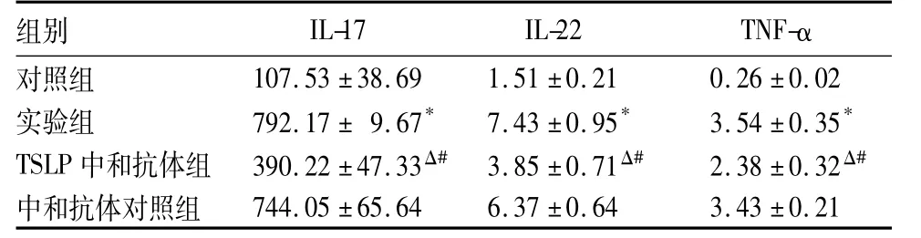
表1 四組培養上清液IL-17、IL-22、TNF-α水平比較( pg/mL,珔x±s)
2.3四組Th17細胞構成比比較對照組、實驗組、TSLP中和抗體組、中和抗體對照組Th17細胞構成比分別為( 0.87±0.22) %、( 9.88±2.16) %、( 3.42 ±1.15) %、( 9.25±1.83) %。與對照組比較,實驗組Th17細胞構成比明顯升高( P<0.01) ;與實驗組比較,TSLP中和抗體組Th17細胞構成比下降( P<0.01) ;與中和抗體對照組比較,TSLP中和抗體組Th17細胞構成比明顯降低( P<0.01)。
3 討論
Th17是近年來發現的T細胞亞群,其分泌的細胞因子主要有IL-17、IL-6、TNF-α及IL-22[4]。研究顯示,Th17細胞及相關細胞因子在AS患者斑塊局部及循環血中大量表達[5]。AS小鼠伴隨著Th17炎癥反應的激活,而敲除IL-17基因可明顯抑制小鼠AS斑塊形成[6,7]。Th17炎癥反應對AS發生有促進作用,而并非是AS炎癥反應的結果或其“伴隨效應”。
研究表明,微生物、Toll樣受體( TLR)配體以及某些細胞因子如IL-1β均可通過NF-κB途徑誘導上皮細胞、纖維細胞、角質細胞及內皮細胞產生誘導型TSLP[8]。不同細胞類型產生的誘導型TSLP所需的刺激原不同[9]。在人氣管上皮細胞,單獨的TNF-α 或IL-1β刺激可以誘導細胞產生誘導型TSLP[10];而在小氣道上皮細胞中,只有TNF-α及IL-1β聯合才可導致誘導型TSLP的產生[11]。因此,不同細胞類型能否誘導產生誘導型TSLP,由細胞所處局部微環境決定。本研究發現,ox-LDL刺激人平滑肌細胞產生TSLP,表明人血管平滑肌細胞所處的ox-LDL微環境是產生誘導型TSLP的決定性因素。TSLP一直被認為是Th2炎癥反應的“總開關”,在特異性皮炎、過敏性哮喘等以Th2炎癥反應為主的自身免疫性疾病中起主要作用。然而近期研究表明,TSLP也可誘導向Th17細胞亞群分化。Tanaka等[12]研究發現,單獨刺激可誘導DC導致效應性T細胞向T2分化,但是TSLP與TLR配體聯合共同刺激誘導DC導致效應性T細胞向Th17分化。本研究發現,ox-LDL誘導產生的誘導型TSLP能夠激活DC,導致Th17細胞分化、Th17細胞因子表達增加,其可能機制為ox-LDL可作為配體與TLR結合,與TSLP聯合共同刺激促使初始T細胞向Th17分化。
本研究發現,與對照組比較,實驗組上清液TSLP水平升高,表明ox-LDL可誘導人平滑肌細胞產生TSLP;抗體中和試驗發現,TSLP中和抗體可顯著降低培養上清液TSLP、IL-17、IL-22水平及Th17細胞構成比,從而阻止DC誘導的Th17細胞分化,進一步表明TSLP在DC誘導的Th17細胞亞群分化機制中發揮重要作用。
綜上所述,ox-LDL誘導人平滑肌細胞產生的TSLP可作用于DC,導致效應性T細胞向Th17分化。結合我們前期研究發現的AS斑塊中TSLP表達升高及Th17在AS中的重要作用,認為TSLP在AS中的重要作用之一可能為促使T細胞分化,具體作用及分子機制尚需進一步研究。
參考文獻:
[1]Ross R.Atherosclerosis-an inflammatory disease[J].N Engl J Med,1999,340( 2) :115-126.
[2]Stemme S,Holm J,Hansson GK.T lymphocytes in human atherosclerotic plaques are memory cells expressing CD45RO and the integrin VLA-1[J].Arterioscler Thromb,1992,12( 2) : 206-211.
[3]Liu X,Li H,Ren X.Signaling cascades initiated by tslp-mediated signals in different cell types[J].Cell Immunol,2012,279: 174-179.
[4]Kolls JK,Lindén A.Interleukin-17 family members and inflammation[J].Immunity,2004,21( 4) : 467-476.
[5]Liu Z,Lu F,Pan H,et al.Correlation of peripheral th17 cells and th17-associated cytokines to the severity of carotid artery plaque and its clinical implication[J].Atherosclerosis,2012,221( 1) : 232-241.
[6]Usui F,Kimura H,Ohshiro T,et al.Interleukin-17 deficiency reduced vascular inflammation and development of atherosclerosis in western diet-induced apoe-deficient mice[J].Biochem Biophys Res Commun,2012,420( 1) : 72-77.
[7]Gao Q,Jiang Y,Ma T,et al.A critical function of th17 proinflammatory cells in the development of atherosclerotic plaque in mice[J].J Immunol,2010,185( 10) : 5820-5827.
[8]Takai T.Tslp expression: Cellular sources,triggers,and regulatory mechanisms[J].Allergol Int,2012,61( 1) : 613-617.
[9]Zhao H,Li M,Wang L,et al.Angiotensin ii induces tslp via an at1 receptor/nf-kappab pathway,promoting th17 differentiation [J].Cell Physiol Biochem,2012,30( 6) : 1383-1397.
[10]Haro H,Komori H,Okawa A,et al.Sequential dynamics of monocyte chemotactic protein-1 expression in herniated nucleus pulposus resorption[J].J Orthop Res,1997,15( 5) : 734-741
[11]Kanamaru Y,Sumiyoshi K,Ushio H,et al.Smad3 deficiency in mast cells provides efficient host protection against acute septic peritonitis[J].J Immunol,2005,174( 7) : 4193-4197.
[12]Tanaka J,Watanabe N,Kido M,et al.Human tslp and tlr3 ligands promote differentiation of th17 cells with a central memory phenotype under th2-polarizing conditions[J].Clin Exp Allergy,2009,39( 1) : 89-100.
收稿日期:( 2015-06-05)
通信作者簡介:梁磊( 1974-),男,主任醫師,博士,主要研究方向為動脈粥樣硬化分子生物學機制。E-mail: ll008@163.com
作者簡介:第一林靜( 1979-),女,主治醫師,博士,主要研究方向為動脈粥樣硬化發病機制。E-mail: linjing.123456789@163.com
基金項目:國家自然科學基金資助項目( 81400333)。
文章編號:1002-266X( 2015)29-0004-03
文獻標志碼:A
中圖分類號:R311.1+44
doi:10.3969/j.issn.1002-266X.2015.29.002