Aire對巨噬細胞極化影響的研究①
2017-02-15 09:43:35朱武飛羅亞東王艷玲
中國免疫學(xué)雜志 2017年1期
朱武飛 羅亞東 趙 博 王艷玲 楊 巍
(吉林大學(xué)基礎(chǔ)醫(yī)學(xué)院免疫學(xué)系,長春130021)
·基礎(chǔ)免疫學(xué)·
Aire對巨噬細胞極化影響的研究①
朱武飛 羅亞東 趙 博 王艷玲②楊 巍③
(吉林大學(xué)基礎(chǔ)醫(yī)學(xué)院免疫學(xué)系,長春130021)
目的:研究自身免疫調(diào)節(jié)因子(Aire)對巨噬細胞極化的影響。方法:分別用LPS、IL-4以及LPS聯(lián)合免疫復(fù)合物刺激小鼠單核巨噬細胞系RAW264.7細胞、穩(wěn)定表達GFP-Aire的RAW264.7細胞(A33-3)細胞和穩(wěn)定表達GFP的RAW264.7細胞(C1-6),使其向M1(LPS)、M2a(IL-4)和M2b(LPS聯(lián)合免疫復(fù)合物)型巨噬細胞極化。通過Real-time PCR檢測各組細胞中M1型巨噬細胞特征分子IL-1α、iNOS和IL-6,M2a型特征分子Arg-1和M2b型特征分子IL-10的表達水平,研究Aire對各種類型巨噬細胞極化的影響。結(jié)果:LPS在 0.5 μg/ml濃度時,RAW264.7細胞中M1型巨噬細胞產(chǎn)物IL-1α、iNOS和IL-6基因表達量最高;而IL-4以及LPS聯(lián)合免疫復(fù)合物的刺激作用有顯著的劑量依賴性,都在濃度最高時RAW264.7細胞中Arg1(M2a)和IL-10(M2b)基因表達量最高。LPS刺激后,A33-3細胞中IL-1α和iNOS表達水平明顯高于C1-6細胞,IL-6則相反;IL-4及LPS聯(lián)合免疫復(fù)合物刺激后,A33-3細胞中Arg1和IL-10的表達水平明顯低于C1-6細胞。結(jié)論:Aire可能促進巨噬細胞向M1極化,同時抑制其向M2a和M2b極化。
自身免疫調(diào)節(jié)因子;巨噬細胞;極化
自身免疫調(diào)節(jié)因子(Aire)是一種轉(zhuǎn)錄因子,人類Aire基因的突變將導(dǎo)致由自身免疫應(yīng)答所介導(dǎo)的、以多器官損傷為特征的一種常染色體隱性遺傳病(Autoimmune polyendocrinopathy candidiasis ectoderm-al dystrophy,APECED),也稱為自身免疫性多腺體綜合征 Ⅰ(Autoimmune polyendocrine syndrome type Ⅰ,APS-Ⅰ)。APECED其典型臨床表現(xiàn)為甲狀旁腺功能減退、Addison′s病和慢性皮膚黏膜念珠菌感染的三聯(lián)征[1]。現(xiàn)有的研究證實Aire主要表達在胸腺上皮細胞,起到維持中樞耐受的作用。然而,對APECED病人易患慢性皮膚黏膜念珠菌感染仍然沒有合理的解釋。有研究發(fā)現(xiàn)Aire也表達于外周巨噬細胞,但其具體功能作用以及與念珠菌感染之間是否存在聯(lián)系尚不清楚。我們前期的研究發(fā)現(xiàn)TLR1、TLR3和TLR8在過表達Aire的小鼠的巨噬細胞系RAW264.7細胞中是上調(diào)的[2,3],提示Aire可能在抗原提呈細胞針對病原體的應(yīng)答中發(fā)揮重要作用。
巨噬細胞作為一類重要的抗原提呈細胞,廣泛分布于機體,最近研究表明巨噬細胞具有明顯的異質(zhì)性,表現(xiàn)為針對不同微環(huán)境應(yīng)答可使其功能狀態(tài)發(fā)生改變。目前認為巨噬細胞可以功能性地極化為M1型(經(jīng)典激活的巨噬細胞)或M2型(非經(jīng)典激活的巨噬細胞)。M1型巨噬細胞由Ⅰ型干擾素(IFN-γ)和微生物代謝產(chǎn)物(LPS)激活,通過產(chǎn)生大量促炎細胞因子如白介素1(IL-1)、IL-6、IL-23、超氧化物陰離子和NO[4,5],具有殺滅細菌、真菌等多種病原體的潛能和介導(dǎo)宿主防御的功能。根據(jù)表達的活化標志不同M2型巨噬細胞又可分成三個亞群:M2a、M2b和M2c。Th2細胞產(chǎn)生的細胞因子IL-4/IL-13可促進M2a巨噬細胞極化,通過產(chǎn)生精氨酸1(Arg1)和細胞外基質(zhì)(ECM)參與組織重構(gòu)。用免疫復(fù)合物或Toll樣受體(TLR)激動劑可誘導(dǎo)M2b極化。M2c則被抗炎物質(zhì)所誘導(dǎo),如糖皮質(zhì)激素或IL-10[6,7]。目前,大多數(shù)研究都集中在巨噬細胞的異質(zhì)性以及M1和M2巨噬細胞的產(chǎn)生條件。然而,調(diào)控M1和M2巨噬細胞極化的分子機制還沒有得到深入的研究。
鑒于Aire突變可導(dǎo)致機體易感染念珠菌,而M1型巨噬細胞又具有抵抗真菌的作用,那么Aire是否可通過參與不同類型巨噬細胞的極化過程,從而在抗真菌感染中發(fā)揮作用有待探究。因此本研究試圖通過觀察Aire對巨噬細胞極化類型的影響,探討其在外周免疫系統(tǒng)如抗真菌感染中的作用。
1 材料與方法
1.1 細胞系 RAW264.7細胞從上海細胞研究所獲得。課題組前期已完成穩(wěn)定轉(zhuǎn)染pEGFPC1/Aire 和pEGFPC1質(zhì)粒至RAW264.7細胞中,分別得到A33-3和C1-6細胞[6],用含10%胎牛血清的RPMI-1640(Gibco,USA)進行培養(yǎng)。
1.2 方法
1.2.1 巨噬細胞亞群的極化 RAW264.7細胞調(diào)濃度至1×10⁶ml-1,吸取細胞懸液加至6孔細胞培養(yǎng)板,每孔2 ml含 10%NCS的RPMI1640,置于37℃、5% CO2培養(yǎng)箱中培養(yǎng)過夜。RPMI1640洗一遍細胞,然后用LPS(Sigma,USA),IL-4(R&D Systems, minneapoils,MN)和LPS聯(lián)合免疫復(fù)合物(OVA和抗OVA IgG)(Sigma,USA)刺激24 h后收細胞。
1.2.2 RT-PCR 從細胞中提取總RNA(如上所述)用TRIzol(Invitrogen,USA)裂解,溶解于DEPC水中。用紫外分光光度計檢測總RNA的A260/A280的比值和濃度。取1 μg RNA為模板,加入隨機引物和AMV逆轉(zhuǎn)錄酶(TaKaRa,Japan)逆轉(zhuǎn)錄成cDNA,反應(yīng)條件:30℃ 10 min,45℃ 30 min,5℃ 5 min,1循環(huán)。cDNA用表1中的引物進行PCR。應(yīng)用ImageMaster VDS(Pharmacia Biotech,China)圖像分析軟件分析光密度值,各種基因的mRNA表達水平均表示為目的基因/β-actin光密度值。
表1 RT-PCR和Real-time PCR所用引物及其擴增片段大小
Tab.1 Primers for RT-PCR and quantitative Real-time PCR and their amplification size
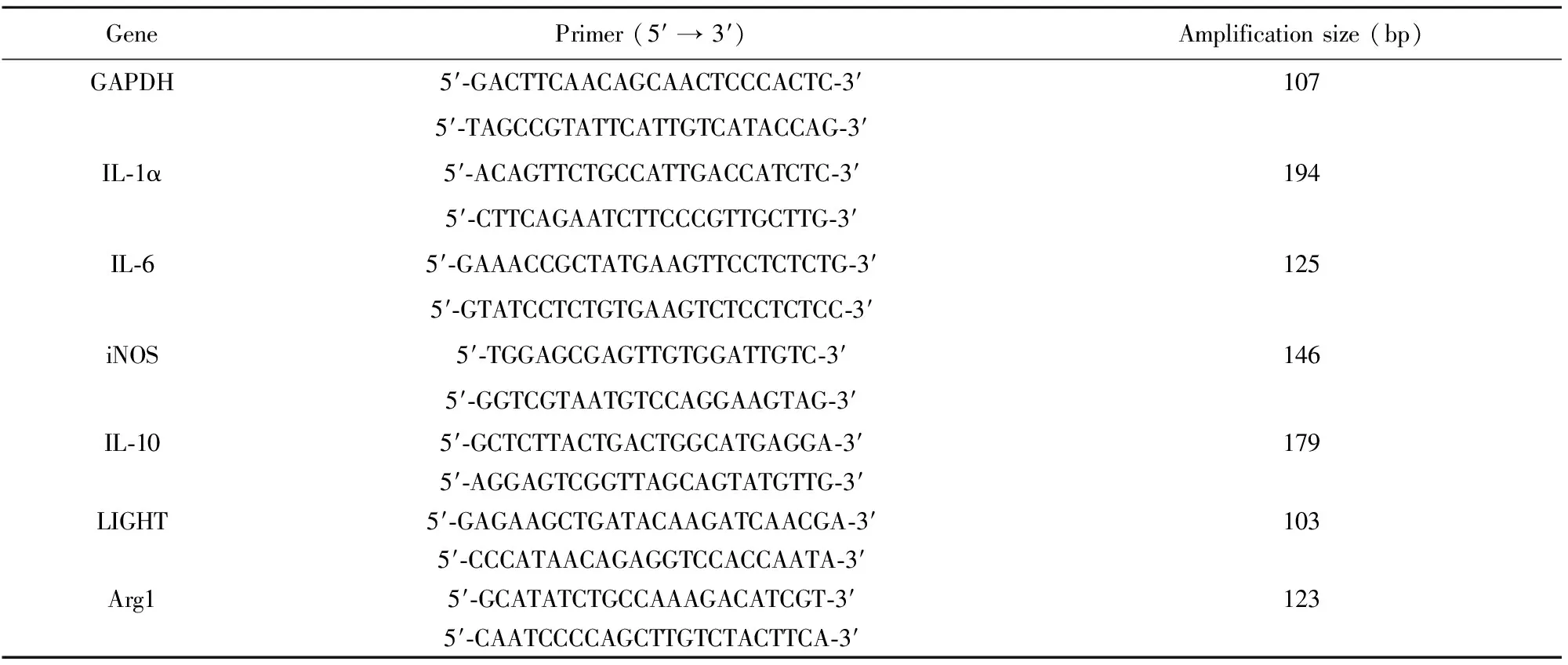
GenePrimer(5′→3′)Amplificationsize(bp)GAPDH5′?GACTTCAACAGCAACTCCCACTC?3′1075′?TAGCCGTATTCATTGTCATACCAG?3′IL?1α5′?ACAGTTCTGCCATTGACCATCTC?3′1945′?CTTCAGAATCTTCCCGTTGCTTG?3′IL?65′?GAAACCGCTATGAAGTTCCTCTCTG?3′1255′?GTATCCTCTGTGAAGTCTCCTCTCC?3′iNOS5′?TGGAGCGAGTTGTGGATTGTC?3′1465′?GGTCGTAATGTCCAGGAAGTAG?3′IL?105′?GCTCTTACTGACTGGCATGAGGA?3′1795′?AGGAGTCGGTTAGCAGTATGTTG?3′LIGHT5′?GAGAAGCTGATACAAGATCAACGA?3′1035′?CCCATAACAGAGGTCCACCAATA?3′Arg15′?GCATATCTGCCAAAGACATCGT?3′1235′?CAATCCCCAGCTTGTCTACTTCA?3′
1.2.3 Real-time PCR 取上述cDNA為模板,以表1中的引物,采用SYBR Premix Ex Taq試劑盒(TaKaRa,Japan)和ABI7300儀器(Life Technol-ogies,USA)進行實時定量PCR反應(yīng),條件為:95℃ 30 s,95℃ 5 s和60℃ 30 s,40循環(huán)。結(jié)果用公式 2-ΔΔCt計算。
2 結(jié)果
2.1 不同的刺激物刺激巨噬細胞的極化 為了確定RAW264.7細胞是否能夠極化為不同類型的巨噬細胞,并明確激活巨噬細胞亞群的各種刺激物的有效濃度,分別采用不同濃度LPS、IL-4和LPS聯(lián)合免疫復(fù)合物來刺激RAW264.7細胞。結(jié)果顯示,LPS濃度在 0.5 μg/ml時,M1的產(chǎn)物IL-1α、IL-6和誘導(dǎo)的一氧化氮合成酶(iNOS)的水平在RAW264.7細胞中明顯地上調(diào)(圖1)。經(jīng)IL-4和LPS聯(lián)合免疫復(fù)合物刺激后,RAW264.7細胞中Arg1和IL-10基因表達呈劑量依賴的方式增加(圖2)。因此確定IL-4在200 pg/ml和0.5 μg/ml LPS聯(lián)合IC3(150 μg/ml OVA和15 μl anti-OVA IgG)分別為M2a和M2b極化的最佳濃度,用于后續(xù)實驗。結(jié)果表明不同刺激物可激活RAW264.7細胞分化為M1或M2型巨噬細胞。
2.2 Aire促進M1型巨噬細胞極化 為了研究Aire是否影響M1型巨噬細胞的極化,采用0.5 μg/ml LPS刺激C1- 6和A33-3細胞, Real-time PCR檢測特異的M1型巨噬細胞相關(guān)標志的表達。結(jié)果顯示LPS刺激后,與C1-6細胞相比,A33-3細胞中iNOS和IL-1α表達增加,而IL-6表達水平降低(圖3)。這些數(shù)據(jù)表明Aire可能促進M1的極化。

圖1 不同濃度的LPS刺激下RAW264.7細胞產(chǎn)生的 IL-1α、IL-6 和 iNOS的表達量Fig.1 Dose effects of IL-1α,IL-6 and iNOS produced by RAW264.7 cells in response to LPSNote:A.The expression of different genes in RAW264.7 cells stimulated with 0,0.5,5.0 or 50 μg/ml LPS (Lanes 1-4);B.The gene/β-actin optical density values from the top electrophoregram were calculated and plotted. All data were obtained from three independent experiments and expressed as the ±s.

圖2 RAW264.7 細胞中Arg-1和IL-10各自對不同刺激物的劑量反應(yīng)Fig.2 Dose effects of Arg-1 and IL-10 produced by RAW264.7 cells in response to different stimuliNote: A. RAW264.7 cells were stimulated with different doses of IL-4:0,10,50,100 or 200 pg/ml (Lanes 1-5);B.RAW264.7 cells were stimulated with 0.5 μg/ml LPS with different doses of immune complexes. Lanes 1-6 represent normal saline,150 μg/ml OVA,LPS+150 μg/ml OVA,LPS+IC.1 (150 μg/ml OVA+2.5 μl anti-OVA IgG),LPS+IC.2 (150 μg/ml OVA+5 μl anti-OVA IgG),LPS+IC.3 (150 μg/ml OVA+15 μl anti-OVA IgG).The bottom graphs were plotted with the gene/β-actin optical density values from the top electropherogram. All data were obtained from three independent experiments and expressed as the ±s.

圖3 LPS刺激對C1-6和A33-3細胞中IL-1α、IL-6和iNOS表達水平的影響Fig.3 Stimulation with LPS affects expression levels of iNOS,IL-1α and IL-6 in C1-6 and A33-3 cellsNote: A. The expression levels of iNOS and IL-1α in C1-6 and A33-3 stimulated with LPS;B. The expression levels of IL-6 in C1-6 and A33-3 stimulated with LPS. All data were obtained from three independent experiments and expressed as the ±s. a,b and c represent comparisons of stimulated C1-6,unstimulated A33-3 and stimulated A33-3 cells,respectively,with unstimulated C1-6 cells.**.P< 0.01.
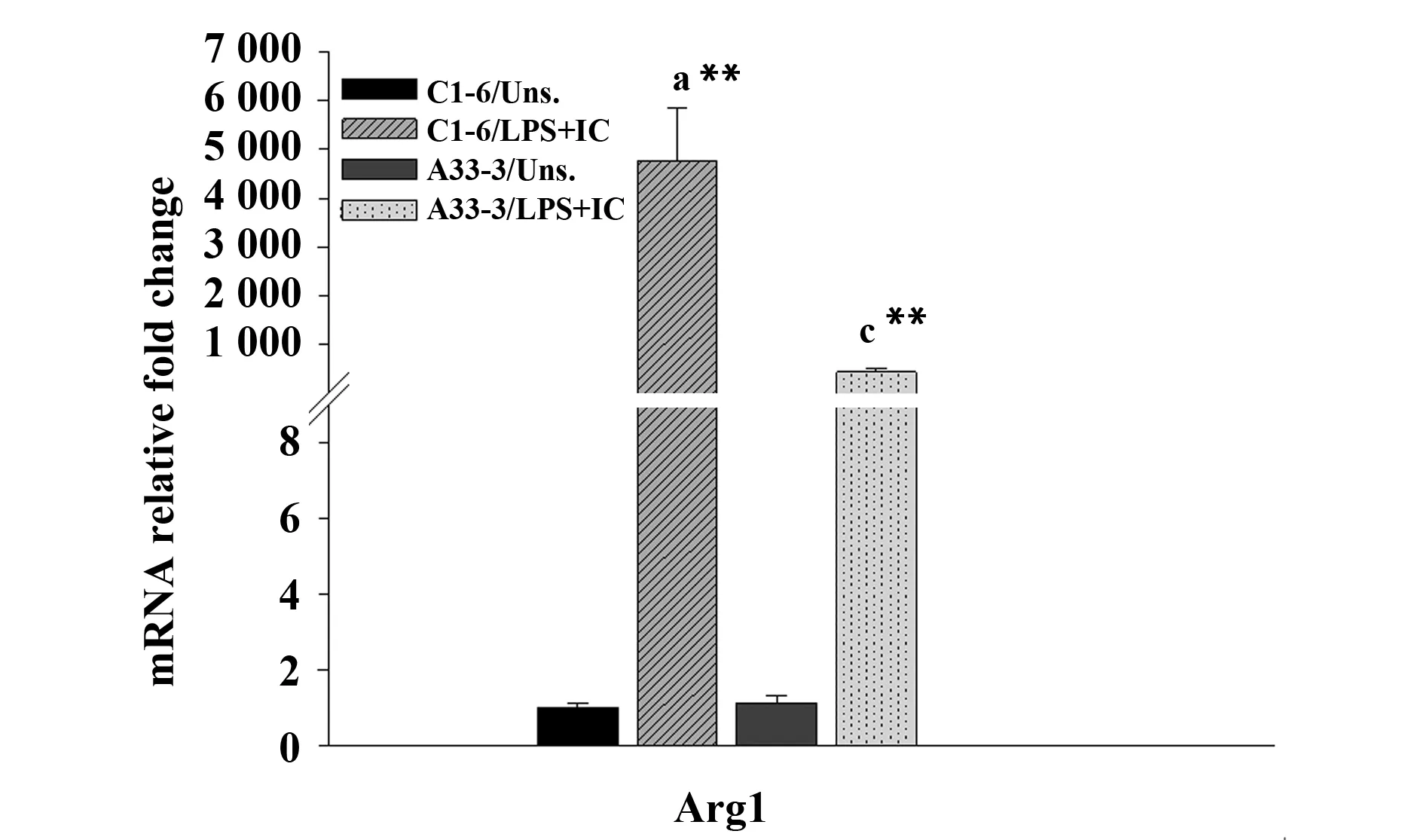
圖4 IL-4刺激對C1-6和A33-3細胞中Arg1表達水平的影響Fig.4 Stimulation with IL-4 affects expression levels of Arg1 in C1-6 and A33-3 cellsNote: All data were obtained from three independent experiments and expressed as the ±s. a and c represent comparisons of stimulated C1-6 and stimulated A33-3 cells,respectively,with unstimulated C1-6 cells. **.P<0.01.
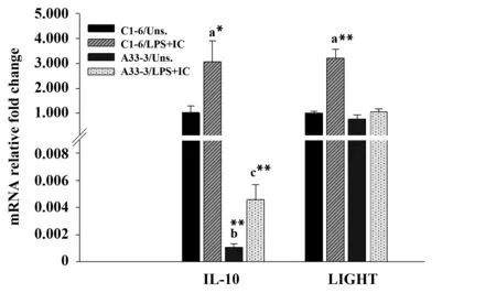
圖5 LPS聯(lián)合免疫復(fù)合物刺激對C1-6和A33-3細胞中IL-10和LIGHT表達水平的影響Fig.5 Stimulation of LPS along with IC effects on expression levels of IL-10 and LIGHT in C1-6 and A33-3 cellsNote: All data were obtained from three independent experiments and expressed as the ±s. a,b and c represent comparisons of stimulated C1-6,unstimulated A33-3 and stimulated A33-3 cells,respectively,with unstimulated C1-6 cells. *.P< 0.05,**.P< 0.01.
2.3 Aire抑制M2a型巨噬細胞極化 為進一步研究Aire對M2a巨噬細胞極化的影響。采用200 pg/ml IL-4刺激后,Real-time PCR檢測結(jié)果發(fā)現(xiàn)相比于C1-6細胞,A33-3細胞中Arg-1基因表達較低(圖4)。說明Aire可能抑制M2a極化。
2.4 Aire抑制M2b型巨噬細胞極化 為明確Aire是否也影響另一種M2型巨噬細胞——M2b的極化。采用0.5 μg/ml LPS聯(lián)合IC3分別刺激A33-3和C1-6細胞,Real-time PCR檢測M2b相關(guān)分子的表達。結(jié)果顯示在有或沒有刺激物時,相比于C1-6細胞A33-3細胞中IL-10的表達均顯著降低。然而,在刺激后A33-3細胞中LIGHT基因的表達并未發(fā)生變化(圖5)。表明Aire可能抑制M2b極化。
3 討論
本研究中,我們發(fā)現(xiàn)不同的誘導(dǎo)劑刺激可使RAW264.7細胞分別極化為M1、M2a和M2b型巨噬細胞。并且相比于C1-6細胞,A33-3細胞中M1型巨噬細胞特異的下游分子IL-1α和iNOS的表達增加,而M2a和M2b型巨噬細胞特異的下游分子表達降低。說明AIRE可能促進M1極化,抑制M2極化。
M1型巨噬細胞的極化發(fā)生在Th1細胞介導(dǎo)的免疫應(yīng)答過程中[8],在清除胞內(nèi)病原體中發(fā)揮重要作用。在感染早期,激活的NK細胞和Th1細胞分泌大量的IFN-γ,為促進M1極化的重要物質(zhì)。其他的細菌產(chǎn)物如LPS或某些細胞因子(TNF和GM-CSF)也可以誘導(dǎo)M1極化。活化的M1型巨噬細胞分泌大量促炎細胞因子(IL-1、IL-6和IL-23)和大量的NO和超氧陰離子,從而增強細胞的殺傷能力和抗感染能力。本研究表明在LPS刺激M1巨噬細胞極化過程中,A33-3細胞中IL-1α和iNOS的mRNA的表達水平顯著高于C1-6細胞,而IL-6的表達水平降低。iNOS可以促進瓜氨酸形成NO,增加巨噬細胞的殺傷和抗感染能力。IL-1在免疫防御中也具有重要作用。另外,過多分泌的IL-6和IL-23也可以引起組織損傷。IL-6和IL-23與Th17細胞的分化和增殖有關(guān)[9-11]。Th17分泌的IL-17可以引起組織中大量多形核白細胞的募集,導(dǎo)致一種炎癥性自身反應(yīng),因此Aire可能通過促進巨噬細胞極化為M1發(fā)揮抗感染作用,同時抑制IL-6分泌所誘導(dǎo)的自身反應(yīng)性炎癥。
M2a巨噬細胞的極化可以被Th2型細胞分泌的IL-4和IL-13所誘導(dǎo)[8]。這些巨噬細胞有較弱的殺傷病原體的能力且不具有提呈外來抗原的能力[12]。然而,它可以產(chǎn)生大量的Arg-1,催化精氨酸變?yōu)轼B氨酸,鳥氨酸是一種多胺和膠原的前體,有助于形成細胞外基質(zhì)(ECM)[13],這些復(fù)合物都具有促進傷口愈合的功能。我們研究發(fā)現(xiàn)IL-4刺激后,A33-3細胞中Arg1的上調(diào)水平較C1-6細胞低,證明Aire可抑制M2a的極化。M2b和M2c都具有免疫調(diào)節(jié)作用,經(jīng)常被稱為調(diào)節(jié)型巨噬細胞。在體外試驗中,IgG免疫復(fù)合物或TLR激動劑刺激下,會產(chǎn)生M2b[14],M2b巨噬細胞高表達免疫抑制性細胞因子IL-10。本研究結(jié)果表明,LPS聯(lián)合OVA和anti-OVA IgG免疫復(fù)合物可以促進巨噬細胞的極化,并且相比于未刺激的細胞,刺激后A33-3和C1-6細胞中IL-10的表達水平明顯增加,但A33-3細胞中IL-10的表達明顯低于C1-6細胞(無論刺激前或刺激后)。表明Aire可能通過降低IL-10的表達抑制M2b巨噬細胞極化。
然而,Aire調(diào)節(jié)巨噬細胞極化的機制尚不明確。最近一項研究證明Micro RNA 155(miR-155)是調(diào)控M1型巨噬細胞極化的關(guān)鍵分子[15]。過表達miR-155可以將抗炎的M2型巨噬細胞轉(zhuǎn)變?yōu)榇傺椎腗1型巨噬細胞。相反,沉默M1中miR-155可使M1型巨噬細胞重新極化為M2型[15]。此外,Macedo等[16]發(fā)現(xiàn)胸腺髓質(zhì)上皮細胞中的Aire可調(diào)控miR-155的表達。因此,我們推測miR-155可能在Aire誘導(dǎo)M1極化過程中發(fā)揮重要作用,但還需要進一步研究證實。
綜上,在巨噬細胞極化為不同亞群的過程中,Aire可促進M1極化,同時抑制M2的極化,有助于機體形成抗感染能力,參與某些病原體(包括真菌)的清除,這也為APECED病人出現(xiàn)的真菌易感提供了一個新的解釋。
[1] Perheentupa J.Autoimmune polyendocrinopathy--candidiasis--ectodermal dystrophy (APECED)[J].Horm Metab Res,1996,28(7):353-356.
[2] Zhu W,Yang W,He Z,etal.Overexpressing autoimmune regulator regulates the expression of toll-like receptors by interacting with their promoters in RAW264.7 cells[J].Cell Immunol,2011,270(2):156-163.
[3] Wu J,Zhu W,Fu H,etal.DNA-PKcs interacts with Aire and regulates the expression of Toll-like receptors in RAW264.7 cells[J].Scand J Immunol,2012,75(5):479-488.
[4] Gordon S,Taylor PR.Monocyte and macrophage heterogeneity[J].Nat Rev Immunol,2005,5 (12):953-964.
[5] Gordon S.The macrophage:past,present and future[J].Eur J Immunol,2007,37(S1):S9-S17.
[6] Gordon S.Alternative activation of macrophages[J].Nat Rev Immunol,2003,3(1):23-35.
[7] Martinez FO,Hel ming L,Gordon S.Alternative activation of macrophages:an immunologic functional perspective[J].Annu Rev Immunol,2009,27:451-483.
[8] Mills CD,Kincaid K,Alt JM,etal.M-1/M-2 macrophages and the Th1/Th2 paradigm[J].J Immunol,2000,164(12):6166-6173.
[9] Bettelli E,Carrier Y,Gao W,etal.Reciprocal developmental pathways for the generation of pathogenic effector TH17 and regulatory T cells[J].Nature,2006,441(7090):235-238.
[10] Zheng SG,Wang J,Horwitz DA.Cutting edge:Foxp3+CD4+CD25+regulatory T cells induced by IL-2 and TGF-beta are resistant to Th17 conversion by IL-6[J].J Immunol,2008,180(11):7112-7116.
[11] Lee Y,Awasthi A,Yosef N,etal.Induction and molecular signature of pathogenic TH17 cells[J].Nat Immunol,2012,13(10):991-999.
[12] Edwards JP,Zhang X,Frauwirth KA,etal.Biochemical and functional characterization of three activated macrophage populations[J].J Leukoc Biol,2006,80(6):1298-1307.
[13] Shiraishi M,Shintani Y,Shintani Y,etal.Alternatively activated macrophages deter mine repair of the infarcted adult murine heart[J].Curr Opin Immunol,2016,126(6):2151-2166.
[14] Gerber JS,Mosser DM.Reversing lipopolysaccharide toxicity by ligating the macrophage Fc gamma receptors[J].J Immunol,2001,166(11):6861-6868.
[15] Cai X,Yin Y,Li N,etal.Re-polarization of tumor-associated macrophages to pro-inflammatory M1 macrophages by microRNA-155[J].J Mol Cell Biol,2012,4(5):341-343.
[16] Macedo C,Evangelista AF,Marques MM,etal.Autoimmune regulator (Aire) controls the expression of microRNAs in medullary thymic epithelial cells[J].Immunobiology,2013,218(4):554-560.
[收稿2016-08-01]
(編輯 張曉舟)
Effects of Aire on macrophage polarization
ZHUWu-Fei,LUOYa-Dong,ZHAOBo,WANGYan-Ling,YANGWei.
DepartmentofImmunology,CollegeofBasicMedicalSciences,JilinUniversity,Changchun130021,China
Objective:To study whether autoimmune regulator (Aire) affects macrophage polarization.Methods: The mouse mononuclear macrophage cell line RAW264.7 cells,stable expressing GFP-Aire protein RAW264.7 cells (A33-3) and stable expressing GFP protein RAW264.7 cells (C1-6) were stimulated with LPS,IL-4 and LPS combined with immune complex respectively to make the macrophage polarize to M1 (LPS),M2a (IL-4) and M2b (LPS with immune complex).To investigate the effects of Aire on the various types of macrophages polarization,the M1 related molecules (IL-1α,iNOS and IL-6),M2a related molecules (Arg-1) and M2b related molecule (IL-10) were detected by Real-time PCR.Results: The expression of IL-1α,IL-6 and inducible nitric oxide synthase (iNOS),the products of M1 macrophages,were significantly upregulated in RAW264.7 cells treated with LPS at 0.5 μg/ml.The expression of Arg-1 and IL-10 mRNA in RAW264.7 cells increased in a dose-dependent manner after stimulation with IL-4 or LPS combined with immune complexes,respectively.M1 macrophage-related marker iNOS and IL-1 increased,whereas IL-6 levels decreased in A33-3 cells compared with C1-6 cells after treatment with LPS.The expression of Arg1 and IL-10 were downregulated in A33-3 cells compared with C1-6 cells after IL-4 and LPS combined with immune complexes stimulation,respectively.Conclusion: Aire may promote M1 macrophage polarization while inhibit M2a and M2b macrophage polarization.
Autoimmune regulator;Macrophage;Polarization
10.3969/j.issn.1000-484X.2017.01.002
①本文受國家自然科學(xué)基金項目(81373127)和吉林省科技廳項目(20160414038GH)資助。
朱武飛(1981年- ),男,博士,主要從事免疫耐受方面的研究。
R392
A
1000-484X(2017)01-0011-05
②吉林大學(xué)基礎(chǔ)醫(yī)學(xué)院病原免疫細胞遺傳學(xué)實驗中心,長春130021。
③通訊作者,E-mail:ywei@jlu.edu.cn。
猜你喜歡
體育科技文獻通報(2022年3期)2022-05-23 13:46:54
美與時代·美術(shù)學(xué)刊(2022年3期)2022-04-27 01:18:15
天津外國語大學(xué)學(xué)報(2021年3期)2021-08-13 08:32:18
遼金歷史與考古(2021年0期)2021-07-29 01:06:54
科技傳播(2019年22期)2020-01-14 03:06:54
火花(2019年12期)2019-12-26 01:00:28
民用飛機設(shè)計與研究(2019年4期)2019-05-21 07:21:24
人大建設(shè)(2019年12期)2019-05-21 02:55:32
汽車工程學(xué)報(2017年2期)2017-07-05 08:13:02
學(xué)苑創(chuàng)造·A版(2015年11期)2016-01-14 09:03:27