視網膜神經節細胞復合體厚度與神經纖維層厚度的相關性及對新生血管性青光眼的影響
2019-10-15 04:31:06熊云帆李琴
中國醫藥導報 2019年36期
熊云帆 李琴
[摘要] 目的 研究視網膜神經節細胞復合體(mGCC)厚度與神經纖維層(pRNFL)厚度的相關性及對新生血管性青光眼(NVG)的影響。 方法 選取2016年5月~2019年5月昆明醫科大學第一附屬醫院接受傅里葉光學相干斷層掃描術(FD-OCT)檢查的NVG患者40例作為NVG組,選取同期接受FD-OCT檢查的正常人群40名作為正常組,比較兩組mGCC厚度、pRNFL厚度,Pearson積距相關分析兩者相關性,并評估其對NVG的診斷效力。 結果 NVG組平均GCC、上方GCC、下方GCC厚度,平均RNFL、顳側RNFL、上方RNFL、鼻側RNFL、下方RNFL厚度均低于正常組,差異均有統計學意義(均P < 0.05)。Pearson積距相關分析顯示,各部位mGCC厚度與pRNFL厚度均呈正相關。且各部位mGCC厚度和pRNFL厚度對NVG均有診斷效力(P < 0.01)。 結論 在NVG診斷中,mGCC厚度可作為pRNFL厚度的一個重要補充手段,值得應用和推廣。
[關鍵詞] 青光眼;新生血管性青光眼;視網膜神經節細胞復合體;視網膜神經纖維層
[中圖分類號] R775 ? ? ? ? ?[文獻標識碼] A ? ? ? ? ?[文章編號] 1673-7210(2019)12(c)-0108-04
The relationship between retinal ganglion cell complex thickness and retinal nerve fiber layer thickness and its effect on neovascular glaucoma
XIONG Yunfan ? LI Qin
Department of Ophthalmology, the First Affiliated Hospital of Kunming Medical University,Yunnan Province, Kunming ? 650000, China
[Abstract] Objective To research the relationship between the thickness of retinal ganglion cell complex (mGCC) and retinal nerve fiber layer (pRNFL) and its effect on neovascular glaucoma. Methods From May 2016 to May 2019, 40 cases with NVG patients receiving fourier domain optical coherence tomography (FD-OCT) examination in the First Affiliated Hospital of Kunming Medical University were selected as NVG group. In the same period, 40 cases with normal persons receiving FD-OCT examination were selected as normal group. The mGCC thickness and pRNFL thickness were compared between two groups. Pearson accumulates correlation analysis was used to analyze the correlation between the mGCC thickness and pRNFL thickness. And the diagnostic effectiveness for NVG was evaluated. Results The thickness of GCC-average, GCC-superior, GCC-inferior and the thickness of RNFL-average, RNFL-temporal, RNFL-superior, RNFL-nasal, RNFL-inferior in NVG group were lower than those in normal group, and the differences were statistically significant (all P < 0.05). Pearson accumulates correlation analysis showed that mGCC thickness at all sites were positively correlated with pRNFL thickness. mGCC thickness and pRNFL thickness at all sites have diagnostic effect on NVG (P < 0.01). Conclusion In the diagnosis of NVG, mGCC thickness can be used as an important supplementary measure of pRNFL thickness, which is worthy of application and promotion.
[Key words] Glaucoma; Neovascular glaucoma; Retinal ganglion cell complex; Retinal nerve fiber layer
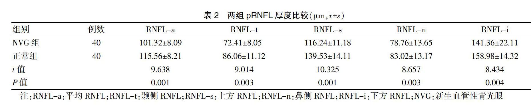
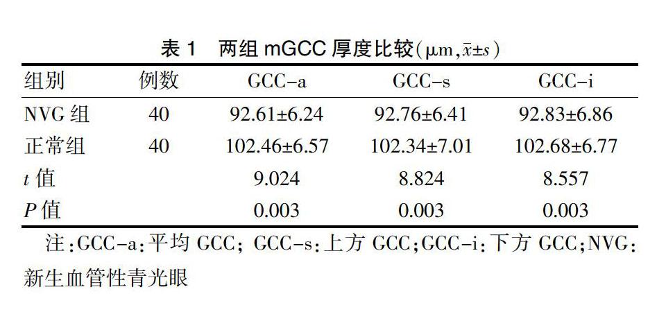
臨床評價NVG的常用手段是RNFL厚度,鮮有學者分析mGCC厚度。為此,本研究分析mGCC厚度與RNFL厚度的相關性及其對NVG的影響。研究顯示,NVG組GCC-a、GCC-s、GCC-i厚度均低于正常組(P < 0.05);NVG組RNFL-a、RNFL-t RNFL-s、RNFL-n、RNFL-i厚度均低于正常組(P < 0.05)。與許暢等[20]研究結果相似,提示mGCC厚度與RNFL厚度均能夠反映NVG患者的病理改變,有助于NVG的盡早發現和診斷。
本研究結果顯示,各部位mGCC與pRNFL厚度均呈正相關。提示將其用于診斷NVG患者具有較好的診斷效率。GCCs胞體盡管分布在大部分區域,但其僅為1層,在視盤周圍有2~3層,但在黃斑區域卻有8~10層。因而對NVG患者黃斑區的mGCC厚度進行檢測非常必要。
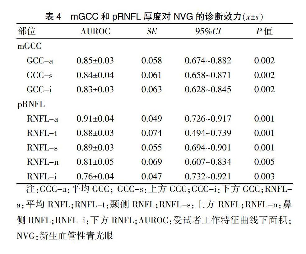
研究結果還顯示,各部位mGCC和pRNFL厚度對NVG均有診斷效力(P < 0.01)。pRNFL診斷效力為pRNFL-a厚度>pRNFL-s厚度>pRNFL-t厚度>pRNFL-n厚度>pRNFL厚度-i;而mGCC各個部位診斷效力相當。與Akcay等[21]研究結果相似。
綜上所述,在NVG診斷中,mGCC厚度可作為pRNFL厚度的一個重要補充手段,值得應用和推廣。
[參考文獻]
[1] ?徐麗娟,Nitter TA,梁遠波,等.視網膜神經節細胞復合體厚度檢測對原發性開角型青光眼的診斷意義[J].眼科,2015,24(1):26-30,35.
[2] ?張紅軍,趙世紅.視網膜黃斑區神經節細胞復合體厚度檢測的研究進展[J].第二軍醫大學學報,2018,39(4):417-421.
[3] ?Guclu H,Gorgulu Y,Gurlu VP,et al. Effects of selective serotonin reuptake inhibitors on macular ganglion cell complex thickness and peripapillary retinal nerve fiber layer thickness [J]. Curr Eye Res,2018,43(4):547-552.
[4] ?厲君.光相干斷層掃描測量視網膜神經節細胞復合體在青光眼中的研究進展[J].中華實驗眼科雜志,2018,36(4):299-304.
[5] ?謝靜,王輝,謝林英.青光眼視網膜神經纖維層厚度變化及其與視野缺損的相關性[J].眼科新進展,2015,35(12):1163-1165,1169.
[6] ?胡雅斌,郭彥,王懷洲,等.正常老年人群黃斑內層視網膜厚度和視盤周圍神經纖維層的改變[J].中華實驗眼科雜志,2018,36(4):274-278.
[7] ?薛博.青光眼疾病中視網膜膠質細胞的作用[J].中華實驗眼科雜志,2016,34(7):649-653.
[8] ?孔偉,余桂國,馮宇,等.毛果蕓香堿治療原發性急性閉角型青光眼的效果及對血流動力學的影響[J].中國醫藥導報,2017,14(6):135-138.
[9] ?宋蕾.原發性閉角型青光眼術后發生惡性青光眼的危險因素分析[J].中國醫藥導報,2016,13(7):51-54.
[10] ?呂建芳,方毅,楊艷蓓,等.原發性閉角型青光眼術后發生惡性青光眼的危險因素分析[J].中外醫療,2016,35(33):99-100.
[11] ?黃萍,王雯倩,石硯,等.貝伐單抗聯合小梁切除術或睫狀體光凝術治療晚期新生血管性青光眼療效比較[J].中華實驗眼科雜志,2015,33(4):362-366.
[12] ?萬道紅,趙強.小梁切除術聯合絲裂霉素和干擾素治療新生血管性青光眼療效觀察[J].國際眼科雜志,2015, 15(1):146-148.
[13] ?王中峰,楊雄里.谷氨酸受體在實驗性青光眼視網膜細胞損傷中的作用[J].生理學報,2016,68(4):483-491.