動態三維超聲造影評價肝腫瘤血流灌注的價值探討
2020-09-02 06:38:48黃增榮
中外醫學研究
2020年16期
黃增榮
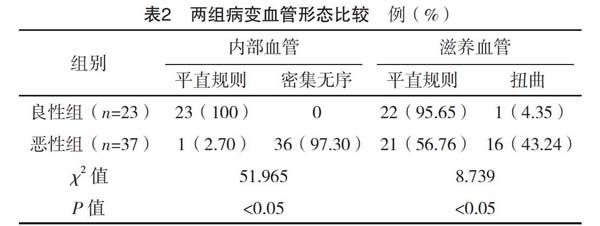

【摘要】 目的:分析動態三維超聲造影(3D-CEUS)評價肝腫瘤血流灌注的價值。方法:選擇2016年10月-2019年10月于筆者所在醫院就診的肝腫瘤患者60例作為本次研究對象,其中良性患者23例(良性組),惡性患者37例(惡性組)。兩組均采取動態三維超聲造影檢查,比較兩組檢查結果。結果:惡性組以整體增強、離心型增強、環狀增強作為主要表現,良性組以向心型增強作為主要表現,兩組增強方式比較差異有統計學意義(P<0.05)。良性組內部血管、滋養血管平直規則率均高于惡性組,差異有統計學意義(P<0.05)。結論:對于肝腫瘤患者而言,采取動態三維超聲造影進行檢查可有效分辨良惡性圖像,包括供血血管、內部小血管的走形、形態等,便于更加清晰直觀地分析肝腫瘤患者的影像學圖像,值得實踐與推廣。
【關鍵詞】 肝腫瘤 血流灌注 動態三維超聲造影
doi:10.14033/j.cnki.cfmr.2020.16.024 文獻標識碼 B 文章編號 1674-6805(2020)16-00-03
The Value of Dynamic Three-dimensional Contrast-enhanced Ultrasound in Evaluating Liver Tumor Blood Perfusion/HUANG Zengrong. //Chinese and Foreign Medical Research, 2020, 18(16): -63
[Abstract] Objective: To analyze the value of dynamic three-dimensional contrast-enhanced ultrasound (3d-CEUS) in the evaluation of liver tumor blood perfusion. Method: From October 2016 to October 2019, 60 patients with liver tumor admitted in our hospital were selected as the study objects, including 23 benign patients (benign group) and 37 malignant patients (malignant group). Both groups were examined by dynamic three-dimensional contrast-enhanced ultrasound. The results of the two groups were compared. Result: The main manifestations of the malignant group were whole enhancement, centrifugal enhancement and annular enhancement, while the main manifestations of the benign group were centripetal enhancement. The difference in the enhancement modes between the two groups was statistically significant (P<0.05). The regular rate of internal blood vessels and nourishing blood vessels in the benign group were higher than those in the malignant group, and the differences were statistically significant (P<0.05). Conclusion: For patients with liver tumor, dynamic three-dimensional contrast-enhanced ultrasound can effectively distinguish the benign and malignant images, including the shape and shape of blood supply vessels and internal small vessels, so as to analyze the imaging images of patients with liver tumors more clearly and intuitively, which is worthy of practice and promotion.
[Key words] Liver tumor Blood perfusion Dynamic three-dimensional contrast-enhanced ultrasound
First-authors address: Meixian District Traditional Chinese Medicine Hospital of Meizhou, Meizhou 514000, China
肝腫瘤是指出現在肝臟部位的腫瘤性病變,且肝臟同樣為腫瘤的好發部位,較為常見的肝腫瘤類型為惡性腫瘤中轉移性腫瘤,而良性腫瘤較為少見[1]。通常情況下,原發性腫瘤主要見于膽管上皮、肝細胞索與血管等部位,轉移性腫瘤多半為轉移性癌。隨著醫學技術的發展進步,超聲造影診斷方式也在不斷地更新換代,并廣泛應用于臨床各種疾病的診斷中,對肝腫瘤患者采取超聲造影方式進行診斷可顯示肝腫瘤在不同時相的血流動力學變化,在此基礎上發展而來的三維超聲造影技術,可對肝腫瘤血管空間結構進行更加清晰地觀察分析,與一般的三維超聲不同,動態三維超聲造影(3D-CEUS)診斷方式不僅僅局限于靜態的呈現,還可進行實時的動態灌注[2]。為了對該項診斷技術進行更加深入地分析判斷,本文將筆者所在醫院收治的60例肝腫瘤患者納入試驗,分析該項診斷方式的應用價值,現報道如下。……
登錄APP查看全文
