敲減SET8表達通過上調p53/DRAM1信號通路促進大鼠血管平滑肌細胞自噬和凋亡*
2022-03-28 02:25:04劉蘭張東雪朱榮芳李暉梁向楠張勝雷白亞玲
中國病理生理雜志 2022年3期
關鍵詞:檢測
劉蘭, 張東雪, 朱榮芳, 李暉, 梁向楠, 張勝雷, 白亞玲
敲減表達通過上調p53/DRAM1信號通路促進大鼠血管平滑肌細胞自噬和凋亡*
劉蘭, 張東雪, 朱榮芳, 李暉, 梁向楠, 張勝雷, 白亞玲△
(河北醫科大學第四醫院腎內科,河北 石家莊 050011)
探討賴氨酸甲基轉移酶SET8(SET domain-containing protein 8)低表達通過p53/DRAM1(DNA damage-regulated autophagy modulator 1)通路調控大鼠血管平滑肌細胞(vascular smooth muscle cells, VSMCs)自噬和凋亡的作用及機制。體外原代培養大鼠VSMCs,敲減表達,將細胞分為3組:正常組、空載體對照組和SET8-shRNA組。流式細胞術檢測細胞凋亡,MTT法檢測細胞活力,Western blot檢測SET8、p53、DRAM1、LC3、Bax和Bcl-2的蛋白水平。過表達,將大鼠VSMCs分為3組:正常組、空載體對照組和DRAM1組。檢測各組細胞活力,凋亡及DRAM1、LC3、Bax和Bcl-2的蛋白水平。敲減表達的同時敲減或表達,觀察LC3、Bax和Bcl-2的蛋白表達情況。(1)敲減表達后,大鼠VSMCs凋亡顯著增多,活力顯著降低(<0.05)。Western blot結果顯示,敲減表達后,SET8和Bcl-2表達顯著降低(<0.05),p53、DRAM1、LC3-II和Bax表達顯著升高(<0.05)。(2)過表達后,大鼠VSMCs凋亡顯著增多(<0.05),活力顯著降低(<0.05)。Western blot結果顯示,過表達后,DRAM1、LC3-II和Bax表達顯著升高(<0.05),Bcl-2表達顯著降低(<0.05)。(3)敲減或表達后,LC3-II和Bax表達降低,Bcl-2表達升高(<0.05)。敲減表達可促進大鼠血管平滑肌細胞發生自噬和凋亡,可能機制是通過上調p53和DRAM1表達,促進自噬蛋白LC3-II及凋亡蛋白Bax表達實現的。
血管平滑肌細胞;SET8蛋白;DRAM1蛋白;自噬;細胞凋亡
血管鈣化是增加心血管疾病發病率和死亡率的重要危險因素[1-2]。血管鈣化的重要機制有細胞的自噬、凋亡、表型轉化等[3-4]。SET8 (SET domain-containing protein 8)是現今發現的唯一能夠特異性催化H4賴氨酸20位的賴氨酸甲基轉移酶,能夠甲基化p53和Twist等非組蛋白,參與調控細胞增殖凋亡等生理學功能[5]。自噬基因(DNA damage-regulated autophagy modulator 1)是p53下游調節自噬和凋亡的基因,2006年由Crighton等[6]提出,可調節原發性腫瘤細胞的自噬和凋亡。作為DRAM依賴的一種自噬凋亡途徑,自噬能夠作為一種細胞死亡程序,誘導肝細胞凋亡及肝損傷[7]。現今SET8和DRAM1在大鼠血管平滑肌細胞(vascular smooth muscle cells, VSMCs)自噬和凋亡中的作用研究均較少,并且機制尚不明確。鑒于此,本項工作以體外培養的大鼠血管平滑肌細胞為研究對象,探討SET8是否通過p53/DRAM1通路調控大鼠血管平滑肌細胞的自噬和凋亡。
材料和方法
1 主要實驗試劑
選取胎牛血清(Gibco);SET8質粒和DRAM1質粒(廣州復能基因有限公司);Lipofectamine? 3000(Invitrogen);SET8抗體(Abcam);DRAM1和LC3抗體(華安生物技術有限公司);Bcl-2和Bax抗體(Abcam);GAPDH抗體(Bioworld)。
2 實驗方法
2.1實驗模型的制備與分組選取河北醫科大學動物實驗中心的健康雄性清潔級SD大鼠(證書號:1305090)6只,8~10周齡,體重80~100 g,做原代培養[8]。取胸主動脈中膜層,剪成小塊,均勻鋪于平底進行培養。取第3~4代細胞,給與刺激。將大鼠VSMCs分為3組:(1)正常normal)組:細胞未轉染;(2)對照(vector)組:轉染濃度為2 μg/L的對照NS-shRNA質粒;(3)SET8-shRNA組:轉染濃度為2 μg/L的SET8-shRNA質粒。檢測細胞自噬及凋亡情況。為驗證DRAM1對VSMCs自噬及凋亡的影響,將大鼠VSMCs分為3組:(1)normal組:細胞未轉染;(2)vector組:轉染對照質粒,濃度為2 μg/L;(3)DRAM1組:轉染DRAM1過表達質粒,濃度為2 μg/L。檢測細胞自噬及凋亡情況。
2.2MTT法檢測細胞活力取第3代大鼠血管平滑肌細胞接種于96孔板中,細胞融合達60%~70%給予刺激,每組設3個復孔。檢測前4 h將5 g/L的MTT 按每孔20 μL加入各檢測孔中,培養4 h,之后棄去培養液,將二甲基亞砜按每孔150 μl加入各檢測孔,室溫振蕩10 min。于酶標儀490 nm波長處測定吸光度()值,記錄結果。
2.3流式細胞術檢測細胞凋亡收集大鼠血管平滑肌細胞,根據試劑說明書進行Annexin V-PE/7-AAD雙重染色,使用流式細胞儀進行檢測,右下象限Annexin V陽性、7-AAD陰性,為早期凋亡細胞;右上象限Annexin V和7-AAD均為陽性,為晚期凋亡細胞。實驗重復3次。
2.4應用 Western blot檢測各指標蛋白的表達提取各組大鼠VSMCs的蛋白質,配制成12%的SDS-聚丙烯酰胺凝膠,測蛋白濃度并取20 μg蛋白質加樣,電泳條件95 V、1.5 h。使用PVDF膜轉膜,轉膜條件95 V、1 h。洗膜3次,牛血清白蛋白(5%)封閉1 h。加Ⅰ抗稀釋液(SET8, 1∶500; DRAM1, 1∶1 000; LC3, 1∶1 000; Bax, 1∶2 000; Bcl-2, 1∶1 000; p53, 1∶2 000; GAPDH, 1∶5 000),4 ℃孵育過夜;次日洗膜,加入II抗稀釋液(1∶5 000),37 ℃孵育1 h。使用蛋白成像系統照膜,實驗重復3次。
2.5共轉染實驗為驗證SET8對p53和DRAM1的調節作用,在大鼠VSMCs中轉染SET8-shRNA,并在轉染SET8-shRNA的同時共轉染p53-shRNA和DRAM1-shRNA,vector作為對照組,觀察VSMCs凋亡和自噬蛋白的表達情況。收集轉染48 h后的各組細胞,提取蛋白,檢測Bax、Bcl-2和LC3蛋白表達。
3 統計學處理
采用SPSS 20.0軟件進行統計學處理。呈正態分布的計量資料以均數±標準差(mean±SD)表示,兩組間比較采用檢驗,多組間的比較采用單因素方差分析,組間兩兩比較采用Student-Newman-Keuls(SNK)檢驗,<0.05為差異有統計學意義。
結果
1 敲減SET8對大鼠VSMCs凋亡的影響
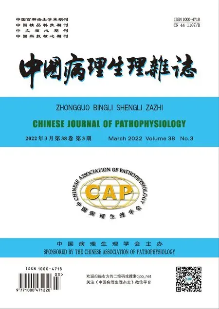
給予敲減表達后,采用Annexin V-PE/7-AAD染色,應用流式細胞儀檢測細胞凋亡的變化情況。結果顯示,與normal組和vector組比較,SET8-shRNA組細胞凋亡顯著增多(<0.05),見圖1。
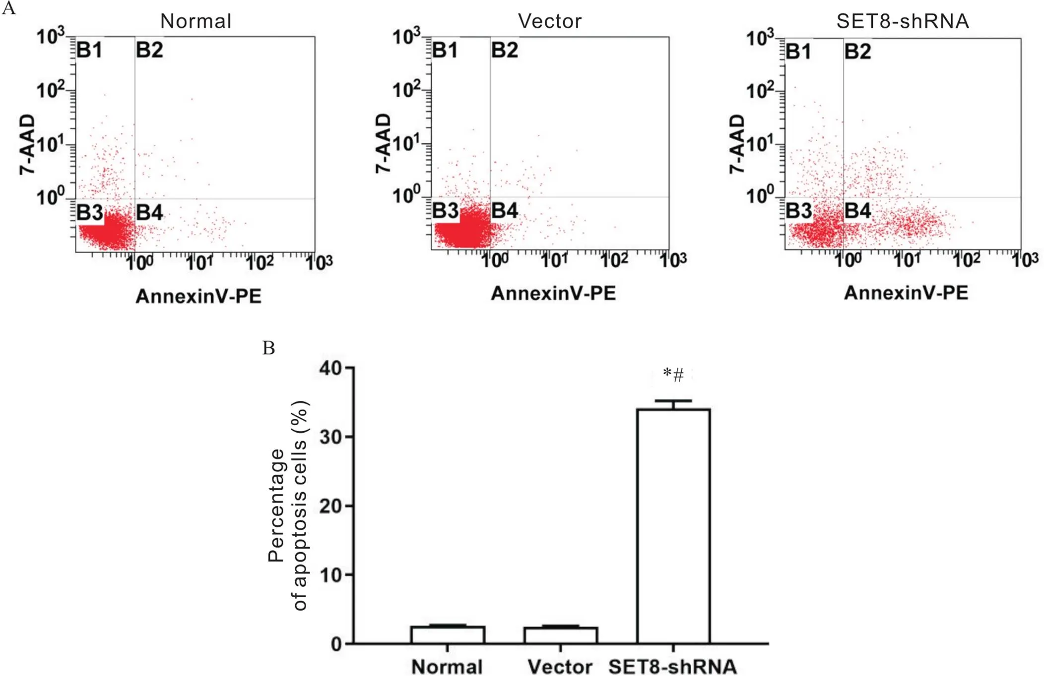
Figure 1.Effect of SET8 knockdown on apoptosis of rat VSMCs. A: flow cytometry results; B: percentage of apoptotic cells. Mean±SD. n=3. *P<0.05 vs normal group; #P<0.05 vs vector group.
2 敲減SET8對大鼠VSMCs活力的影響
MTT結果顯示,與normal組和vector組比較,SET8-shRNA組細胞活力于12 h、24 h、36 h和48 h均顯著降低(<0.05);normal組與vector組比較,差異無統計學意義(>0.05),見圖2。
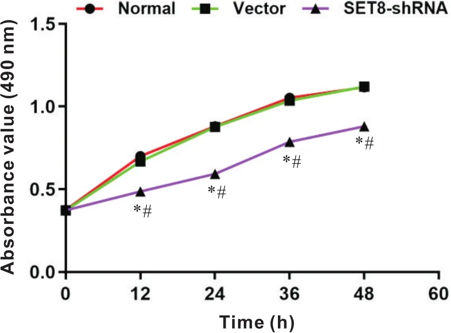
Figure 2.Effect of SET8 knockdown on rat VSMCs viability. Mean±SD. n=3. *P<0.05 vs normal group; #P<0.05 vs vector group.
3 敲減SET8表達后對各組大鼠VSMCs p53、DRAM1、LC3、Bcl-2和Bax表達的影響
Western blot結果顯示,與normal組和vector組比較,SET8-shRNA組SET8和Bcl-2蛋白相對表達量顯著降低,p53、DRAM1、LC3-II和Bax蛋白相對表達量顯著升高(<0.05),見圖3。

Figure 3.Effect of SET8 knockdown on the protein expression of p53, DRAM1, LC3, Bcl-2 and Bax in rat VSMCs in each group. A: Western blot results for each protein; B: relative protein expression. Mean±SD. n=3. *P<0.05 vs normal group; #P<0.05 vs vector group.
4 過表達DRAM1對大鼠VSMCs凋亡的影響
流式細胞術結果顯示,與normal組和vector組比較,DRAM1組細胞凋亡顯著增多(<0.05),見圖4。
Figure 4.Effect of overexpression of DRAM1 on apoptosis of rat VSMCs. A: flow cytometry results; B: percentage of apoptotic cells. Mean±SD. n=3. *P<0.05 vs normal group; #P<0.05 vs vector group.
5 DRAM1對大鼠VSMCs活力的影響
MTT結果顯示,與normal組和vector組比較,DRAM1組細胞活力于12 h、24 h、36 h和48 h均顯著降低(0.05);normal組與vector組比較,差異無統計學意義(0.05),見圖5。
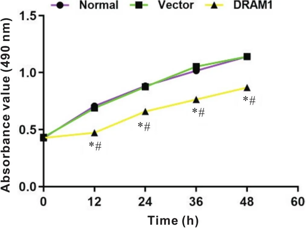
Figure 5.Effect of overexpression of DRAM1 on the viability of rat VSMCs. Mean±SD. n=3. *P<0.05 vs normal group; #P<0.05 vs vector group.
6 DRAM1過表達對各組大鼠VSMCs LC3、Bcl-2和Bax表達的影響
Western blot結果顯示,與normal組和vector組比較,DRAM1組Bcl-2蛋白相對表達量顯著降低,DRAM1、LC3-II和Bax蛋白相對表達量顯著升高(<0.05),見圖6。
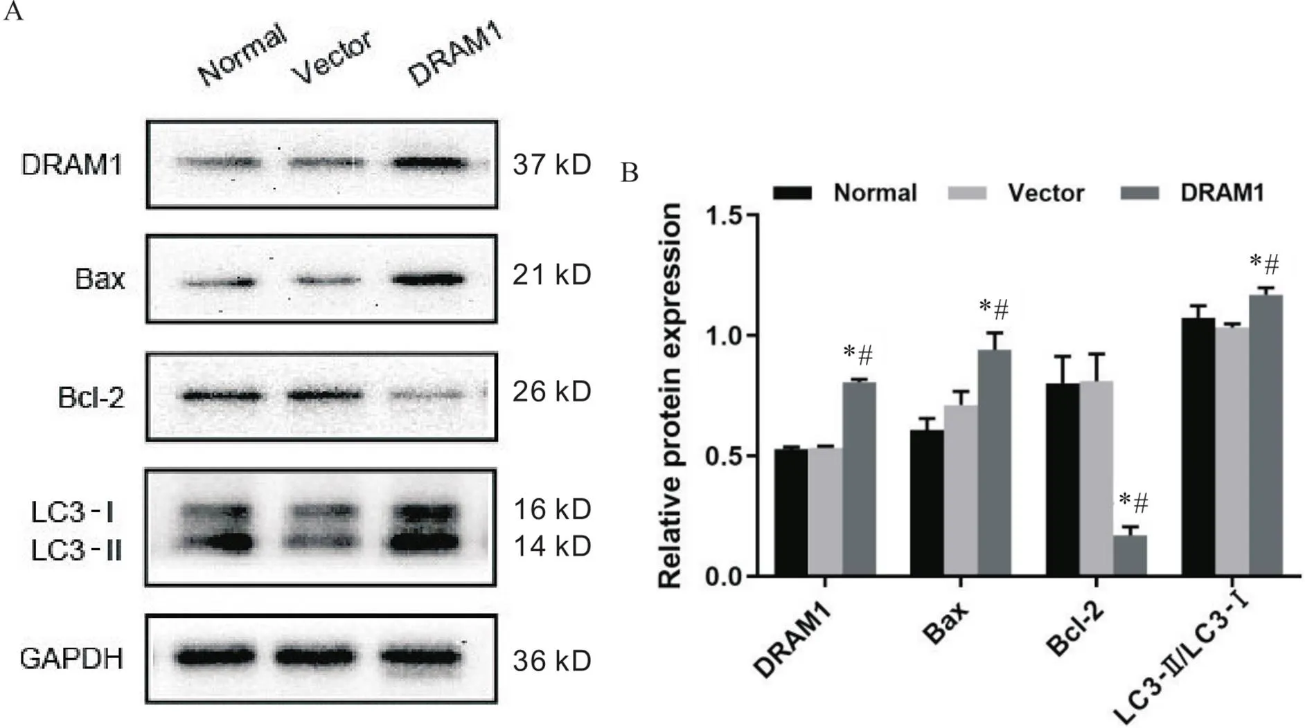
Figure 6.Effects of overexpression of DRAM1 on the expression of LC3, Bcl-2 and Bax proteins in rat VSMCs. A: Western blot results for each protein; B:r elative protein expression. Mean±SD. n=3. *P<0.05 vs normal group; #P<0.05 vs vector group.
7 SET8-shRNA與p53-shRNA和DRAM1-shRNA共轉染
與vector組比較,將SET8-shRNA轉染至VSMCs后,Bax及LC3-II蛋白表達增高,Bcl-2蛋白表達顯著降低(0.05);與SET8-shRNA組比較,SET8-shRNA與p53-shRNA共同轉染VSMCs后,Bax及LC3-II蛋白表達降低,Bcl-2蛋白表達顯著升高(0.05);與SET8-shRNA組比較,SET8-shRNA與DRAM1-shRNA共同轉染VSMCs后,Bax及LC3-II蛋白表達降低,Bcl-2蛋白表達升高(0.05),見圖7。
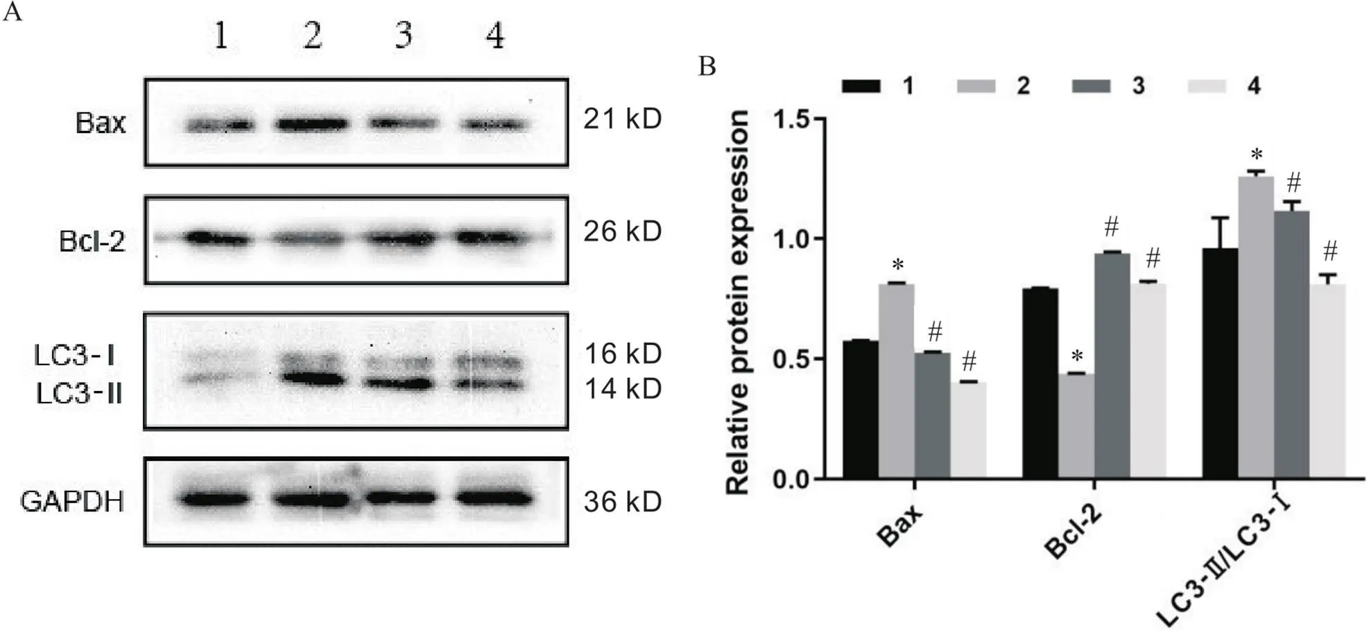
Figure 7.Effects of co-transfection of SET8-shRNA with p53-shRNA or DRAM1-shRNA on the expression of Bax, Bcl-2 and LC3 proteins. A: Western blot results for each protein; B: relative protein expression. 1: vector group; 2: SET8-shRNA group; 3: SET8-shRNA+p53-shRNA group; 4,SET8-shRNA+DRAM1-shRNA group. Mean±SD. n=3. *P<0.05 vs vector group; #P<0.05 vs SET8-shRNA group.
討論
血管鈣化是心血管疾病死亡率增高的獨立危險因素[9]。而細胞的凋亡、自噬和表型轉化等是引起血管鈣化的重要機制[10-11]。參與細胞凋亡和自噬的通路有多種。本項工作以體外培養的大鼠血管平滑肌細胞為研究對象,探討SET8調控大鼠血管平滑肌細胞自噬和凋亡的發生機制。研究結果顯示SET8低表達可誘導大鼠血管平滑肌細胞發生自噬與凋亡,其可能機制是通過促進p53及DRAM1表達實現的。
賴氨酸甲基轉移酶SET8可以特異性單甲基化抑癌基因的382賴氨酸位點(K382me1),能夠抑制的靶基因轉錄,進而影響細胞的增殖、凋亡等生物學功能[12-13]。而抑癌基因參與細胞生存和分裂的控制,對細胞凋亡、自噬和細胞周期有重大的影響[14-15]。Zhang等[12]顯示敲減表達可通過促進p53表達進而促進胃癌細胞發生凋亡。也有研究顯示在血管平滑肌細胞凋亡中SET8也起重要作用[16]。但SET8在大鼠血管平滑肌細胞凋亡和自噬中的具體作用機制尚不明確,因此本研究以體外培養大鼠血管平滑肌細胞為模型,通過敲減表達,觀察大鼠血管平滑肌細胞凋亡情況及自噬蛋白DRAM1和LC3-II等蛋白變化。本研究結果顯示敲減表達后,p53、DRAM1和LC3-II表達上調,促凋亡蛋白Bax表達升高,抑凋亡蛋白Bcl-2表達降低,大鼠血管平滑肌細胞增殖能力降低,凋亡增多。提示敲減后,可通過上調p53、DRAM1、LC3-II和Bax表達,抑制Bcl-2的表達,進而促進大鼠血管平滑肌細胞發生凋亡。
有研究顯示過表達能夠上調自噬蛋白LC3-II表達,進而誘導癌細胞發生自噬[17]。也有研究表明DRAM1能夠上調促凋亡蛋白Bax表達,進而促進宮頸癌細胞凋亡[18]。而Bax和Bcl-2是一對正負凋亡調節因子,Bax起促進凋亡作用,Bcl-2起抗凋亡作用[19]。因此,我們推測DRAM1可能通過調控Bax、Bcl-2和LC3,進而誘導大鼠血管平滑肌細胞發生自噬和凋亡。本研究以體外培養大鼠血管平滑肌細胞為模型,給予過表達,觀察大鼠血管平滑肌細胞凋亡及自噬情況。結果顯示給予過表達后,自噬相關蛋白LC3-II表達升高,促凋亡蛋白Bax表達升高,抑凋亡蛋白Bcl-2表達降低,大鼠血管平滑肌細胞凋亡增多,增殖顯著減少。結果提示,DRAM1可通過上調自噬蛋白LC3-II和凋亡蛋白Bax表達,抑制Bcl-2的表達,進而促進大鼠血管平滑肌細胞發生自噬和凋亡。2006年Crighton等[6]提出,是下游調節自噬的重要基因。編碼238個氨基酸,是調控自噬和凋亡的溶酶體膜蛋白[20-21]。為驗證SET8是否通過p53和DRAM1通路調控大鼠血管平滑肌細胞自噬和凋亡,給予抑制表達的同時抑制和表達,結果顯示與只抑制表達組相比,抑制同時抑制或表達后,Bax和LC3-II顯著降低,Bcl-2顯著升高。
綜上所述,SET8低表達可以促進大鼠血管平滑肌細胞發生自噬和凋亡,可能機制是通過調節p53/DRAM1表達,促進自噬蛋白LC3-II和凋亡蛋白Bax表達,抑制抗凋亡蛋白Bcl-2的表達,進而引起大鼠血管平滑肌細胞發生了自噬和凋亡。
[1] Yamada S, Giachelli CM. Vascular calcification in CKD-MBD: roles for phosphate, FGF23, and Klotho[J]. Bone, 2017, 100:87-93.
[2] Disthabanchong S, Srisuwarn P. Mechanisms of vascular calcification in kidney disease[J]. Adv Chronic Kidney Dis, 2019, 26(6):417-426.
[3] Liu R, Leslie KL, Martin KA. Epigenetic regulation of smooth muscle cell plasticity [J]. Biochim Biophys Acta, 2015, 1849(4):448-453.
[4]周薇, 徐金升, 白亞玲, 等. miR-30b在高磷誘導的大鼠血管平滑肌細胞鈣化中的作用[J]. 中國病理生理雜志, 2021, 37(1):54-59.
Zhou W, Xu JS, Bai YL, et al. Role of miR-30b in calcification of rat vascular smooth muscle cells induced by high phosphorus[J]. Chin J Pathophysiol, 2021, 37(1):54-59.
[5] Yang C, Wang K, Zhou Y, et al. Histone lysine methyltransferase SET8 is a novel therapeutic target for cancer treatment[J]. Drug Discov Today, 2021, 26(10):2423-2430.
[6] Crighton D, Wilkinson S, O'Prey J, et al. DRAM, a p53-induced modulator of autophagy, is critical for apoptosis[J]. Cell, 2006, 126(1):121-134.
[7] Xu J, Zang Y, Liu D, et al. DRAM is involved in hypoxia/ischemia-induced autophagic apoptosis in hepatocytes[J]. Aging Dis, 2019, 10(1):82-93.
[8] Cui L, Bai Y, Zhang J, et al. Effects of extracellular acid stimulation on rat vascular smooth muscle cell in gas6/Axl or PI3K/Akt signaling pathway[J]. Clin Exp Hypertens, 2016, 38(5):451-456.
[9] Kakani E, Elyamny M, Ayach T, et al. Pathogenesis and management of vascular calcification in CKD and dialysis patients[J]. Semin Dial, 2019, 32(6):553-561.
[10] Gu Q, Li F, Ge S, et al. CDC20 knockdown and acidic microenvironment collaboratively promote tumorigenesis through inhibiting autophagy and apoptosis[J]. Mol Ther Oncolytics, 2020, 17:94-106.
[11] Grootaert MOJ, Moulis M, Roth L, et al. Vascular smooth muscle cell death, autophagy and senescence in atherosclerosis[J]. Cardiovasc Res, 2018, 114(4):622-634.
[12] Zhang X, Peng Y, Yuan Y, et al. Histone methyltransferase SET8 is regulated by miR-192/215 and induces oncogene-induced senescence via p53-dependent DNA damage in human gastric carcinoma cells[J]. Cell Death Dis, 2020, 11(10):937.
[13] Watts AJ, Storey KB. Hibernation impacts lysine methylation dynamics in the 13-lined ground squirrel, ictidomys tridecemlineatus[J]. J Exp Zool A Ecol Integr Physiol, 2019, 331(4):234-244.
[14] Kang R, Kroemer G, Tang D. The tumor suppressor protein p53 and the ferroptosis network[J]. Free Radic Biol Med, 2019, 133:162-168.
[15]王美月, 賀燕婷, 于潔, 等. p53在層流剪切應力下對EPCs分化及功能的影響[J]. 中國病理生理雜志, 2021, 37(2):225-231.
Wang MY, He YT, Yu J, et al. Role of p53 in differentiation and function of EPCs under laminar shear stress[J]. Chin J Pathophysiol, 2021, 37(2):225-231.
[16] 張東雪, 高少輝, 張勝雷. SET8介導AKT信號通路在調控高磷誘導的血管平滑肌細胞鈣化中的作用[J]. 中華腎臟病雜志, 2020, 36(3):214-220.
Zhang D, Gao S, Zhang S. SET8 regulates high-phosphorus-induced vascular smooth muscle cell calcification via AKT signaling pathway[J]. Chin J Nephrol, 2020, 36(3):214-220.
[17] Lu T, Zhu Z, Wu J, et al. DRAM1 regulates autophagy and cell proliferation via inhibition of the phosphoinositide 3-kinase-Akt-mTOR-ribosomal protein S6 pathway[J]. Cell Commun Signal, 2019, 17(1):28.
[18] Zhang X, Wang Y, Zhao A, et al. Long non-coding RNA LINC00511 accelerates proliferation and invasion in cervical cancer through targeting miR-324-5p/DRAM1 axis[J]. Onco Targets Ther, 2020, 13:10245-10256.
[19] Zhang Y, Yang X, Ge X, et al. Puerarin attenuates neurological deficits via Bcl-2/Bax/cleaved caspase-3 and Sirt3/SOD2 apoptotic pathways in subarachnoid hemorrhage mice[J]. Biomed Pharmacother, 2019, 109:726-733.
[20] Hu W, Chen S, Thorne RF, et al. TP53, TP53 target genes (DRAM, TIGAR), and autophagy[J]. Adv Exp Med Biol, 2019, 1206:127-149.
[21] Lee HY, Oh SH. Autophagy-mediated cytoplasmic accumulation of p53 leads to apoptosis through DRAM-BAX in cadmium-exposed human proximal tubular cells[J]. Biochem Biophys Res Commun, 2021, 534:128-133.
(責任編輯:盧萍,李淑媛)
Knockdown ofpromotes autophagy and apoptosis of rat vascular smooth muscle cells by up-regulating p53/DRAM1 signaling pathway
LIU Lan, ZHANG Dong-xue, ZHU Rong-fang, LI Hui, LIANG Xiang-nan, ZHANG Sheng-lei, BAI Ya-ling△
(,,050011,)
To investigate whether lysine methyltransferase SET domain-containing protein 8 () knockdown regulates the autophagy and apoptosis of rat vascular smooth muscle cells (VSMCs) through p53/DNA damage-regulated autophagy modulator 1 (DRAM1) signaling pathway.Rat VSMCs were divided into 3 groups: normal group, vector group and SET8-shRNA group. Apoptosis was detected by flow cytometry, viability was detected by MTT, and the protein levels of SET8, p53, DRAM1, LC3, Bax and Bcl-2 were detected by Western blot. VSMCs of rats were divided into 3 groups: normal group, vector group and DRAM1 overexpression group. Cell viability, apoptosis and protein levels of DRAM1, LC3, Bax and Bcl-2 were detected.The protein expressions of LC3, Bax and Bcl-2 were observed in VSMCs of rats following suppression oforwhile interfering with.The apoptosis of VSMCs was increased significantly and viability was decreased significantly afterknockdown(<0.05). Western blot showed that the expressions of SET8 and Bcl-2 were decreased, and the expressions of p53, DRAM1, LC3-II and Bax were increased significantly afterknockdown(<0.05). Apoptosis of VSMCs was increased significantly and viability was decreased significantly after overexpression of(<0.05). Western blot showed that the expressions of DRAM1, LC3-II and Bax were increased and the expression of Bcl-2 was decreased after overexpression of DRAM1 (<0.05). The expressions of LC3 and Bax were decreased and the expression of Bcl-2 was increased after inhibiting the expression ofor(<0.05).Autophagy and apoptosis in vascular smooth muscle cells of rats were promoted by knockdown ofThe possible mechanism is to promote the expression of autophagy protein LC3-II and apoptotic protein Bax by upregulating the expressions of p53 and DRAM1.
Vascular smooth muscle cells; SET8; DRAM1; Autophagy; Apoptosis
R363.2; R543
A
10.3969/j.issn.1000-4718.2022.03.006
1000-4718(2022)03-0427-07
2021-08-20
2022-01-04
[基金項目]河北省醫學科學研究重點課題(No. 20180513; No. 20190702)
Tel: 15081150811; E-mail: snbyl@163.com
猜你喜歡
中國設備工程(2022年12期)2022-07-11 04:33:00
中學生數理化·七年級數學人教版(2021年6期)2021-11-22 07:50:58
中學生數理化·七年級數學人教版(2021年6期)2021-11-22 07:50:58
中學生數理化·七年級數學人教版(2021年6期)2021-11-22 07:50:58
中學生數理化·七年級數學人教版(2020年12期)2021-01-18 06:57:46
中學生數理化·七年級數學人教版(2020年12期)2021-01-18 06:57:46
中學生數理化·七年級數學人教版(2019年9期)2019-11-25 07:34:36
中學生數理化·七年級數學人教版(2019年9期)2019-11-25 07:34:34
中學生數理化·七年級數學人教版(2019年12期)2019-05-21 02:53:50
中學生數理化·七年級數學人教版(2019年12期)2019-05-21 02:53:48