應用納米球刻蝕法在自組裝膜修飾的硅表面生成中尺度的網狀蛋白層
2017-05-12 06:58:02SCHLERETHAndrewNOOMUNAPanaeGAOPei
物理化學學報 2017年4期
SCHLERETHAndrew NOOMUNAPanae GAO Pei
應用納米球刻蝕法在自組裝膜修飾的硅表面生成中尺度的網狀蛋白層
SCHLERETHAndrew NOOMUNAPanae GAO Pei*
(Department of Chemistry,Eastern Kentucky University,521 Lancaster Ave,Richmond 40475,KY,USA)
在自組裝膜修飾的硅表面制備有序的蛋白陣列是研發生物傳感器的先決條件之一,因此如何產生有序的表面蛋白陣列一直是生物醫藥研究方向的前沿。本研究通過應用納米球刻蝕法在氧化的10-烯基十一烷基三氯硅烷自組裝膜修飾的硅表面生成了網狀結構溶菌酶蛋白層。網孔的大小(從納米到微米級別)由表面沉積的納米球的尺寸來調控。我們利用原子力顯微鏡和熒光顯微鏡對樣品表面進行了詳細表征。結果表明:這種新方法比傳統的通過掃描探針在固體表面修飾而聚集溶菌酶蛋白的方法更快捷簡便,而且它能夠在相對大的硅表面形成網狀蛋白層。此外,網孔表面附著具有強吸附活性的羧酸基團層,它可以通過靜電吸引或者共價結合來吸附液相中的第二種蛋白分子。
納米球刻蝕法;溶菌酶;網狀陣列;自組裝膜;原子力顯微鏡;熒光顯微鏡
1 Introduction
Nanoscale protein patterns have the potential to be used in many fields including enzyme catalysis,biosensors,tissue engineering,diagnostic protein tips,and pharmacology1-5.However, precisely and easily immobilizing protein molecules onto a specific place on a surface while retaining their native biological functionality is difficult and challenging.Although there are lots of surface patterning techniques such as microcontact printing (μCP)6,nanoimprinting7,8,dip-pen lithography9,nanografting10-13and conducting tip AFM(atomic force microscopy)writing14,15, they either lack the surface coverage or a specialized stamp is required in the patterning of protein arrays on surfaces.To meet the nanometer precision and achieve high throughput,particle lithography has been investigated and applied for various nanoscale patterning purposes on surfaces16-19.Cai and Ocko20have demonstrated a simple nanosphere based lithography method to direct the assembly of proteins into arrays over macroscopic surface area.In this method,the self-assembly of a nanosphere monolayer on a solid surface provides a simple and effective mask to prevent the deposition and modification directly beneath the nanospheres.Therefore,in the subsequent surface processing,only the interstitial area between nanospheres is exposed,and thus a hexagonally patterned surface will be formed as nanospheres are later removed.In this study,he utilized both nanosphere lithography and silane chemistry to chemically pattern surfaces with a regular array islands over cm sample regions.These chemically patterned regions are used as a template to selectively adsorb lysozyme on carboxylic acid-terminated islands and not on the interstitial regions between the islands.In this manner,positivetoned protein nanoarrays are fabricated on the surface.Based on this method,we also developed a kind of negative-toned nanoarrays by selectively adsorbing protein molecules directly on the interstitial areas between the nanospheres on the carboxylic acid terminated(UTSox)silane monolayer coated silicon surface. Compared with Cai′s approach,this method involves fewer steps and has higher reproducibility.
To finish the fabrication,firstly,the self-assembled undecyltrichlorosilane(UTS)monolayer was grown and chemically converted into an oxidized undecyltrichlorosilane(UTSox)layer on the silicon surface,which generated a layer that coated the substrate with carboxylic acid-terminated functional groups for the subsequent fabrication.Furthermore,polystyrene nanospheres form an ordered monolayer on the hydrophilic UTSox coated surface upon vaporization from a solution.This two dimensional crystal layer can provide a simple and effective mask to prevent the deposition and modification directly beneath the nanospheres. Therefore,in the subsequent surface processing,only the interstitial areas between nanospheres are exposed for the adsorption of lysozyme molecules from the solution.When the nanospheres were removed from the surface,negative-toned protein patterns were fabricated on the UTSox monolayer coated silicon surface. Therefore,we demonstrated a simpler nanosphere based lithography method to assemble protein patterns ranged from nanometer to sub-millimeter over macroscopic surface area.The similar negative-toned nanoarrays have been reported by Liu21and Kingshott22groups,respectively.However,extra steps of plasma polymerization and covalent grafting were needed and all these requirements limit it from being a generic patterning method for other protein patterns.
In comparison,nanosphere lithography requires less steps and is more reliable to be applied on many biological molecules fabrication23-25and other different purposes26,27.Additionally,the method in this paper will provides an effective means to immobilize more than one protein on the same surface,which is necessary for the development of biosensors that can detect multiple analytes,especially in cancer diagnosis and monitoring,since most types of cancer involve multiple biomarkers.
2 Materials and methods
2.1 Materials
10-undecenyltricholorosilane(UTS)was purchased from Gelest (United States).Toluene(99.8%),hydrogen peroxide(30%), sulfuric acid(98%),and 1-pyrenyldiazomethane(PDAM)were purchased from Sigma-Aldrich(United States).Silicon(100) wafers(Virginia Semiconductors from United States,Nitrogen doped,30 Ω·cm resistivity)were polished to ultraflat grade with a root-mean-square(rms)roughness smaller than 0.5 nm.
2.2 Instrumentation
The chemical pattern characterization was performed with the Agilent(United States)PicoPlus AFM in an environmental chamber.The patterns were characterized in the tapping mode with MikroMasch(United States)NSC-14 tips,which have a typical natural frequency of 150 kHz and a force constant of 5 N· m-1.The UTS film thickness and quality were examined with an Angstrom Advance PhE 101 ellipsometer(United States)and a Varian Excalibur 3100 Fourier transformed infrared spectrometer equipped with a semiconductor cooled DTGS detector(United States).The IR spectra of UTS and oxidized UTS films on silicon wafer were taken at 4 cm-1resolution with 4096 scans.The fluorescent images were acquired using Nikon Eclipse 80i microscope(United States).
2.3 Preparation of self-assembled 10-undecenyltricholorosilane(UTS)film
The silicon wafer was boiled in the piranha solution(1 part of 30%hydrogen peroxide solution in 3 parts of 98%sulfuric acid) at 125°C for 20 min.After rinsing in de-ionized water and drying in a stream of nitrogen,the wafer was dipped in a 5 mmol·L-1UTS toluene solution overnight.A monolayer of closely packed UTS film was formed after the overnight soaking in the UTS toluene solution.The UTS film was examined with the Agilent PicoPlus 3000 AFM,the Varian Exalibur FT-3100FT-IR spectrometer and the PhE-101 ellipsometer.Results fromAFM showed that the UTS film was an ultra-flat surface with an RMS roughness smaller than 0.2 nm.Over the AFM scanner′s range(80μm×80 μm),the height variation was smaller than 5 nm.The UTS surface is a featureless homogenous surface.The infrared spectroscopyresults demonstrated the successful conversion from the terminal double bond on the UTS silane surface into a carboxylic acidterminated C10 silane(referred to in the following as UTSox) film.The ellipsometry results indicated that the UTS film thickness was(1.5±0.3)nm,which was consistent with the published data28.
2.4 Formation of PDAM labelled oxidized 10-undecenyltricholorosilane(UTSox)patterns between nanospheres on the surface
Apiece of 1 cm×1 cm UTS layer coated wafer was incubated in the oxidizing solution(0.01 mol·L-1NaIO4,5×10-4mol·L-1KMnO4,in 0.05 mol·L-1Na2CO3buffer)for 10 h at 40°C29.To remove remnant permanganate or MnO2,the sample was then rinsed with deionized water and followed by 1%hydrazine for 1 min.As prepared,the wafer surface is hydrophilic,suggesting that the existence of the carboxylic acid-terminated UTSox layer on the surface.Finally,the sample was dried in a stream of nitrogen. Then,1.5 μL of 1%900 nm polystyrene nanosphere aqueous solution was spread onto the dried UTSox layer coated silicon wafer.The wafer was covered and left to dry for 24 h at room temperature.The nanospheres acted as a mask which inhibited the subsequent adsorption in the regions covered by them.Furthermore,the sample was then dipped into 3 mmol·L-11-pyrenyldiazomethane(PDAM)methanol solution to start the reaction. After 10 hours of incubation,the sample was rinsed and ultrasonicated in methanol.When the sample was dried with a steam of Nitrogen gas,it was checked with Nikon Eclipse 80i Microscope.The experimental procedure follows Scheme 1 with the use of 3 mmol·L-11-pyrenyldiazomethane methanol solution instead of fluorescein isothiocyanate(FITC)tagged lysozyme solution.
2.5 Fabrication of Lysozyme patterns on UTSox layer coated Si(100)wafer
In this study,the protein lysozyme was used since it is readily available and its structure and properties are well-known.Lysozyme molecules could be immobilized on the carboxylic acidterminated OTSpd and UTSox patterns through the Coulomb attraction30,31.The carboxylic acid-terminated silane surface has an estimated pKa(acid dissociation constant)of 4.932.Lysozyme is positively charged in a pH 7 buffer solution since it has an isoelectric point of 1133.Thus,during the incubation in the pH 7 buffer solution,the positively charged lysozyme molecules will selectively adsorb on the negatively charged carboxylic acid terminated areas based on the electrostatic interactions.
A carboxylic acid terminated chemical patterned surface was generated through the usage of nanosphere monolayer mask along with silane chemistry.The experimental procedure is illustrated in Scheme 1.A layer of nanosphere mask was assembled on a carboxylic acid terminated UTSox monolayer.Then,the dried nanosphere-coated wafer was incubated in 5 μmol·L-1FITC tagged lysozyme solution in a 25 mmol·L-1pH=7,HEPES buffer for another 12 h.The sample was then dried with a burst of compressed nitrogen,and subsequently rinsed with a 25 mmol·L-1pH 7 HEPES buffer solution.The excessive solution was removed by touching a paper towel to the edge of the substrate,and the surface was investigated by using a Nikon Eclipse 80i Microscope immediately after the sample was dried.When the nanospheres were further removed through ultrasonication in water,the sample surface was imaged by atomic force microscopy(AFM).
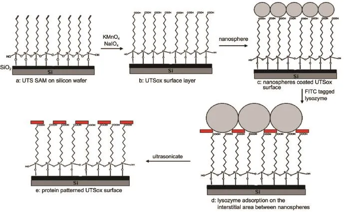
Scheme 1 Procedure for preparation of large-scale negative-toned protein patterns through nanosphere lithography
3 Results and discussion
3.1 Self-assembled UTSox monolayer on silicon surface
In the Brewster angle IR spectra of the UTS wafer before and after oxidization,we saw that the 3080 and 1645 cm-1peaks of the vinyl group disappear and a new peak at 1710 cm-1appears,which corresponds to the carboxylic acid groups.It can be concluded that the double bonds were successfully converted into carboxylic acid groups after the above oxidation process,which is consistent with the published results29,34.The Brewster angle IR spectra of the UTS monolayer and UTSox monolayer could be found in the supporting information of my published paper30.
In order to optimize the preparation conditions to obtain the best quality patterned surfaces,the exposed UTSox patterns areas on the mask coated surface were firstly investigated.It is known that 1-pyrenyldiazomethane(PDAM)can specifically react with the carboxylic acid group to form an ester product and generate fluorescence upon excitation35,36.The schematic equation about this reaction is illustrated in Scheme 2.Therefore,PDAM can be used as a fluorescence-labeling reagent to identify the chemical properties of UTSox surface.After 10 h of incubation in 3 mmol· L-1PDAM methanol solution and dried with a steam of Nitrogen gas,the binding of carboxylic acid-terminated UTSox layer with PDAM was directly visualized by fluorescent microscopy.The fluorescence image in Fig.1a shows the existence of orderly arranged nanospheres on the surface and the strong fluorescence signal emitted by the PDAM molecules.Since the surface was coated by a monolayer of nanospheres,the PDAM molecules did not attach to the parts of surface that are directly underneath the nanospheres.Therefore,the green color on surface indicated the binding of PDAM with carboxylic acid groups in the interstitial area between nanospheres,and dark color areas indicated where nanospheres had occupied the surface.This could also be clearly identified from Fig.1b,which showed the zoomed blue circled area in Fig.1a.Since the control experiment showed that the fluorescence signal from the un-labeled protein was insignificant for the exposure time we used,it can be concluded that the fluorescence signal generated in the fluorescence image is from the FITC-tagged protein molecules accumulated in the interstitial areas between the nanospheres.
3.2 Fabrication of lysozyme patterns on UTSox surface
After verifying the effectiveness of each step in the protein patterning scheme,analysis of the final protein pattern was conducted.By using a relatively slow solution evaporation rate,a reasonably uniform monolayer of the polystyrene nanospheres was obtained on the entire 1 cm×1 cm wafer.The fluorescence microscope shows nanospheres are well ordered on the surface. This uniformity is key in ensuring that the protein pattern is also uniform.When the fluorescein isothiocyanate(FITC)tagged lysozyme was deposited onto a wafer which had a nanosphere mask present,the resulting pattern showed that the nanospheres were effective in acting as a mask for the surface underneath the nanospheres.The images captured by the florescence microscope of the patterned FITC tagged lysozyme can be seen in Fig.2.
Fig.2a demonstrated the UTSox surface contained both polystyrene nanospheres and FITC tagged lysozyme molecules under the UV light.In Fig.2a,the nanospheres appear to have a dark color and the FITC tagged lysozyme gave off a light green color. We can see that nanospheres were orderly arranged on the surface, and FITC tagged lysozyme molecules accumulated in the interstitial areas between nanosphere.Fig.2b shows the same surface after the nanospheres on the substrate were removed by the ultrasonication in water,and the image was captured under the whitelight instead.In comparison,the dark dots in Fig.2b signified where nanospheres had occupied the surface,and the lighter regions of the pattern simply correspond with higher concentrations of the lysozyme relative to the other regions.Upon analysis,the protein pattern utilizing the polystyrene nanospheres was very uniform and distinct.This uniformity encompassed large areas of space on the silicon wafer(up to 1 cm).
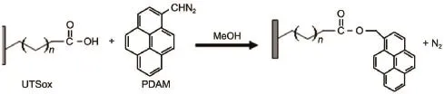
Scheme 2 Schematic reaction of PDAM with carboxylic acid group on OTSpd and UTSox
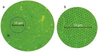
Fig.1 (a)Fluorescence microscope image of the UTSox monolayer dyed with PDAM;(b)the zoomed area of PDAM labelled UTSox patterns in the blue circle in image a
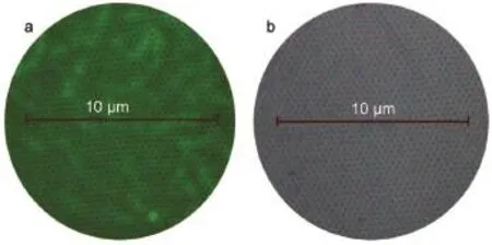
Fig.2 (a)Image of the patterned UTSox coated silicon wafer under UV light;(b)image of the same area under white light after removing the nanospheres
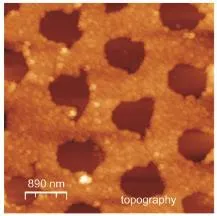
Fig.3 AFM tapping mode topographic image of lysozymepatterns on the UTSox silane surface through Scheme 1
In Fig.3,we showed an AFM image of lysozyme patterned surface after the polystyrene nanospheres were removed by the ultrasonication in water.All the round brown colored holes on the images represent the areas where polystyrene nanospheres had occupied,and the yellow colored mesh show the accumulation of protein patterns.Each hole is roughly 900 nm in diameter,which is consistent with the size of the nanospheres we applied on the surface.
To provide further proof that the pattern on the surface are from the lysozyme,additional control measurements were carried out. In the first experiment,a buffer solution without lysozyme was applied to the polystyrene nanosphere coated UTSox surface. AFM scans showed no patterns after the nanospheres were removed.
4 Conclusions
In summary,we have developed a method to fabricate negativetoned mesoscale protein patterns by incorporating nanosphere lithography and surface silane chemistry.This method has some incomparable advantages such as simplicity,mild environment and high throughout.Moreover,by using different sized polystyrene nanosphere,the area density of the protein array can be varied.These promising results indicate that this method could be easily adopted for the fabrication of wafer sized different protein patterns with mesoscale,which is the first step in the production of various biosensors.Furthermore,the negative-toned protein patterns surface with carboxylic acid group terminated surface as background can be used to assemble another type of protein molecules on the same surface.The surface with two different protein patterns is necessary for the biosensor to detect multiple analyzes in practice.
(1) Rosi,N.L.;Mirkin,C.A.Chem.Rev.2005,105,1547. doi:10.1021/cr030067f
(2) Ngunjiri,J.;Garno,J.C.Anal.Chem.2008,80,1361. doi:10.1021/ac086049l
(3) Christman,K.L.;Enriquez-Rios,V.D.;Maynard,H.D.Soft Matter 2006,2,928.doi:10.1039/b611000b
(4) Cornell,B.A.;BraachMaksvytis,V.L.B.;King,L.G.;Osman, P.D.J.;Raguse,B.;Wieczorek,L.;Pace,R.J.Nature 1997, 387,580.doi:10.1038/42432
(5) Liu,W.;Li,Y.;Yang,B.Sci.China Chem.2013,56,1087. doi:10.1007/s11426-013-4909-6.
(6) Kane,R.S.;Takayama,S.;Ostuni,E.;Ingber,D.E.; Whitesides,G.M.Biomaterials 1999,20,2363.doi:10.1016/ S0142-9612(99)00165-9
(7) Chou,S.Y.;Krauss,P.R.;Renstrom,P.J.J.Vac.Sci.Technol.B 1996,14,4129.doi:10.1116/1.588605
(8) Hoff,J.D.;Cheng,L.J.;Meyhofer,E.;Guo,L.J.;Hunt,A.J. Nano Lett.2004,4,853.doi:10.1021/nl049758x
(9) Lee,K.B.;Park,S.J.;Mirkin,C.A.;Smith,J.C.;Mrksich,M. Science 2002,295,1702.doi:10.1126/science.1067172
(10)Cheung,C.L.;Camarero,J.A.;Woods,B.W.;Lin,T.W.; Johnson,J.E.;De Yoreo,J.J.J.Am.Chem.Soc.2003,125, 6848.doi:10.1021/ja034479h
(11) Liu,G.Y.;Amro,N.A.Proc.Natl.Acad.Sci.U.S.A.2002,99, 5165.doi:10.1073/pnas.072695699
(12)Case,M.A.;McLendon,G.L.;Hu,Y.;Vanderlick,T.K.; Scoles,G.Nano Lett.2003,3,425.doi:10.1021/nl025795h
(13) Kenseth,J.R.;Harnisch,J.A.;Jones,V.W.;Porter,M.D. Langmuir 2001,17,4105.doi:10.1021/La0100744
(14) Pavlovic,E.;Oscarsson,S.;Quist,A.P.Nano Lett.2003,3,779. doi:10.1021/nl025795h
(15)Gu,J.H.;Yam,C.M.;Li,S.;Cai,C.Z.J.Am.Chem.Soc. 2004,126,8098.doi:10.1021/ja048405x
(16) Saner,C.K.;Lu,L.;Zhang,D.H.;Garno,J.C.Nanotechnol. Rev.2015,4,129.doi:10.1515/ntrev-2015-0002
(18) Lin,W.F.;Swartz,L.A.;Li,J.R.;Liu,Y.;Liu,G.Y.J.Phys. Chem.C 2013,117,23279.doi:10.1021/jp406239d.
(19) Taylor,Z.R.;Keay,J.C.;Sanchez,E.S.;Johnson,M.B.; Schmidtke,D.W.Langmuir 2012,28,9656.doi:10.1021/ la300806m
(20) Cai,Y.G.;Ocko,B.M.Langmuir 2005,21,9274.doi:10.1021/ la051656e
(21)Garno,J.C.;Amro,N.A.;Wadu-Mesthrige,K.;Liu,G.Y. Langmuir 2002,18,8186.doi:10.1021/la020518b
(22) Singh,G.;Griesser,H.J.;Bremmell,K.;Kingshott,P.Adv. Funct.Mater.2011,21,540.doi:10.1002/adfm.201001340
(23) Bognar,J.;Szucs,J.;Dorko,Z.;Horvath,V.;Gyurcsanyi,R.E. Adv.Funct.Mater.2013,23,4703.doi:10.1002/adfm.201300113
(24) Park,S.J.Korean Phys.Soc.2015,67,706.doi:10.3938/ jkps.67.706
(25) Dixit,C.K.;Kumar,A.;Kaushik,A.Biochem.Biophys.Res. Commun.2012,423,473.doi:10.1016/j.bbrc.2012.05.144
(26) Sun,P.;Xu,L.;Zhao,W.M.Acta Phys.Sin.2008,56,1951. doi:10.7498/aps.57.1951
(27) Zhou,Z.T.;Yang,L.;Yao,J.Acta Phys.Sin.2013,62,188104. doi:10.7498/aps.62.188104
(28) Tillman,N.;Ulman,A.;Schildkraut,J.S.;Penner,T.L.J.Am. Chem.Soc.1988,110,6136.doi:10.1021/Ja00226a031
(29)Wasserman,S.R.;Tao,Y.T.;Whitesides,G.M.Langmuir 1989, 5,1074.doi:10.1021/La00088a035
(30) Gao,P.;Cai,Y.G.Langmuir 2008,24,10334.doi:10.1021/ la801020b
(31) Gao,P.;Cai,Y.G.Ultramicroscopy 2009,109,1023. doi:10.1016/j.ultramic.2009.03.023
(32) Gershevitz,O.;Sukenik,C.N.J.Am.Chem.Soc.2004,126, 482.doi:10.1021/Ja037610u
(33) Koehler,J.A.;Ulbricht,M.;Belfort,G.Langmuir 1997,13, 4162.doi:10.1021/La970010m
(34) Faucheux,N.;Schweiss,R.;Lutzow,K.;Werner,C.;Groth,T. Biomaterials 2004,25,2721.doi:10.1016/j. biomaterials.2003.09.069
(35) Nimura,N.;Kinoshita,T.;Yoshida,T.;Uetake,A.;Nakai,C. Anal.Chem.1988,60,2067.doi:10.1021/Ac00170a017
(36) Cai,Y.G.Langmuir 2009,25,5594.doi:10.1021/la9004483
Mesoscale Protein Patterning on a Self-Assembled Monolayer Coated Silicon Surface through Nanosphere Lithography
SCHLERETHAndrew NOOMUNAPanae GAO Pei*
(Department of Chemistry,Eastern Kentucky University,521 Lancaster Ave,Richmond 40475,KY,USA)
The patterning and immobilization of protein molecules onto functionalized silicon substrate through surface silane chemistry is of interest because protein patterning is an important prerequisite for the development of protein-based diagnostics in biological and medicinal fields.As a model system,mesoscale netty lysozyme arrays were assembled on oxidized undecyltrichlorosilane(UTSox)monolayer coated silicon surface through nanosphere lithography.The size of the arrays ranged from nanometer to micrometer can be easily adjusted by changing the size of nanospheres applied on the surface.By using nanosphere lithography,we are capable of fabricating a regular array of protein islands over centimeter sample regions.The created lysozyme protein patterns were characterized by atomic force microscopy(AFM)and fluorescence microscope,respectively.The analysis has demonstrated that this newly established approach offers a faster and more reliable process to fabricate netty protein arrays over large areas compared to conventional scanning-probe based fabrication methods.Furthermore,the carboxylic acid-terminated layer on surfaces is particularly effective for immobilizing protein molecules through either electrostatic interactions or covalent attachment via imine bonds.Therefore, the negative-toned protein structure on the surface with carboxylic acid groups coated on the bare areas makes it possible to fabricate two types of protein molecules on one surface.
Nanosphere lithography;Lysozyme;Netty array;Self-assembled monolayer;Atomic force microscopy;Fluorescence microscope
O647
Ye,X.;Qi,L.Nano Today 2011,6,608.
10.1016/j. nantod.2011.10.002
doi:10.3866/PKU.WHXB201701032
Received:October 31,2016;Revised:January 2,2017;Published online:January 3,2017.
*Corresponding author.Email:pei.gao@eku.edu;Tel:+1-859-622-1982.
The project was supported by National Institute of General Medical Sciences of the National Institutes of Health,USA(P20GM103436)and National Science Foundation,USA(3048111570-15-153).美國國立衛生研究院(P20GM103436)和美國國家科學基金會(3048111570-15-153)資助項目
