調節性樹突細胞對皮膚移植小鼠IL-10+調節性B細胞的影響
2013-08-02 03:56:43魏玉香周文強肖漓鄭德華齊幟蔡明錢葉勇于濤廖珂石炳毅
解放軍醫學雜志 2013年4期
關鍵詞:小鼠
魏玉香,周文強,肖漓,鄭德華,齊幟,蔡明,錢葉勇,于濤,廖珂,石炳毅
調節性樹突細胞對皮膚移植小鼠IL-10+調節性B細胞的影響
魏玉香,周文強,肖漓,鄭德華,齊幟,蔡明,錢葉勇,于濤,廖珂,石炳毅
目的探討吞噬了供者凋亡淋巴細胞的未成熟樹突細胞(imDC)對皮膚移植受體小鼠外周血IL-10+CD19+調節性B細胞(Breg)比例及移植物存活時間的影響。方法以C57BL/6小鼠作為受者,BALB/c小鼠為供者,建立小鼠皮膚移植模型。分離C57BL/6小鼠骨髓細胞,經小鼠重組白細胞介素4(IL-4)和粒-巨噬細胞集落刺激因子(GM-CSF)共同誘導,制備并培養imDC。分離BALB/c小鼠脾淋巴細胞(SP),經光化學照射方法(PUVA)處理,得到供者小鼠脾淋巴細胞(PUVA-SP)。在體外將PUVA-SP與C57BL/6小鼠骨髓來源的imDC共同培養,得到PUVA-SP DCs。根據受體小鼠術前接受的靜脈輸注成分將其隨機分為4組(n=14):PUVA-SP DC組、imDC組、成熟樹突細胞(maDC)組和PBS對照組。于手術前7d分別從尾靜脈注入1×l06個(0.2ml)PUVA-SP DC、imDC、maDC或0.2ml PBS。于移植術后觀察受體小鼠的移植物存活時間、外周血IL-10+CD19+Breg比例及IL-10的表達情況。結果移植術后,受體小鼠外周血IL-10+CD19+Breg細胞占CD19+B細胞的比例在PUVA-SP DC組為7.48%,明顯高于imDC組(4.12%)、maDC組(3.01%)和PBS對照組(2.37%)。PUVA-SP DC組小鼠血清中IL-10表達水平為58.2±0.9ng/ml,與maDC組(20.1±1.6ng/ml)、imDC組(26.2±1.3ng/ml)及PBS對照組(19.0±0.6ng/ml)比較顯著升高(P<0.01)。PUVA-SP DC組移植物存活時間為62.3±2.6d,顯著長于maDC組(20.7±1.9d)、imDC組(12.1±1.0d)和PBS對照組(11.0±1.3d,P<0.01)。結論移植術前輸注PUVA-SP DCs可顯著延長移植物存活時間,增加受者體內IL-10的表達水平,誘導產生較多分泌IL-10的調節性B細胞。
樹突細胞;皮膚移植;調節性B細胞
樹突細胞(dendritic cells,DC)作為體內最有效的抗原呈遞細胞,不但具有免疫激活作用,而且在一定狀態下可誘導免疫耐受[1-2]。本課題組前期制備了一種未成熟的樹突狀細胞(immature dendritic cells,imDC),在吞噬了異基因供體大鼠凋亡淋巴細胞后仍然維持不成熟狀態,由于采用了光化學照射的方法(如PUVA)處理異基因供體大鼠淋巴細胞促使其發生凋亡,故將這種imDC命名為PUVA-SP DC。給移植受者輸注這種PUVA-SP DC能誘導受者T細胞的免疫低反應性,使受者CD4+CD25+Foxp3+Treg比例明顯增加,移植物存活時間明顯延長[3],提示其具有負向免疫調節作用。B細胞除了能通過產生抗體在體液免疫中發揮中心作用以外,還具有免疫調節作用。為了探討皮膚移植受體小鼠接受具有獨特調節功能的PUVA-SP DC會對小鼠體內B細胞產生何種效應,本實驗建立了小鼠皮膚移植模型,研究負載供者PUVA-SP的受者imDC對移植小鼠產生分泌IL-10的調節性B細胞的影響。
1 材料與方法
1.1 實驗動物 BALB/c(H-2d)、C57BL/6(H-2Kb)近交系小鼠,4~6周齡,雄性,SPF級,由北京維通利華實驗動物有限公司提供。
1.2 實驗器材與試劑 長波紫外線(UVA)光源為北京電光源研究所研制。紫外線照射儀購自北京師范大學。autoMACS細胞分選儀為德國Miltenyi Biotec產品。FACS Calibur流式細胞儀為美國Bacton Dickinson產品。小鼠重組白細胞介素4(rmIL-4)和重組粒-巨噬細胞集落刺激因子(rmGM-CSF)購自Peprotech公司。小鼠淋巴細胞分離液為天津市TBD生物技術發展中心產品。胎牛血清、RPMI 1640培養基均購自PAA Laboratories。脂多糖(LPS)、光敏劑8-甲氧基補骨脂素(8-methoxypsoralen,8-MOP)購于Sigma公司。抗CD11c 磁珠購自Miltenyi Biotec公司。熒光素標記的單抗CD19、IL-10以及同型對照抗體購自BD PharMingen公司。細胞因子IFN-γ、IL-10 ELISA檢測試劑盒為R&D公司產品。
1.3 C57BL/6小鼠未成熟樹突細胞(imDC)和成熟樹突細胞(maDC)的培養 骨髓來源的小鼠樹突細胞的培養參考Inaba等[4]的方法并稍作改進:頸椎脫位法處死C57BL/6小鼠,無菌取股骨和脛骨,用注射器反復沖洗,收集得到骨髓細胞懸液,1 000×g離心5min,棄上清,加入2ml無菌Tris-NH4Cl溶液裂解紅細胞,1 000×g離心5min,棄上清。洗滌后的細胞用含10% 胎牛血清(FCS)、20ng/ml rmGM-CSF、10ng/ml rmIL-4的RPMI 1640培養基懸浮,分至6孔培養板中,每孔加入約4ml培養基,細胞密度2×10⁶/ml。細胞在37℃、含5%CO2的孵箱中培養48h后,輕輕吹洗除去懸浮細胞,僅保留疏松貼壁細胞,加入新鮮的含10% FCS、20ng/ml rmGM-CSF、10ng/ml rmIL-4的RPMI 1640培養基,取5~6d時的未成熟imDC在低濃度LPS(10ng/ml)刺激下繼續培養至第8天以達到成熟狀態。用吸管輕輕吹打后收集所有懸浮細胞,無菌條件下用抗CD11c磁珠進行分選,可得到實驗所需骨髓來源的小鼠imDC和maDC。
1.4 BALB/c小鼠脾淋巴細胞懸液(SP)的制備及PUVA處理 頸椎脫位法處死BALB/c小鼠,無菌切取脾臟,移入4℃ PBS中,剪碎后經100目篩網充分研磨,200目篩網過濾去除殘渣,獲單細胞懸液,采用淋巴細胞分離液Ficoll密度梯度法,以1 600r/min離心20min,取單個核細胞層,加RPMI 1640液重復洗滌2次,并懸浮細胞,置塑料平皿中,在37℃、5%CO2孵箱中孵育30min后,除去貼壁的單核細胞,獲得懸浮的脾淋巴細胞,臺盼藍染色法計算活細胞百分率,活細胞>90%用于實驗。
采用8-MOP聯合A波段長波紫外線(UVA)照射的體外光化學方法(psoralen+UVA treatment for eczema,psoriasis,graft-versus-host disease,PUVA)處理鼠脾淋巴細胞。向上述脾淋巴細胞中加入200ng/ml 8-MOP,在37℃、5%CO2、飽和濕度孵箱中孵育20min,然后在距離UVA光源20cm處直接照射9min,照射強度2J/cm2。照射結束后,用RPMI 1640重復洗滌2次;用含10%FCS的RPMI 1640完全培養液懸浮細胞,在37℃、5%CO2、飽和濕度孵箱中繼續培養過夜,即得到PUVA處理后的供者小鼠脾淋巴細胞(PUVA-SP)。
1.5 吞噬經PUVA處理的異基因淋巴細胞的樹突細胞(PUVA-SP DC)的制備 在DC培養的第6天,加入經PUVA處理的小鼠脾淋巴細胞(PUVA-SP),以PUVASP:DC=5:1的比例,共培養16h,去除懸浮細胞,即可得到吞噬PUVA-SP的imDC,稱為PUVA-SP DC。
1.6 小鼠皮膚移植排斥模型的建立 以BALB/c小鼠為供體,C57BL/6小鼠為受體。供體鼠剪毛后,浸入0.5%新潔爾滅溶液中消毒15min,取軀干部全層皮膚,清除皮下組織,制成直徑18mm的圓形皮片,保存于4℃生理鹽水中備用。受體鼠在戊巴比妥鈉(60mg/kg)麻醉下,剪去背部相應面積的皮膚,徹底止血后植入供皮皮片。
1.7 實驗分組及處理 在皮膚移植前將受體鼠分為4組(n=14):PUVA-SP DC組、imDC組、maDC組和PBS對照組,根據分組在移植手術前7d經尾靜脈分別注射1×l06個PUVA-SP DC、imDC、maDC或等體積(0.2ml)PBS。
1.8 外周血細胞因子檢測 于術后6d取尾靜脈血,分離血清,ELISA法檢測受者外周血中IL-10和IFN-γ濃度,具體操作按試劑盒說明書進行。
1.9 IL-10+CD19+調節性B(Breg)細胞比例的檢測對CD19+B細胞進行染色,計算IL-10+CD19+Breg細胞的比例。染色參考Yanaba等[5]的方法并稍作改進:將純化的外周血淋巴細胞(2×10⁶/ml)重懸于RPMI 1640培養基,加入刺激劑LPS(10μg/ml)、佛波酯(PMA,50ng/ml)、離子霉素(ionomycin,500ng/ml),同時加入布雷菲德菌素(abrefeldin A,10μg/ml),置37℃、5%CO2、飽和濕度的培養箱中培養6h,收集細胞,使用BD Cytofix/CytopermTM試劑盒進行細胞內染色,具體操作按試劑盒說明書進行,最后應用流式細胞儀對IL-10+CD19+Breg細胞進行計數。
1.10 統計學處理 應用SPSS 15.0軟件進行統計分析,計量資料以±s表示,組間比較采用單因素方差分析。P<0.05為差異有統計學意義。
2 結 果
2.1 受體鼠血清IL-10的表達水平 PUVA-SP DC組、maDC組、imDC組和PBS對照組小鼠血清IL-10水平分別為58.2±0.9、20.1±1.6、26.2±1.3和19.0±0.6ng/ml。maDC組與PBS對照組比較差異無統計學意義(P>0.05)。imDC組略高于PBS對照組和maDC組,但差異仍無統計學意義(P>0.05)。PUVASP DC組較PBS對照組、maDC組及imDC組比較顯著升高,差異有統計學意義(P<0.01)。
2.2 輸注PUVA-SP DC對受體鼠分泌IL-10 B細胞比例的影響 由圖1可見,PUVA-SP DC 組小鼠外周血分泌IL-10的B細胞比例為7.48%,明顯高于maDC組(3.01%)、imDC組(4.12%)和PBS對照組(2.37%)。maDC組和imDC組小鼠分泌IL-10 B細胞的比例略高于PBS對照組。
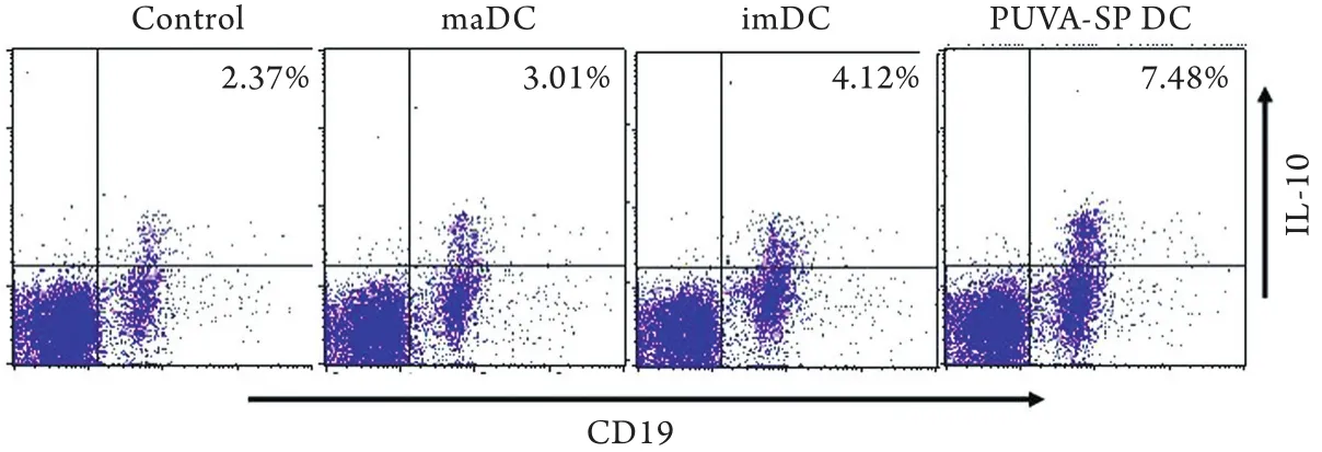
圖1 小鼠外周血IL-10+CD19+Breg細胞占CD19+B細胞的比例Fig. 1 Proportion of IL-10+CD19+Breg cells among CD19+B cells in the peripheral blood of mice
2.3 輸注PUVA-SP DC對受體小鼠移植皮片存活的影響 PUVA-SP DC組小鼠移植皮片存活期為62.3±2.6d,顯著長于maDC組(20.7±1.9d)、imDC組(12.1±1.0d)和PBS對照組(11.0±1.3),差異有統計學意義(P<0.01)。輸注maDC、imDC組移植皮片存活時間略長于PBS對照組,但差異無統計學意義(P>0.05,圖2)。
3 討 論
本課題組前期采用PUVA處理外周血淋巴細胞,并促進imDC吞噬PUVA處理后的淋巴細胞,結果發現,PUVA可促使大量大鼠淋巴細胞發生早期凋亡,而大鼠DC吞噬了凋亡的異基因供體大鼠淋巴細胞后,仍能維持其不成熟狀態,命名為PUVA-SP DC,其表面分子CD80、CD86、MHC-Ⅱ(OX6)呈低水平表達[3]。給移植受者輸注這些DC后,能誘導受者T細胞的免疫低反應性,使受者CD4+CD25+Foxp3+Treg比例明顯增加,移植物存活時間明顯延長[3]。由此,我們認為PUVA-SP DC具有負向免疫調節功能。
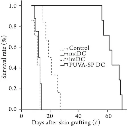
圖2 輸注不同種類DC對小鼠同種異體皮膚移植存活時間的影響Fig.2 Effect of infusing different types of DC on the survival time of skin allograft in mice
多種免疫效應細胞及其表達的免疫相關分子的相互作用所構成的免疫網絡是免疫識別與免疫調控、免疫應答與免疫耐受的基礎。免疫系統通過刺激免疫應答和誘導免疫耐受這兩種互補的功能維持機體的穩態。在此動態過程中,DC與周圍很多免疫細胞存在相互作用,彼此精細調節[6]。近年來,相關研究主要集中在DC和T細胞的相互作用產生的免疫激活或免疫耐受方面[7-9]。B細胞是免疫系統中的主要細胞,能通過產生抗體在體液免疫中發揮核心作用。最近研究表明,與T細胞一樣,B細胞也具有異質性,根據其分泌細胞因子的不同,可分為調節性B細胞和效應性B細胞亞群[10-11],其中調節性B細胞主要分泌IL-10或TGF-β1等免疫負向調節因子。2006年,Mizoguchi和Bhan[12]首次使用調節性B細胞的概念來定義具有負向調控功能的B細胞。隨后,Evans等[13]發現了過渡2型邊緣帶前體(transitional 2 marginal zone precursor,T2 MZP) Bregs。T2 MZP Bregs通過產生IL-10抑制CD4+T細胞分化及效應T細胞的活性,從而抑制過度的炎癥反應[13-14]。2008年,有研究者發現產生IL-10的B細胞亞群能夠抑制T細胞增殖及T細胞介導的炎癥反應,他們將B10細胞回輸至CD19-/-小鼠體內,發現原本增強的CD4+T細胞依賴性超敏反應恢復至正常水平,并證明B細胞的抗原特異性是通過IL-10介導的抑制功能實現的[5,15]。IL-10是體內重要的免疫負向調控分子,可通過下調主要組織相容性抗原、共刺激分子及抑制多種促炎細胞因子的分泌而誘導耐受性DC、調節性T細胞和Th2細胞的生成,從而直接或間接抑制淋巴細胞的活化,使其在促炎因子的作用下仍能誘導異基因抗原特異性免疫耐受,降低免疫抑制劑的非特異性免疫抑制所造成的感染及腫瘤擴散風險。本研究結果表明,在移植術前輸注PUVA-SP DC可顯著延長移植物存活時間,在促使受者體內產生較多負向免疫調節因子IL-10的同時,誘導體內產生較多分泌IL-10的Bregs。該結果有助于更好地理解PUVA-SP DC參與免疫調控的機制,豐富調節性樹突細胞的負向免疫調控功能,為體外光化學療法應用于器官移植免疫排斥的治療提供理論基礎。
[1] Beriou G, Moreau A, Cuturi MC. Tolerogenic dendritic cells: applications for solid organ transplantation[J]. Curr Opin Organ Transplant, 2012, 17(1): 42-47.
[2] Shi BY. Review the effects of regulatory dendritic cells in the transplantation inmmunity[J]. Natl Med J Chin, 2011, 91(8): 505-507.[石炳毅. 重新審視調節性樹突細胞在移植免疫調節中的作用[J]. 中華醫學雜志, 2011, 91(8): 505-507.]
[3] Zheng DH, Dou LP, Wei YX, et al. Uptake of donor lymphocytes treated with 8-methoxypsoralen and ultraviolet A light by recipient dendritic cells induces CD4+CD25+Foxp3+regulatory T cells and down-regulates cardiac allograft rejection[J]. Biochem Biophys Res Commun, 2010, 395(4): 540-546.
[4] Inaba K, Inaba M, Romani N, et al. Generation of large numbers of dendritic cells from mouse bone marrow cultures supplemented with granulocyte/macrophage colony-stimulating factor[J]. J Exp Med, 1992, 176(6): 1693-1702.
[5] Yanaba K, Bouaziz JD, Haas KM, et al. A regulatory B cell subset with a unique CD1dhiCD5+phenotype controls T celldependent inflammatory responses[J]. Immunity, 2008, 28(5): 639-650.
[6] Shi BY. Effects of regulatory network of immunocytes in the transplantation immunity[J]. Natl Med J Chin, 2011, 91(44): 3154-3157.[石炳毅. 調節性免疫細胞網絡在移植免疫中的作用[J]. 中華醫學雜志, 2011, 91(44): 3154-3157.]
[7] Zhang DH, Lu H, Shi BY, et al. Induction effects of dendritic cells loaded with PUVA-treated allogeneic cells on alloantigenspecific CD4+CD25+Foxp3+T regulatory cells in vitro[J]. Med J Chin PLA, 2010, 35(5): 543-546.[鄭德華, 陸宏, 石炳毅, 等.吞噬PUVA處理的同種異基因細胞的樹突細胞對抗原特異性CD4+CD25+Foxp3+調節T細胞的體外誘導作用[J]. 解放軍醫學雜志, 2010, 35(5): 543-546. ]
[8] Zheng DH, Shi BY, Zou YP, et al. Effect of a novel negative immunoregulatory dendritic cell on Th1/Th2 balance of cardiac allograft recipient rats[J]. Med J Chin PLA, 2009, 34(6): 715-718.[鄭德華, 石炳毅, 鄒一平, 等. 負向免疫調節樹突細胞對心臟移植大鼠Th1/Th2類細胞因子的影響[J]. 解放軍醫學雜志, 2009, 34(6): 715-718.]
[9] 鄭智茵, 陳均法, 沈建平, 等. 青蒿琥酯誘導凋亡U937細胞負載樹突細胞后介導的免疫應答[J]. 中國實用內科雜志, 2007, 27(Suppl 1): 86-87.[Zheng ZY, Chen JF, Shen JP, et al. Dendritic cell-induced immune response after phagocytizing Artesunate-leading apoptotic U937 cells[J]. Chin J Pract Intern Med, 2007, 27(Suppl 1): 86-87.]
[10] Shi BY, Xiao L, Gao Y. Identification and exploration of function of Tim-1+CD19+regulatory B cells in peripheral blood in recipients of kidney transplantation[J]. Natl Med J Chin, 2011, 91(48): 3388-3392.[石炳毅, 肖漓, 高鈺. Tim-1+CD19+調節性B細胞在腎移植受者外周血的鑒定與功能研究[J]. 中華醫學雜志, 2011, 91(48): 3388-3392.]
[11] Wojciechowski W, Harris DP, Sprague F, et al. Cytokineproducing effector B cells regulate type 2 immunity to H. polygyrus[J]. Immunity, 2009, 30(3): 421-433.
[12] Mizoguchi A, Bhan AK. A case for regulatory B cells[J]. JImmunol, 2006, 176(2): 705-710.
[13] Evans JG, Chavez-Rueda KA, Eddaoudi A. Novel suppressive function of transitional 2 B cells in experimental arthritis[J]. J Immunol, 2007, 178(12): 7868-7878.
[14] Blair PA, Chavez-Rueda KA, Evans JG, et al. Selective targeting of B cells with agonistic anti-CD40 is an efficacious strategy for the generation of induced regulatory T2-like B cells and for the suppression of lupus in MRL/lpr mice[J]. J Immunol, 2009, 182(6): 3492-3502.
[15] Watanabe R, Ishiura N, Nakashima H, et al. Regulatory B cells (B10 cells) have a suppressive role in murine lupus: CD19 and B10 cell deficiency exacerbates systemic autoimmunity[J]. J Immunol, 2010, 184(9): 4801-4809.
Effect of regulatory dendritic cells on IL-10-producing regulatory B cells of skin allograft in mice
WEI Yu-xiang, ZHOU Wen-qiang, XIAO Li, ZHENG De-hua, QI Zhi, CAI Ming, QIAN Ye-yong, YU Tao, LIAO Ke, SHI Bing-yi*
Organ Transplantation Institute, Beijing Key Organ Transplantation and Immune Regulation Laboratory, 309 Hospital of PLA, Beijing 100091, China
*
, E-mail: shibingyi@medmail.com.cn
The work was supported by the China Postdoctoral Science Fund (200902680 and 20080441308), the Special Fund from Beijing Science & Technology Commission (Z111102055311086) and the National Natural Science Youth Foundation of China (81102242)
ObjectiveTo investigate the effect of recipient immature dendritic cells (imDCs) loaded with PUVA-treated donor apoptotic splenic lymphocytes (PUVA-SP DC) on IL-10+CD19+regulatory B cells (Breg) and the survival duration of skin allograft in mice.MethodsBone marrow-derived DCs of C57BL/6 mice were obtained from bone marrow cells by co-culturing with recombinant mouse IL-4 and GM-CSF. Spleen lymphocytes (SP) of BALB/c mice were isolated and prepared as PUVA- SP by treating the cells with 8-methoxypsoralen plus ultraviolet A irradiation. The bone marrow-derived imDCs of C57BL/6 mice were co-cultured with PUVA-SP of BALB/c mice to obtain PUVA-SP DCs. The skin allograft model was then established. Animals were randomly grouped according to different pretreatments as follows: the control group was iv. introduction of PBS (0.2ml) alone 7 days before skin transplantation, the PUVA-SP DC group
an iv. injection of PUVA-SP DCs, the maDC (mature DC) group received recipient maDCs, and the imDC group was given recipient imDCs. Mice were monitored daily from day 6 after transplantation for signs of rejection of skin graft. The recipients′ peripheral blood serum samples were then collected and the level of cytokines were measured by using ELISA kits. The survival time of skin allograft was evaluated every day. The expression of IL-10+CD19+regulatory B cells was analyzed by flow cytometry.ResultsAfter transplantation, the proportion of IL-10+CD19+Breg in the peripheral blood of PUVA-SP DC group was 7.48%, which was obviously higher than that of imDC group (4.12%), maDC group (3.01%) and control group (2.37%). The serum level of cytokine IL-10 in PUVA-SP DC group was 58.2±0.9ng/ml, and it was significantly higher than that in maDC group (20.1±1.6ng/ml), imDC group (26.2±1.3ng/ml) and control group (19.0±0.6ng/ ml, P<0.01). The survival time of allograft in PUVA-SP DC group was 62.3±2.6d, and it was markedly longer than that in maDC group (20.7±1.9d), imDC group (12.1±1.0d) and control group (11.0±1.3d, P<0.01).ConclusionsAdministration of PUVASP DCs, in the absence of an immunosuppressant, may significantly delay allograft rejection. This effect is associated with upregulation of circulating regulatory B cells with preferential IL-10 secretion.
dendritic cells; skin transplantation; B-lymphocytes, regulatory
R392.4
A
0577-7402(2013)04-0274-05
2013-01-09;
2013-03-04)
(責任編輯:李恩江)
中國博士后基金(200902680,20080441308);北京市科技專項2011階梯計劃項目(Z111102055311086);國家自然科學基金青年基金(81102242)
魏玉香,醫學博士。主要從事器官移植免疫方面的研究
100091 北京 解放軍309醫院全軍器官移植研究所,器官移植與免疫調節北京市重點實驗室(魏玉香、周文強、肖漓、鄭德華、齊幟、蔡明、錢葉勇、于濤、廖珂、石炳毅)
石炳毅,E-mail:shibingyi@medmail.com.cn
猜你喜歡
興趣閱讀·興趣作文與閱讀(低年級)(2025年8期)2025-08-18 00:00:00
東方少年·布老虎畫刊(2023年8期)2023-08-01 15:45:12
科學大眾(2021年6期)2021-07-20 07:42:44
科學(2020年3期)2020-11-26 08:18:30
學苑創造·A版(2020年9期)2020-10-13 09:41:02
娃娃樂園·綜合智能(2019年3期)2019-04-03 09:17:36
中成藥(2018年2期)2018-05-09 07:19:34
小學生學習指導(低年級)(2017年10期)2017-10-10 01:00:05
湖北師范大學學報(自然科學版)(2015年2期)2016-01-10 08:41:55
云南中醫學院學報(2014年3期)2014-07-31 18:57:34