多光子發光的稀土上轉換納米顆粒在生物光子學中的研究進展*
2013-11-10 11:13:28詹求強趙宇翔
激光生物學報 2013年1期
關鍵詞:生物
詹求強,劉 靜,趙宇翔,張 欣
(華南師范大學華南先進光電子研究院光及電磁波研究中心,廣東廣州 510006)
0 前言
隨著生物醫學的快速發展,光學生物成像技術在其中發揮的作用越來越重要,而熒光成像因為其高分辨率、靈敏、快速的特點得到了非常廣泛的研究和應用。目前的熒光標記物主要是有機染料熒光團、量子點(quantum dot,QD)、金屬納米顆粒、上轉換納米顆粒(upconversion nanoparticles,UCNPs)、碳納米顆粒等。這其中,有機染料熒光團使用的最早也最廣泛,但其光穩定性差,不能長時間連續觀察,且吸收和發光譜線寬;半導體量子點光穩定性好,輻射譜線窄,但其可能存在的生物毒性和化學不穩定性使其不能進一步在生物熒光成像領域得到應用。
UCNPs作為一種近期快速發展的熱門熒光標記物,在某些方面比有機染料熒光團和量子點有明顯的優勢。圖1給出了UCNPs和QD這兩種材料在生物應用上論文數量的比較,兩條曲線有著相似的走向,可以看出UCNPs的熱門程度不亞于QD,上轉換文章數量的快速增長正說明了這一材料的巨大潛力。UCNPs主要指摻雜了稀土(rare earth,RE)元素后能夠受兩個或多個低頻光子激發而輻射出一個高頻光子的納米級大小的粒子,它具有光穩定性高、發射光譜譜線窄、熒光壽命長、化學穩定性高等優點。而且上轉換材料可以完全消除自發的背景熒光,能獲得超高的成像對比度。同時,由于是多光子發光過程,它還有著非常好的成像分辨率。在近紅外光(near infrared,NIR)激發下UCNPs可發射出近紅外光,具有較好的光學穿透深度,并且對生物組織也幾乎沒有損傷。以上這些優勢都使UCNPs在生物光子學領域有巨大的應用可能。
1 光學特性
1.1 UCNPs的激發、熒光光譜及發光原理
上轉換發光納米材料是一種在納米晶體基質中摻雜RE離子(以Er3+,Tm3+,Ho3+和Yb3+為主)的復合納米材料,RE離子普遍具有多能級結構(圖2),在這幾種RE離子中,Yb3+充當敏化離子(sensitizer)角色,而其他的RE離子充當活化離子(activator)角色。整個上轉換發光過程包括敏化離子-活化離子之間的能量傳遞、敏化離子-敏化離子和激活離子-激活離子之間的能量轉移(圖2)。在這些能量轉移過程中會有雙光子和三光子過程,分別激發出綠光、紅光和藍光。上轉換的發光波長幾乎覆蓋了紫外、可見、以及紅外光區的光譜范圍[1,2]。
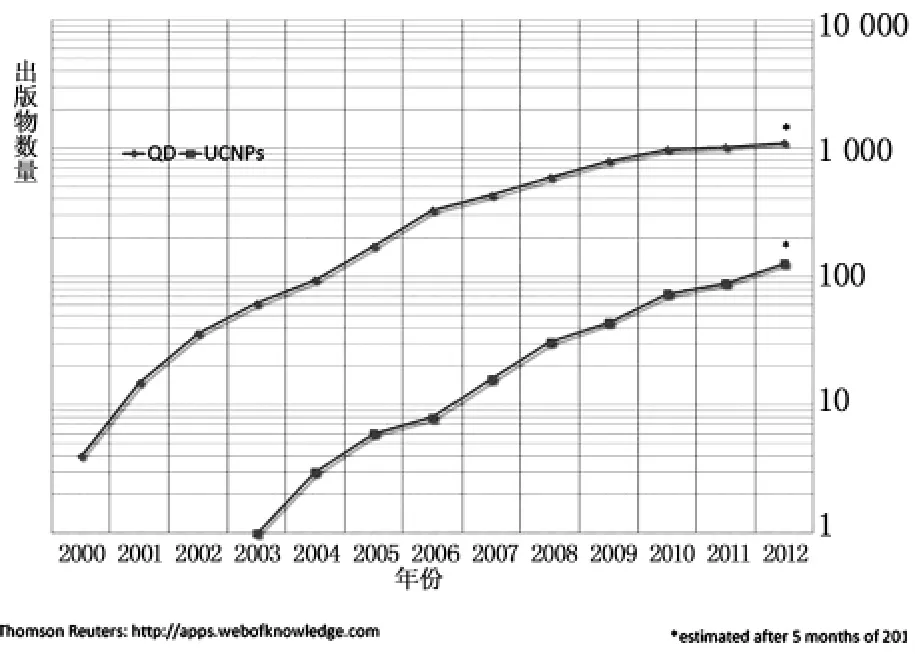
圖1 近年來發表有關QD和UCNPs在生物應用研究中的論文數量比較Fig.1 A quantitative comparison of research activity in bio-applications of QD and UCNPs,measured as number of research articles per year
關于多種RE離子間能量轉移傳遞上轉換發光的過程機制,可以分為激發態吸收(excited state absorption,ESA)、能量傳遞上轉換(energy transfer upconversion,ETU)、交叉弛豫(cross relaxation,CR)、合作敏化上轉換(cooperative sensitization upconversion,CSU)和光子雪崩(photon avalanche,PA)等五種[2-4]。上轉換發光納米材料的發光機制主要是基于能量傳遞上轉換過程,其他幾個能量傳遞過程也存在,但發生幾率很小。
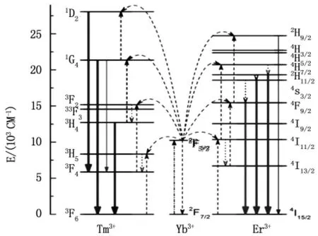
圖2 敏化離子Yb3+與活化離子Er3+,Tm3+的能級結構以及他們之間通過能量轉移實現上轉換發光過程的能級躍遷機制2Fig.2 Energy level structure and proposed UC mechanisms of the Yb3+,Er3+/Tm3+co-doped UCNPs
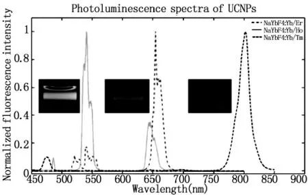
圖3 三種不同UCNPs:NaYbF4:Yb3+/Ho3+(綠),NaFbF4:Yb3+/Er3+(紅),NaFbF4:Yb3+/Tm3+(藍)的發光光譜和發光照片5Fig.3 Photoluminescence spectra of NaYbF4:Yb3+/Er3+(red emission),NaYbF4:Yb3+/Ho3+(green emission),and NaYbF4:Yb3+/Tm3+(blue emission).The insets show the visible photoluminescence imaging of the UCNP colloidal suspension
1.2 UCNPs光學性質
與傳統的熒光標記物不同,UCNPs可以用近紅外光激發而不是紫外光,從而明顯的減小了生物樣品的光致損傷,同時增大了激發光的穿透深度。這種上轉換激發所采用的反斯托克斯機制可以去除自發熒光進行探測,從而帶來非常好的信噪比并且改善探測器的靈敏度。同時上轉換過程存在真實的直接能級,相比普通的雙光子發光現象有著更高的效率,在低功率密度照射下可以產生生物研究中所需要的穩定適中的光強。UCNPs可以用小巧、廉價、低功率的近紅外激光器激發,重要的是在連續照射下不會產生閃爍、光漂白和光化學降解[6-8]。UCNPs的另一個優勢是它包含窄而清晰的發射峰(半高線寬,FWHM<12 nm),具有較長的熒光壽命(μs~ ms)[9,10]。
2 UCNPs的化學合成及修飾方法
2.1 UCNPs的組成材料
上轉換納米粒子一般由無機主基質和摻雜在主基質晶格中的鑭系離子組成,具體可參考圖5,摻雜的離子又分為敏化劑和活化劑。
研究上轉換所用到的主基質很多,理想的主基質有低的晶格聲子能量,這樣將使非輻射損失最小化,同時主基質的晶格也要和摻雜離子相匹配以達到最好的摻雜效果。本文主要介紹的主基質為NaYF4。
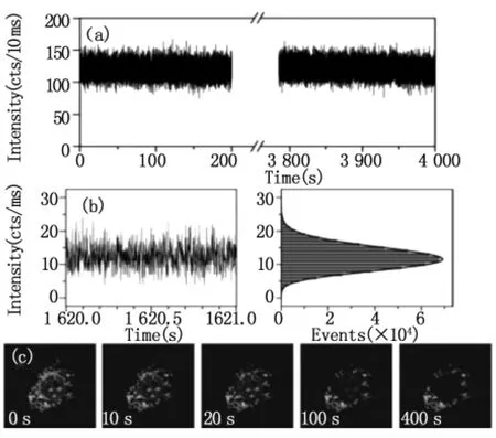
圖4 UCNPs沒有光致漂白和光閃爍現象:(a)單UCNPs在長時間探測后有光穩定性;(b)UCNPs的時間分辨輻射;(c)分別用405,543和980 nm連續激光在1.6,0.13和19 mW下同時激發得到的焦面成像[6-8]Fig.4 Nonbleaching and nonblinking behavior of UCNPs:(a)Photostability of a single UCNP under longtime monitoring;(b)Time-resolved emission of UCNP,suggesting no on/off behavior;(c)Simultaneous excitations was provided by CW lasers at 405,543,and 980 nm with powers of approximately 1.6,0.13,and 19 mW in the focal plane,respectively
理論上大部分的RE離子都可以進行上轉換發射,但在低強度激發功率下,只有少數的RE離子可以進行上轉換發射,如Er3+,Ho3+和Tm3+用來做為活化劑。它們有著階梯狀的能級結構,可以被近紅外光激發產生上轉換發光。活化劑的含量都相對較低(一般<2 mol%),這樣以減小交叉弛豫的能量損失。
為了提高上轉換發光效率并且使其在在近紅外區有足夠大的吸收截面,通常我們還需要一個敏化劑和活化劑共同摻雜。Yb3+相比于其他的鑭系元素離子,在900-1000 nm附近有一個較大的吸收截面,它作為敏化劑經常與Er3+,Ho3+和Tm3+共同摻雜來提高上轉換發光效率(圖2)。
2.2 合成方法
合適的尺寸和均一的形狀是上轉換納米粒子應用于生物成像的前提。目前為止,有報道過多種合成高質量UCNPs的方法,包括有共沉淀法[12]、水熱合成法[13]、熱分解法5、溶膠-凝膠法以及微乳液的合成方法。而在這些方法中,水熱合成法和熱分解法是最為常用來合成均一的、疏水性的納米粒子的方法。本綜述也主要介紹這兩個方法。
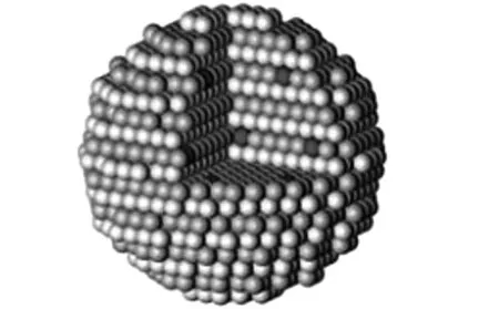
圖5 圖示為晶體主基質和包含有鑭系離子的上轉換納米粒子示意圖(藍色及白色為主基質分子,紅色為摻雜的離子)11Fig.5 Schematic illustration of UC nanoparticles composed of a crystalline host and lanthanide dopant ions embedded in the host lattice
2.2.1 水熱合成法 用水熱合成法合成上轉換納米粒子可以很好的控制納米粒子的大小和形狀,反應是在密閉的高溫高壓環境中進行的(如圖6)。一般在溶液中混合RE元素前驅體和氟化物前驅體,然后密閉在高壓容器中加熱反應。經常用到的RE元素前驅體為硝酸鹽、氯化物和氧化物,而較典型的氟化物前驅體為HF,NH4F,NaF和 NH4HF2,乙二胺四乙酸(EDTA)、溴化十六烷基三甲基銨硝酸鹽(CTAB)、油酸(OA)和檸檬酸三鈉鹽(TSC)為經常用到的表面活性劑。Li[13-16]等人報道了一種簡單的相轉變及分離的方法(LSS),用這種LSS方法成功合成了不同主基質、晶體結構、大小和形狀的納米粒子,如,BaY2F8,Ba2YF7,Ba2YbF7。Zhang[20]等人報道了一種新穎的容易使用的水熱合成法合成純的六方晶型NaYF4:Yb,Er/Tm納米粒子,此方法合成的納米粒子形狀可控而且上轉換發光強。
2.2.2 熱分解法 在熱分解過程中,三氟乙酸鹽被用作前驅體來分解得到相應的金屬氟化物。Yan[21,22]等人創新性地提出了用 OA/OM/1-十八烯(ODE)與三氟乙酸鹽共熱的方法,以達到熱分解法需要的高溫(300℃),并在反應中調節前驅體的比例、反應的溫度和時間、協調溶劑的屬性,來控制納米晶體的相、形態、大小,從而提高上轉換發光效率。但在熱反應的過程中不可避免地會產生一些有毒的副產物,如三氟乙酸醋酸酐(CF3CO)2O,三氟乙酰氟CF3CF2COF,羰基二氟化物 COF2和四氟乙烯C2F4,Wei[23]等人報道了一個更好的熱分解方法,通過RE-油酸鹽和NaF混合作為前驅體進行熱分解得到α-和β-NaYF4UCNPs。Chow[24]等人提出油胺(OM)是唯一能使UCNPs從四方晶型轉變為六方晶型的溶劑,Cohen[25]等人用以上控制條件及晶相的方法合成了低于10 nm的六方晶型的UCNPs。

圖6 UCNPs水熱合成法的步驟示意圖Fig.6 The schematic diagram of hydro(solvo)thermal method of UCNPs
2.3 UCNPs的表面修飾
通過以上方法合成的UCNPs一般為疏水性的,而要用于標記生物發光,則需要通過表面修飾使其轉變為親水性的。到目前為止報道的方法很多,主要有配體交換、配體去除、靜電層層自裝法、配體氧化法、層層疊加法、聚合物包覆法、硅烷化及主客體相互作用法(表1)。
3 UCNPs發光效率優化
3.1 材料合成優化
對于上轉換材料而言,設計一個符合要求的晶體結構是提高發光效率的關鍵性因素,這就需要考慮如何控制材料的晶相、粒度及離子摻雜等因素。
3.1.1 主基質優化 在熱分解和水熱法合成過程中,立方相的NaYF4首先形成,當能量達到一定值時,就能克服晶體相變所需的自由能,立方相就能向六方相轉變[22,25]。(1)延長時間有助于克服立方相的 NaYF4向六方相轉變的自由能[22,52]。如圖 7 所示,在我們的試驗中,Gd3+摻雜的NaYF4:20%Yb3+/30%Gd3+/2%Er3+納米顆粒半小時后顆粒呈不規則球狀,一小時后樣品呈非常規則的的六方相。(2)調控Na+、RE3+及Y-之間的摩爾比例。在大多數情況下,六方相的NaYF4不如立方相的NaYF4穩定,采用高的 Na+/RE3+和 F-/RE3+比例有利于六方相的NaYF4形成[22,53-55]。(3)配體調制相變。油胺可以調制NaYF4的晶相由立方相向六方相轉變[24,25]。
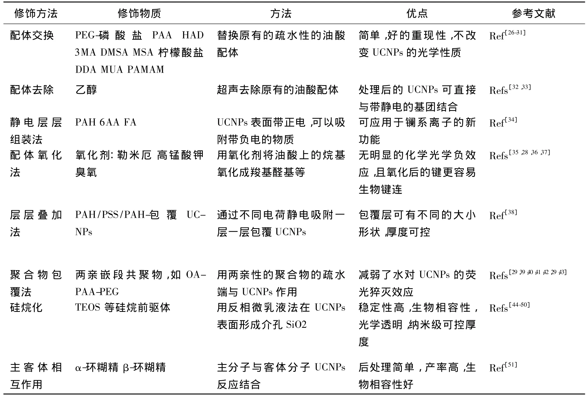
表1 UCNPs的表面修飾方法Tab.1 Methods of surface functionalization of hydrophobic UCNPs
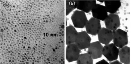
圖7 利用溶膠-凝膠法分別烘烤(a)半小時、(b)1小時后得到的NaYF4:20%Yb3+/30%Gd3+/2%Er3+的透射電鏡圖Fig.7 TEM images of using sol-gel method after baking NaYF4:20%Yb3+/30%Gd3+/2%Er3+for(a)0.5 h,(b)1 h
UCNPs的粒徑越小,比表面積就越大,意味著更多的缺陷和配體將存在于表面,會降低上轉換發光效率。同時,由于顆粒大小會極大影響生物組織對顆粒的吸收及在生物組織中的分布,因此上轉換納米材料顆粒不能太大,否則就不適用于生物成像,故合成足夠小的納米顆粒的同時保持足夠的發光亮度是我們面臨的一個挑戰。研究者們通過改變表面活性劑(油酸和油胺)的濃度、Y3+/F-比例以及反應溫度來調控合成直徑從4.5 nm到15 nm大小的蛋白質尺寸的六方相NaYF4[25],成功合成了小顆粒高亮度的UCNPs,這一進展會拓展和深化UCNPs的生物應用,特別是單分子成像中。
3.1.2 離子摻雜優化 如圖8所示,上轉換發光材料的發光效率和摻雜RE離子的種類、RE離子間的摩爾比例有很大關系,發光強度最大的為Y3+:Yb3+:Tm3+(Er3+)=78:20:2。Li+摻雜到氧化物或氟化物上轉換納米材料中能增強上轉換發光[56-61],Ho3+摻雜離子能增強Tm3+的近紅外上轉換發光[62]。
3.1.3 表面包覆保護殼層優化 在上轉換納米發光材料表面包覆上保護殼層,可以減少晶體缺陷、保護光學活性的離子,從而減少非輻射能量損失[63]。比較無殼層的BaGdF5:Yb3+/Er3+和包覆了活性殼層的BaGdF5:Yb3+/Er3+發光強度,發現包覆了活性殼層的納米顆粒的發光強度較之無殼的有幾百倍的增加[64]。此外,將摻雜離子用一種層狀結構隔離,能抑制焠滅效應,有望使上轉換發光效率得到增強[44,65,66]。
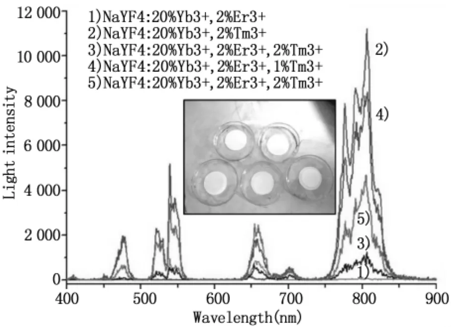
圖8 水熱法合成的5個不同樣品的上轉換發光光譜及其對應的圓薄片樣品圖Fig.8 Hydro(solvo)thermal method synthesis of 5 different samples of upconversion luminescence spectrum and its corresponding circular wafer sample diagram
3.2 表面等離子對輻射增強
眾所周知,金屬結構獨特的表面等離子特性可以用來增強來自臨近熒光團(有機染料和無機量子點)的熒光[67,68]。同樣的,強局部場下的表面等離子共振也能用來增強上轉換輻射效率。Zhang[69]等人和Sudheendra[70]等人成功的將納米金顆粒吸附在UCNPs表面實現了上轉換輻射調制[71,72]。一種特制980 nm輻射金等離子表面對能明顯增強來自納米線層的從近紅外到可見光的上轉換發光[73]。對于有摻雜的上轉換納米顆粒和金屬納米顆粒的優化設計來說,單納米顆粒表面等離子增強機理的研究是很重要的。然而,使用金屬納米顆粒增強上轉換輻射進行生物探測仍面臨很大的挑戰,如復雜的實驗過程和苛刻的實驗條件。
目前利用表面等離子對輻射的金屬增強上轉換主要集中在玻璃復合材料和薄膜上,科學家們將銀或金的納米顆粒附著在UCNPs上可以增強上轉換強度。Yan[74]等人第一次通過一種直接集合的方法將六方形NaYF4:Yb,Er納米顆粒和銀納米線配對觀察到了增強的上轉換輻射。紅光輻射(650 nm)比綠光輻射(550 nm)增強因數更大,并且可以使用聚集了銀顆粒的銀島得到進一步的增強。Schietinger[75]等人用一個光學和原子力顯微鏡相結合的設備觀察并控制單 NaYF4:Yb,Er納米顆粒與金納米球(60 nm)的距離,發現綠光輻射的增強是4.8,而紅光輻射下是2.7。Kennedy[72]等人發現了一種新型的光子材料,它是一種有非定型金殼層(4-8 nm)的立方NaYF4:Yb,Er/Tm納米晶體。在這種有等離子金殼層的NaYF4:Yb,Tm納米顆粒下觀察到的從近紅外到可見光的上轉換增強達到了8。Duan[71]等人通過六方形NaYF4:Yb,Tm和金納米顆粒的等離子接觸調制了上轉換輻射,并且在452-476 nm的范圍內得到了超過150%的輻射強度增強,而647 nm處只有50%左右的增強。最近,Qin[76]等人通過將金納米顆粒(10 nm)吸附在NaYF4:Yb,Tm表面,觀察到對于這種納米復合材料上轉換輻射在波長291 nm和345 nm分別增強了73.7和109.0。
3.3 波長優化
在它的所有應用中,激發光都是采用的975 nm附近(如980 nm)的連續激光,但生物體內的主要成分--水在這個波長附近吸收非常的強,這樣就降低了成像深度并造成了明顯的熱效應[77]。因此,對于上轉換效率在波長方面的優化也有它的必要性,2011年 Zhan[27]等人在國際上首次提出了915 nm光作為上轉換材料的新激發光源波長,在生物成像中具有更深的成像深度和更低的熱損傷。使用同樣強度的500 mWcm-2的915 nm和980 nm連續脈沖激光照射3 min后,溫度分別達到32.2℃和45℃。
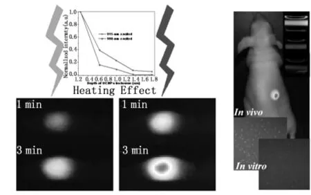
圖9 優化上轉換成像中的激發模式實現低熱無損、深度組織成像Fig.9 The optimization of conversion imaging excitation mode to realize low thermal condition,depth tissue imaging
4 UCNPs的生物應用
UCNPs生物成像由于具有無自發熒光、大的反斯托克斯位移、窄的發射峰、高耐光漂白、無光閃爍、探測深度大以及高的空間分辨率等優良的光學特性,因此近幾年來很多人致力于UCNPs的生物光子學應用。目前在細胞成像、動物成像、生物傳感、漫射光成像、光動力治療(PDT)、多模式成像(MRI/PET)等方面有眾多應用。
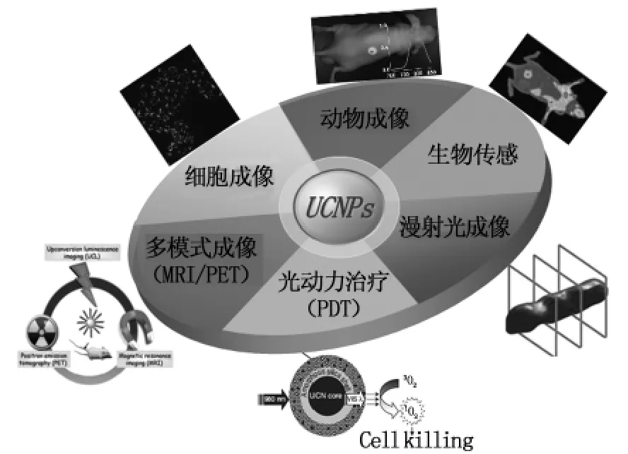
圖10 UCNPs的生物應用Fig.10 The biological application of UCNPs
4.1 細胞成像
近幾年來,隨著上轉換納米粒子性能的改進,基于UCNPs的顯微技術被廣泛應用于高分辨和高對比的離體細胞的成像。
在無抗體標記的細胞中,用UCNPs吸附在細胞膜上或被細胞內吞來進行成像。最近,Jin[78]等人制備了三種類型的聚合物包覆的UCNPs,發現了相比于中性和帶負電荷的聚合物,用帶正電荷的PEI包覆的UCNPs可以增強細胞對UCNPs的攝取能力。2008年 Nyk[79]等人成功將 MSA-UCNPs應用到標記Panc 1細胞進行高對比成像(圖11)。
相比于無抗體標記的細胞成像,有抗體標記的成像會有更多優點,尤其是在腫瘤細胞靶向成像中有更廣泛的應用。表面功能化的UCNPs通過生物分子識別與癌細胞特異性相結合,2009年 Wang[80]等人提出用anti-CEA8于UCNPs結合來進行細胞成像(圖12)。Zhan[27]等人最近報道了一系列對比可控實驗,說明了將NaYF4:Yb3+/Er(Ho)3+與anti-CEA8的抗體相結合,可以特異性地與Hela癌細胞膜結合,從而進行成像。
4.2 動物成像
UCNPs的快速發展使得其實現了在活體中的成像,在早期的研究中,包括皮下注射和UCNPs在生物中的分布成像。Salthouse等[81]人研究了在老鼠的尾巴靜脈注射UCNPs后的積累,結果表明PEG-UCNPs沒有首先靶向積累在小鼠的肝、脾等處,另外長時間觀察發現在活體樣品中注射UCNPs 7天后,還可以觀察到體內有其存在。
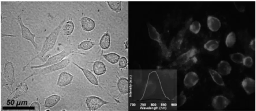
圖11 用UCNPs處理的Panc1細胞的透射成像(左)和光致發光成像(右)。插圖為局部細胞(紅色)和背景(黑色)的光致發光光譜圖[79]Fig.11 In vitro transmission(left)and PL(right)images of Panc 1 cells treated with UCNPs.Inset shows localized PL spectra taken from cells(red)and background(black)
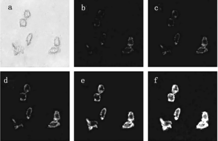
圖12 用兔子的anti-CEA8與UCNPs結合后與He-La細胞培養進行亮場成像(a),和用不同功率980 nm NIR激光激發(b)100,(c)300,(d)500,(e)700,和(f)900 mW[80]Fig.12 Fluorescence imaging of HeLa cells after incubated with rabbit anti-CEA8 Ab-conjugated UCNPs in bright field(a),and excited by a 980 nm NIR laser with different excitation powers:(b)100,(c)300,(d)500,(e)700,and(f)900 mW
由于淋巴排水系統是癌細胞新陳代謝的一個重要途徑,但淋巴結的復雜微小結構使得很難對它進行識別,用NaYF4:Yb,Er和NaYF4:Yb,Tm納米粒子作為熒光探針,Kobayashi[82]等人第一次提出了無內源熒光的小鼠的淋巴結雙顏色成像。同樣的問題也出現在血管成像中,疾病中血管變化及血管功能紊亂,血管成像能提供血管的數量和間距、血管系統的滲透性及血管的異常,Zhang等[83]人報道了用硅烷包覆的NaYF4:Yb,Er UCNPs來動態跟蹤小鼠耳朵的成肌細胞來對血管成像。
到目前為止,在小動物中的成像做了多方面研究,而針對于腫瘤的成像在腫瘤的診斷和治療方面是非常重要的。目前,UCNPs結合葉酸、抗體和多肽能用于活體腫瘤靶向成像。F.Y.Li[84]小組近期提出了一個環肽(c(RGDFK))修飾的NaYF4:Yb,Er,Tm,并用 PEG修飾(UCNP-RGD),來靶向成標記αvβ3-過量表達的老鼠腫瘤(人類惡性膠質瘤U87MG)(圖13)。
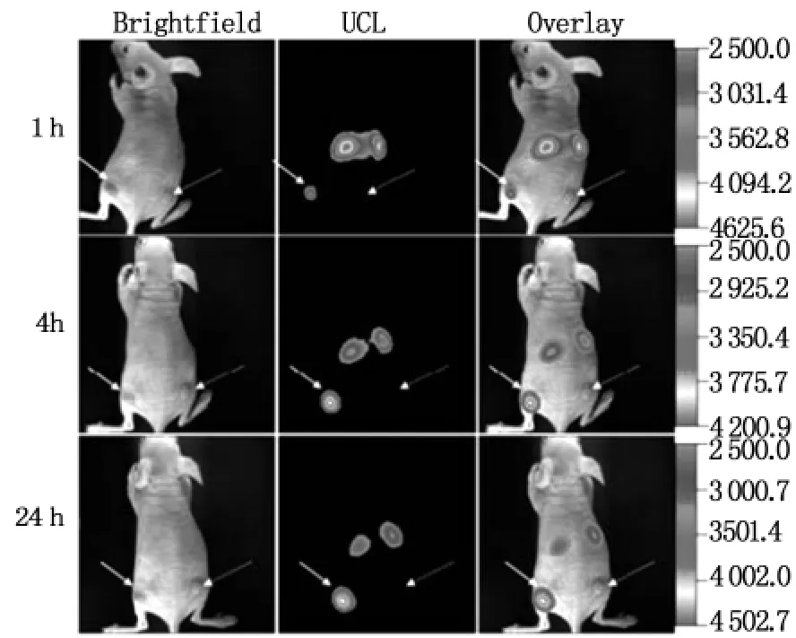
圖13 依據時間的上皮U87MG腫瘤上轉換發光(左后腿,短箭頭指出)和MCF-7腫瘤(右后腿,長箭頭指出)在無胸腺的裸鼠上實驗,在經脈注射UCNP-RGD 24 h 后[84]Fig.13 Time-dependent in vivo upconversion luminescence imaging of subcutaneous U87MG tumor(left hind leg,indicated by short arrows)and MCF-7 tumor(right hind leg,indicated by long arrows)borne by athymic nude mice after intravenous injection of UCNPRGD over a 24 h period
4.3 生物傳感
作為波爾茲曼分布的一個性質,UCNPs的不同發射譜帶的相對強度會依賴于周圍的環境。由于這個原因,UCNPs被提出作為納米溫度傳感器。最近,基于UCNPs的光學溫度傳感器被應用于細胞內部溫度的探測,Vetrone等[85]人提出了用綠色上轉換發光的NaYF4:Yb3+,Er3+納米粒子,用于Hela宮頸癌細胞的溫度傳感。
作為最有效的生物傳感工具,基于熒光能量共振轉移(FRET)的方法能用于檢測生物親和性相互作用并使生物分子在納米級范圍內產生改變。當UCNPs被用作供體分子時,可能會產生許多新的應用可能性,這就是發光能量共振轉移(LRET)。Soukka等[86,87]人在基于上轉換納米粒子的 LRET傳感上做了很多研究,他們提出了一個新穎的上轉換LRET傳感技術及其在多方面的潛在應用可能,如在血清中對E2(17β雌二醇)免疫測定、酶活性實驗、雙參數DNA雜交試驗。
4.4 漫射光層析成像
漫射光成像已經廣泛應用在了小動物和人體組織的成像探測上。在人體上,漫射光成像,已經用于探測乳腺癌腫瘤,腦活動和腦新陳代謝[88-93]。然而,由于內源組織自發背景熒光、光在組織中傳遞的隨機性等問題的存在,漫射光成像得到的成像效果相對不是很好。現有的一些改善成像質量的方法設備復雜、計算繁多。而上轉換激發完全去除自發熒光,由圖14我們可以看出即使很弱的自發背景熒光也會給成像精度產生嚴重的影響[94],同時上轉換激發不需要復雜的儀器設備,而較大的反斯托克斯位移也使激發光和發射光易分開。相比傳統的有機染料(下轉換發光過程),上轉換納米材料的非線性性質可以獲得更高的分辨率,這也是UCNPs在漫射光成像上的一大優勢。
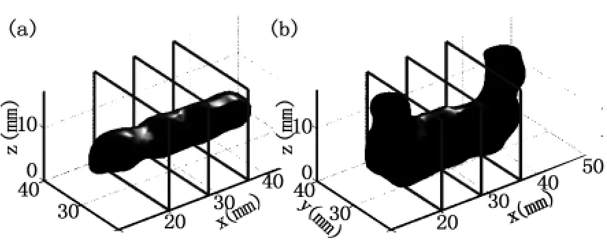
圖14 一個組織圖像中兩個圓柱形熒光目標的熒光漫射光成像重構圖 (a)使用UCNPs進行的重構;(b)使用羅丹明6G進行的重構[94]Fig.14 LDOT reconstruction of two cylindrical luminescent targets in a tissue phantom.Reconstruction using(a)UCNPs;(b)Rhodamine 6G
4.5 輔助光動力治療
在臨床醫學上對癌癥的診斷和治療中,成像和治療是不可分開的。不同于化學療法、放射性療法和外科手術,光動力治療(photodynamic therapy,PDT)作為一個治療癌癥的技術,是被高能量光激發光敏劑產生活性氧類(ROS)來殺死病變細胞。在癌癥組織中,有望能用980 nm的連續激光激發UCNPs穿透深層組織達到治療效果。Austin[95]首次提出了用三層的NaYF4:Yb,Er與卟啉共同定位的光動力治療。Zhang等人在 2009[96]年和 2010 年[97]分別報道了 NaYF4:Yb,Er@nSiO2@mSiO2和 NaYF4:Yb,Er@mSiO2納米粒子將 ZnPc包含在介孔的 SiO2中,在500 mW 980 nm的連續激光下激發5 min,就可以從介孔SiO2中釋放活性氧,達到PDT的治療效果。
在最近的研究工作中,Zhang等[83]人又用葉酸(FA)和PEG修飾的UCNPs分別與不做修飾及只用PBS來治療小鼠作為對比(圖15),可以看到PDT的明顯治療效果。
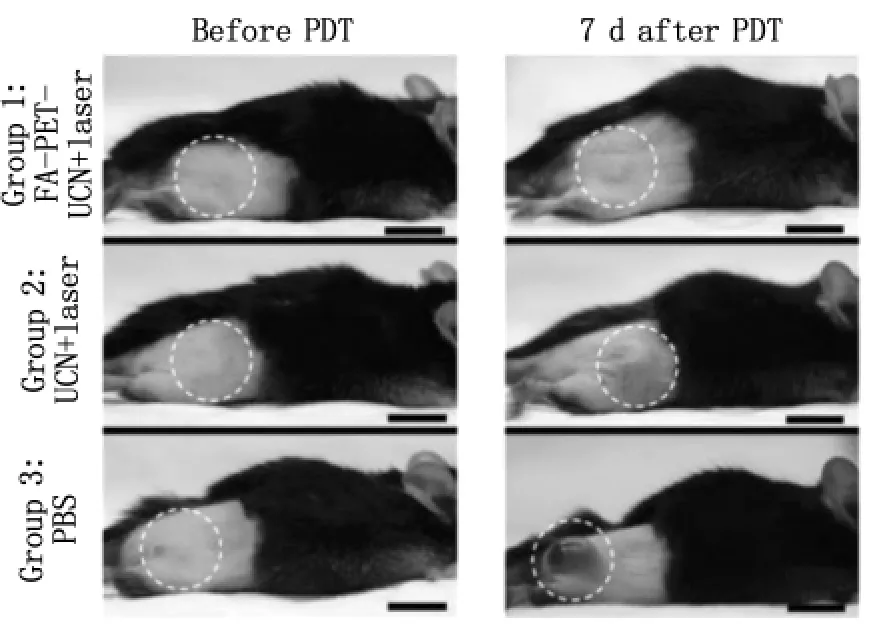
圖15 照片為每組1-3只小鼠分別靜脈注射FAPEG-UCNs、未修飾的UCNP和PBS,照片顯示了注射PDT前(0 d)和注射后(7 d)的腫瘤大小的改變(用虛線圓圈標出)。標尺10 mm[83]Fig.15 Representative gross photos of a mouse from each group 1-3 intravenously injected with FA-PEGUCNs,unmodified UCNs or PBS showing the change in tumor size(highlighted by dashed white circles)before(0 d)and 7 d after PDT treatment.Scale bars,10 mm
4.6 多模式成像
目前,分子多模式成像的技術主要集中在X射線計算機斷層掃描技術(CT)、核磁共振成像(MRI)、單光子發射計算機斷層成像術(SPECT)、正電子發射斷層成像術(PET)和超聲成像等[99-101]。近年來,大量的光致發光材料的出現,催生了一大批研究者對于促進在多模式造影劑存在時分子光學成像研究的興趣[102-103],但明亮和穩定的多模式造影劑難以找到,發光探針在多模式造影劑上容易降解和漂白。然而,UCNPs很穩定,不存在漂白和老化現象,且多模式成像可以通過修飾晶體主體材料實現。漫射光成像,即使利用UCNPs,其空間分辨率依然不高[104];CT和MRI具有高的分辨率,卻難以得到所需的信息;PET能提供手術前的一些詳細數據,卻不能很好適用于術后。主要討論的是,基于UCNPs的多模式成像,如磁-光學成像、核-光學成像、CT-三模式成像。
5 總結與展望
文章綜述了上轉換發光納米粒子的合成方法、光學特性、發光效率的優化及在生物方面的應用。作為新一代生物發光標記材料,UCNPs展現了許多優點,例如毒性小、無背景熒光、化學穩定性高、光穩定性好、光穿透能力強等。近幾年里對UCNPs的生物應用研究急劇增加,展現了上轉換納米粒子的潛在應用價值。
但是盡管UCNPs有著快速強勁的發展,它仍然有著較低的發光效率,表面修飾有待改進,PDT的效率仍然不高。在未來的工作中,我們需要進一步優化材料合成,得到發光效率更高的納米粒子。同時也要優化激發模式,發展上轉換成像的專業儀器,推動該領域更多更好的研究工作的展開。最后重要的是研究要向臨床應用發展,能達到確實診斷、治療疾病的目的。
[1]DIEKE G H.Spectra and energy levels of rare earth ions in crystals[M].1968,Medium:X;Size:Pages:409.
[2]WANG F,LIU X.Recent advances in the chemistry of lanthanide-doped upconversion nanocrystals[J].Chemical Society Reviews,2009,38(4):976-989.
[3]SUYVER J F,AEBISCHER A,BINER D,et al.Novel materials doped with trivalent lanthanides and transition metal ions showing near-infrared to visible photon upconversion[J].Optical Materials,2005,27(6):1111-1130.
[4]AUZEL F.Upconversion and Anti-Stokes processes with f and d ions in solids[J].Chemical Reviews,2003,104(1):139-174.
[5]MAI H X,ZHANG Y W,SUN L D,et al.Size-and phase-controlled synthesis of monodisperse NaYF4:Yb,Er nanocrystals from a unique delayed nucleation pathway monitored with upconversion spectroscopy[J].The Journal of Physical Chemistry C,2007,111(37):13730-13739.
[6]FENG W,X F,WANG M,et al.Multicolour PEI/NaGdF4:Ce3+,Ln3+nanocrystals by single-wavelength excitation[J].Nanotechnology,2007,18(2):025701.
[7]YU M,LI F,CHEN Z,et al.Laser scanning up-conversion luminescence microscopy for imaging cells labeled with rare-earth nanophosphors[J].Analytical Chemistry,2009,81(3):930-935.
[8]WU S,HAN G,MILLIRON D J,et al.Non-blinking and pho-tostable upconverted luminescence from single lanthanide-doped nanocrystals[J].Proceedings of the National Academy of Sciences,2009,106(27):10917-10921.
[9]NIKOOBAKHT B,BURDA C,BRAUN M,et al.The quenching of CdSe quantum dots photoluminescence by gold nanoparticles in solution[J].Photochemistry and Photobiology,2002,75(6):591-597.
[10]CHEN C L,KUO L R,CHANG C L,et al.In situ real-time investigation of cancer cell photothermolysis mediated by excited gold nanorod surface plasmons[J].Biomaterials,2010,31(14):4104-4112.
[11]WANG F,BANERJEE D,LIU Y,et al.Upconversion nanoparticles in biological labeling,imaging,and therapy[J].Analyst,2010,135(8):1839-1854.
[12]YI G,LU H,ZHAO S,et al.Synthesis,characterization,and biological application of size-controlled nanocrystalline NaYF4:Yb,Er infrared-to-visible up-conversion phosphors[J].Nano Letters,2004,4(11):2191-2196.
[13]WANG L,LI Y.Controlled synthesis and luminescence of lanthanide doped NaYF4nanocrystals[J].Chemistry of Materials,2007,19(4):727-734.
[14]WANG X,ZHUANG J,PENG Q,et al.A general strategy for nanocrystal synthesis[J].Nature,2005,437(7055):121-124.
[15]WANG X,ZHUANG J,PENG Q,et al.Hydrothermal synthesis of rare-earth fluoride nanocrystals[J].Inorganic Chemistry,2006,45(17):6661-6665.
[16]WANG L,LI Y.Na(Y1.5Na0.5)F6 single-crystal nanorods as multicolor luminescent materials[J].Nano Letters,2006,6(8):1645-1649.
[17]ZHANG F,WAN Y,YU T,et al.Uniform nanostructured arrays of sodium rare-earth fluorides for highly efficient multicolor upconversion luminescence[J].Angewandte Chemie International Edition,2007,46(42):7976-7979.
[18]WANG L,LI P,LI Y.Down-and up-conversion luminescent nanorods[J].Advanced Materials,2007,19(20):3304-3307.
[19]HE HU,Z C,TIANYE CAO,et al.Hydrothermal synthesis of hexagonal lanthanide-doped LaF3 nanoplates with bright upconversion luminescence[J].Nanotechnology,2008,19(37):375702.
[20]ZHANG,Z L A Y.An efficient and user-friendly method for the synthesis of hexagonal-phase NaYF4:Yb,Er/Tm nanocrystals with controllable shape and upconversion fluorescence[J].Nanotechnology,2008,19(24):345606.
[21]SI R,ZHANG Y W,YOU L P,et al.Rare-earth oxide nanopolyhedra,nanoplates,and nanodisks[J].Angewandte Chemie International Edition,2005,44(21):3256-3260.
[22]MAI H X,ZHANG Y W,SI R,et al.High-quality sodium rare-earth fluoride nanocrystals:controlled synthesis and optical properties[J].Journal of the American Chemical Society,2006,128(19):6426-6436.
[23]WEI Y,LU F,ZHANG X,CHEN D.Synthesis of oil-dispersible hexagonal-phase and hexagonal-shaped NaYF4:Yb,Er nanoplates[J].Chemistry of Materials,2006,18(24):5733-5737.
[24]YI G S,CHOW G M.Synthesis of hexagonal-phase NaYF4:Yb,Er and NaYF4:Yb,Tm nanocrystals with efficient up-conversion fluorescence[J]. Advanced Functional Materials,2006,16(18):2324-2329.
[25]OSTROWSKI A D,CHAN E M,GARGAS D J,et al.Controlled synthesis and single-particle Imaging of bright,sub-10 nm lanthanide-doped upconverting nanocrystals[J]. ACS Nano,2012,6(3):2686-2692.
[26]ZHOU J,YU M,SUN Y,et al.Fluorine-18-labeled Gd3+/Yb3+/Er3+co-doped NaYF4nanophosphors for multimodality PET/MR/UCL imaging[J].Biomaterials,2011,32(4):1148-1156.
[27]ZHAN Q Q,QIAN J,LIANG H,et al.Using 915 nm laser excited Tm3+/Er3+/Ho3+-Doped NaYbF4upconversion nanoparticles for in vitro and deeper in vivo bioimaging without overheating irradiation[J].ACS Nano,2011,5(5):3744-3757.
[28]NACCACHE R,VETRONE F,MAHALINGAM V,et al.Controlled synthesis and water dispersibility of hexagonal phase NaGdF4:Ho3+/Yb3+nanoparticles[J].Chemistry of Materials,2009,21(4):717-723.
[29]BUDIJONO S J,SHAN J,YAO N,et al.Synthesis of stable block-copolymer-protected NaYF4:Yb3+,Er3+up-converting phosphor nanoparticles[J].Chemistry of Materials,2009,22(2):311-318.
[30]KUMAR R,NYK M,OHULCHANSKYY T Y,et al.Combined optical and MR bioimaging using rare earth Ion doped NaYF4nanocrystals[J]. Advanced Functional Materials,2009,19(6):853-859.
[31]SHEN J,SUN L D,ZHANG Y W,et al.Superparamagnetic and upconversion emitting Fe3O4/NaYF4:Yb,Er hetero-nanoparticles via a crosslinker anchoring strategy[J].Chemical Communications,2010,46(31):5731-5733.
[32]WANG M,LIU J L,ZHANG Y X,et al.Two-phase solvothermal synthesis of rare-earth doped NaYF4upconversion fluorescent nanocrystals[J].Materials Letters,2009,63(2):325-327.
[33]BOGDAN N,VETRONE F,OZIN G A,et al.Synthesis of ligand-free colloidally stable water dispersible brightly luminescent lanthanide-doped upconverting nanoparticles[J].Nano Letters,2011,11(2):835-840.
[34]LIU Q,SUN Y,LI C,et al.18F-labeled magnetic-upconversion nanophosphors via rare-earth cation-assisted ligand assembly[J].ACS Nano,2011,5(4):3146-3157.
[35]CHEN G Y,LIU H C,SOMESFALEAN G,et al.Enhancement of the upconversion radiation in Y2O3:Er3+nanocrystals by codoping with Li+ ions[J].Applied Physics Letters,2008,92(11):113114-113114-3.
[36]CHEN G,SOMESFALEAN G,LIU Y,et al.Upconversion mechanism for two-color emission in rare-earth-ion-doped ZrO_{2}nanocrystals[J].Physical Review B,2007,75(19):195204.
[37]ZHOU J,SUN Y,DU X,et al.Dual-modality in vivo imaging using rare-earth nanocrystals with near-infrared to near-infrared(NIR-to-NIR)upconversion luminescence and magnetic resonance properties[J].Biomaterials,2010,31(12):3287-3295.
[38]WANG L,YAN R,HUO Z,et al.Fluorescence resonant energy transfer biosensor based on upconversion-luminescent nanoparticles[J].Angewandte Chemie International Edition,2005,44(37):6054-6057.
[39]WANG Z L,HAO J,CHAN H L W,et al.Simultaneous synthesis and functionalization of water-soluble up-conversion nanoparticles for in vitro cell and nude mouse imaging[J].Nanoscale,2011,3(5):2175-2181.
[40]WANG C,TAO H,CHENG L,et al.Near-infrared light induced in vivo photodynamic therapy of cancer based on upconversion nanoparticles[J].Biomaterials,2011,32(26):6145-6154.
[41]CHENG L,YANG K,LI Y,et al.Facile preparation of multifunctional upconversion nanoprobes for multimodal imaging and dual-targeted photothermal therapy[J].Angewandte Chemie International Edition,2011,50(32):7385-7390.
[42]PEDRONI M,PICCINELLI F,PASSUELLO T,et al.Lanthanide doped upconverting colloidal CaF2 nanoparticles prepared by a single-step hydrothermal method:toward efficient materials with near infrared-to-near infrared upconversion emission[J].Nanoscale,2011,3(4):1456-1460.
[43]LI P,PENG Q,LI Y.Dual-Mode luminescent colloidal spheres from monodisperse rare-earth fluoride nanocrystals[J].Advanced Materials,2009,21(19):1945-1948.
[44]QIAN H S,ZHANG Y.Synthesis of hexagonal-phase core shell NaYF4nanocrystals with tunable upconversion fluorescence[J].Langmuir,2008,24(21):12123-12125.
[45]LI Z,WANG L,WANG Z,et al.Modification of NaYF4:Yb,Er@SiO2 nanoparticles with gold nanocrystals for tunable green-to-red upconversion emissions[J].The Journal of Physical Chemistry C,2011,115(8):3291-3296.
[46]QIAN H S,GUO H C,HO P C L,et al.Mesoporous-silicacoated up-conversion fluorescent nanoparticles for photodynamic therapy[J].Small,2009,5(20):2285-2290.
[47]WANG M,MI C,ZHANG Y,et al.NIR-Responsive silicacoated NaYbF4:Er/Tm/Ho upconversion fluorescent nanoparticles with tunable emission colors and their applications in immunolabeling and fluorescent imaging of cancer cells[J].The Journal of Physical Chemistry C,2009,113(44):19021-19027.
[48]GAI S,YANG P,LI C,et al.Synthesis of magnetic,up-conversion luminescent,and mesoporouscore-shell-structured nanocomposites as drug carriers[J].Advanced Functional Materials,2010,20(7):1166-1172.
[49]LI X,KAO F J,CHUANG C C,et al.Enhancing fluorescence of quantum dots by silica-coated gold nanorods under one-and two-photon excitation[J].Opt Express,2010,18(11):11335-11346.
[50]ZHANG P,STEELANT W,KUMAR M,et al.Versatile photosensitizers for photodynamic therapy at infrared excitation[J].Journal of the American Chemical Society,2007,129(15):4526-4527.
[51]LIU Q,CHEN M,SUN Y,et al.Multifunctional rare-earth self-assembled nanosystem for tri-modal upconversion luminescence/fluorescence /positron emission tomography imaging[J].Biomaterials,2011,32(32):8243-8253.
[52]ZENG J H,SU J,LI Z H,et al.Synthesis and upconversion luminescence of hexagonal-phase NaYF4:Yb,Er3+phosphors of controlled size and morphology[J].Advanced Materials 2005,17(17):2119-2123.
[53]JU,J S A Y.A single-step synthesis and the kinetic mechanism for monodisperse and hexagonal-phase NaYF4:Yb,Er upconversion nanophosphors[J].Nanotechnology,2009,20(27):275603.
[54]GHOSH P,PATRA A.Tuning of crystal phase and luminescence properties of Eu3+doped sodium yttrium fluoride nanocrystals[J].The Journal of Physical Chemistry C,2008,112(9):3223-3231.
[55]WANG Z,TAO F,YAO L,et al.Selected synthesis of cubic and hexagonal NaYF4crystals via a complex-assisted hydrothermal route[J].Journal of Crystal Growth,2006,290(1):296-300.
[56]CHEN G,LIU H,LIANG H,et al.Upconversion emission enhancement in Yb3+/Er3+-Codoped Y2O3 nanocrystals by tridoping with Li+ions[J].The Journal of Physical Chemistry C,2008,112(31):12030-12036.
[57]WANG H Q,NANN T.Monodisperse upconverting nanocrystals by microwave-assisted synthesis[J].ACS Nano,2009,3(11):3804-3808.
[58]BAI Y,WANG Y,YANG K,et al.Enhanced upconverted photoluminescence in Er3+and Yb3+codoped ZnO nanocrystals with and without Li+ions[J].Optics Communications,2008,281(21):5448-5452.
[59]BAI Y,WANG Y,YANG K,et al.The Effect of Li on the spectrum of Er3+in Li-and Er-Codoped ZnO nanocrystals[J].The Journal of Physical Chemistry C,2008,112(32):12259-12263.
[60]BAI Y,YANG K,WANG Y,et al.Enhancement of the upconversion photoluminescence intensity in Li+ and Er3+codoped Y2O3 nanocrystals[J].Optics Communications,2008,281(10):2930-2932.
[61]CHENG Q,SUI J,CAI W.Enhanced upconversion emission in Yb3+and Er3+codoped NaGdF4nanocrystals by introducing Li+ions[J].Nanoscale,2012,4(3):779-784.
[62]WANG L,QIN W,LIU Z,et al.Improved 800 nm emission of Tm3+sensitized by Yb3+and Ho3+in NaYF4nanocrystals un-der 980 nm excitation[J].Opt Express,2012,20(7):7602-7607.
[63]WANG F,WANG J,LIU X.Direct evidence of a surface quenching effect on size-dependent luminescence of upconversion nanoparticles[J]. Angewandte Chemie,2010,122(41):7618-7622.
[64]YANG D,LI C,LI G,et al.Colloidal synthesis and remarkable enhancement of the upconversion luminescence of BaGdF5:Yb3+/Er3+nanoparticles by active-shell modification[J].Journal of Materials Chemistry,2011,21(16):5923-5927.
[65]WANG F,DENG R,WANG J,et al.Tuning upconversion through energy migration in core-shell nanoparticles[J].Nat Mater,2011,10(12):968-973.
[66]VETRONR F,NACCACHE R,MAHALINGAM V,et al.The active-core/active-shell approach:a strategy to enhance the upconversion luminescence in lanthanide-doped nanoparticles[J].Advanced Functional Materials,2009,19(18):2924-2929.
[67]POMPA P P,MARTIRADONNA L,TARRE A D,et al.Metal-enhanced fluorescence of colloidal nanocrystals with nanoscale control[J].Nat Nano,2006,1(2):126-130.
[68]BARDHAN R,GRADY N K,COLE J R,et al.Fluorescence enhancement by Au nanostructures:nanoshells and nanorods[J].ACS Nano,2009,3(3):744-752.
[69]ESTEBAN R,LAROCHE M,GREFFET J J.Influence of metallic nanoparticles on upconversion processes[J].Journal of Applied Physics,2009,105(3),033107-033107-10.
[70]MERTENS H,POLMAN A.Plasmon-enhanced erbium luminescence[J].Applied Physics Letters,2006,89(21):211107-211107-3.
[71]ZHANG H,LI Y,IVANOV I A,et al.Plasmonic modulation of the upconversion fluorescence in NaYF4:Yb/Tm hexaplate nanocrystals using gold nanoparticles or nanoshells[J].Angewandte Chemie,2010,122(16):2927-2930.
[72]SUDHEENDRA L,ORTALAN V,DEY S,et al.Plasmonic enhanced emissions from cubic NaYF4:Yb:Er/Tm nanophosphors[J].Chemistry of Materials,2011,23(11):2987-2993.
[73]PAUDEL H P,ZHONG L,BAYAT K,et al.Enhancement of near-infrared-to-visible upconversion luminescence using engineered plasmonic gold surfaces[J].The Journal of Physical Chemistry C,2011,115(39):19028-19036.
[74]FENG W,SUN L D,YAN C H.Ag nanowires enhanced upconversion emission of NaYF4:Yb,Er nanocrystals via a direct assembly method[J].Chemical Communications,2009,0(29):4393-4395.
[75]SCHIETINGER S,AICHELE T,WANG H Q,et al.Plasmonenhanced upconversion in single NaYF4:Yb3+/Er3+codoped nanocrystals[J].Nano Letters,2009,10(1):134-138.
[76]LIU N,QIN W,QIN G,et al.Highly plasmon-enhanced upconversion emissions from Au@[small beta]-NaYF4:Yb,Tm hybrid nanostructures[J].Chemical Communications,2011,47(27):7671-7673.
[77]KOU L,LABRIE D,CHYLEK P.Refractive indices of water and ice in the 0.65-to 2.5-μm spectral range[J].Appl Opt,1993,32(19):3531-3540.
[78]JIN J,GU Y J,MAN C W Y,et al.Polymer-Coated NaYF4:Yb3+,Er3+upconversion nanoparticles for charge-dependent cellular imaging[J].ACS Nano,2011,5(10):7838-7847.
[79]NYK M,KUMAR R,OHULCHANSKYY T Y,et al.High contrast in vitro and in vivo photoluminescence bioimaging using near infrared to near infrared up-conversion in Tm3+and Yb3+doped fluoride nanophosphors[J].Nano Letters,2008,8(11):3834-3838.
[80]WANG M,MI C C,WANG W X,et al.Immunolabeling and NIR-excited fluorescent imaging of HeLa cells by using NaYF4:Yb,Er upconversion nanoparticles[J].ACS Nano,2009,3(6):1580-1586.
[81]SALTHOUSE C,HILDERBRANDil S,WEISSLEDER R,et al.Design and demonstration of a small-animal up-conversion imager[J].Opt Express,2008,16(26):21731-21737.
[82]KOBAYASHI H,KOSAKA N,OGAWA M,et al.In vivo multiple color lymphatic imaging using upconverting nanocrystals[J].Journal of Materials Chemistry,2009,19(36):6481-6484.
[83]IDRIS N M,GNANASAMMANDHAN M K,ZHANG J,et al.In vivo photodynamic therapy using upconversion nanoparticles as remote-controlled nanotransducers[J].Nat Med,2012,18(10):1580-1585.
[84]XIONG L,CHEN Z,TIAN Q,et al.High contrast upconversion luminescence targeted imaging in vivo using peptide-labeled nanophosphors[J].Analytical Chemistry,2009,81(21):8687-8694.
[85]VETRONE F,NACCACHE R,ZAMARRóN A,et al.Temperature sensing using fluorescent nanothermometers[J].ACS Nano,2010,4(6):3254-3258.
[86]KUNINGAS K,RANTANEN T,UKONAHO T,et al.Homogeneous assay technology based on upconverting phosphors[J].Analytical Chemistry,2005,77(22):7348-7355.
[87]RANTANEN T,J?RVENP?? M L,VUOJOLA J,et al.Fluorescence-quenching-based enzyme-activity assay by using photon upconversion[J].Angewandte Chemie International Edition,2008,47(20):3811-3813.
[88]BOHM T D,GRIFFIN S L,DELUCA J P M,et al.The effect of ambient pressure on well chamber response:monte carlo calculated results for the HDR 1000 Plus[J].Medical Physics,2005,32(4):1103-1114.
[89]CORLU A,CHOE R,DURDURAN T,et al.Three-dimensional in vivo fluorescence diffuse optical tomography of breast cancer in humans[J].Opt Express,2007,15(11):6696-6716.
[90]LEFF D,WARREN O,ENFIED L,et al.Diffuse optical imaging of the healthy and diseased breast:A systematic review[J].Breast Cancer Research and Treatment,2008,108(1):9-22.
[91]A P Gibson,J C H A S R A.Recent advances in diffuse optical imaging[J].Physics in Medicine and Biology,2005,50(4):R01-R43.
[92]LI A,MILLER E L,KILMER M E,et al.Tomographic optical breast imaging guided by three-dimensional mammography[J].Appl Opt,2003,42(25):5181-5190.
[93]FRANCESCHINI M A,BOAS D A.Noninvasive measurement of neuronal activity with near-infrared optical imaging[J].NeuroImage,2004,21(1):372-386.
[94]XU C T,AXELSSON J,ANDRSSON ENGELS S.Fluorescence diffuse optical tomography using upconverting nanoparticles[J].Applied Physics Letters,2009,94(25):251107-251107-3.
[95]UNGUN B,PRUD'HOMME R K,BUDIJON S J,et al.Nanofabricated upconversion nanoparticles for photodynamic therapy[J].Opt Express,2009,17(1):80-86.
[96]YANG L W,HAN H L,ZHANG Y Y,et al.White emission by frequency Up-Conversion in Yb3+-Ho3+-Tm3+triply doped hexagonal NaYF4nanorods[J].The Journal of Physical Chemistry C,2009,113(44):18995-18999.
[97]GUO H,QIAN H,IDRIS N M,et al.Singlet oxygen-induced apoptosis of cancer cells using upconversion fluorescent nanoparticles as a carrier of photosensitizer[J].Nanomedicine:Nanotechnology,Biology and Medicine,2010,6(3):486-495.
[98]ASHBURNER J,FRISTON K.Multimodal image coregistration and partitioning-A unified framework[J].NeuroImage,1997,6(3):209-217.
[99]CAI W,CHEN X.Multimodality molecular imaging of tumor angiogenesis[J].Journal of Nuclear Medicine,2008,49(Suppl 2):113S-128S.
[100]MAES F,COLLIGON A,VANDERMEULEN D,et al.Multimodality image registration by maximization of mutual information[J].Medical Imaging,IEEE Transactions on,1997,16(2):187-198.
[101]PETERS T,DAVEY B,MUNGER P,et al.Three-dimensional multimodal image-guidance for neurosurgery[J].Medical Imaging,IEEE Transactions on Medial Imaging,1996,15(2):121-128.
[102]TROMBERG B J,POGUE B W,PAUSEN K D,et al.Assessing the future of diffuse optical imaging technologies for breast cancer management[J].Medical Physics,2008,35(6):2443-2451.
[103]ZHANG Q,BRUKILACCHIO T J,LI A,et al.Coregistered tomographic x-ray and optical breast imaging:initial results[J].Journal of Biomedical Optics,2005,10(2):024033-024033.
[104]XU C T,SVENMARKER P,LIU H,et al.High-resolution fluorescence diffuse optical tomography developed with nonlinear upconverting nanoparticles[J].ACS Nano,2012,6(6):4788-4795.
猜你喜歡
天天愛科學(2022年9期)2022-09-15 01:12:54
天天愛科學(2022年4期)2022-05-23 12:41:48
當代水產(2022年3期)2022-04-26 14:26:56
科學大眾(2021年9期)2021-07-16 07:02:54
軍事文摘(2020年20期)2020-11-28 11:42:50
航空世界(2020年10期)2020-01-19 14:36:20
小學科學(學生版)(2018年3期)2018-04-18 12:34:19
知識經濟·中國直銷(2017年10期)2017-11-07 02:39:52
知識經濟·中國直銷(2017年3期)2017-04-16 03:08:01
文學少年(有聲彩繪)(2017年2期)2017-03-06 08:10:54