非小細胞肺癌中ETS-1與E-cadherin、N-cadherin、Vimentin表達相關性的研究△
2015-07-26 03:07:36周立莉葛新國蔡茂懷唐峰朱建峰孫林李霞劉菊林王彩蓮
癌癥進展 2015年5期
周立莉 葛新國 蔡茂懷 唐峰 朱建峰 孫林 李霞 劉菊林 王彩蓮
鹽城市第二人民醫院/鹽城市腫瘤醫院1腫瘤內科,2腫瘤外科,3病理科,江蘇鹽城224003 4東南大學醫學院附屬中大醫院腫瘤科,江蘇南京210009
非小細胞肺癌中ETS-1與E-cadherin、N-cadherin、Vimentin表達相關性的研究△
周立莉1,4葛新國2蔡茂懷1#唐峰1朱建峰3孫林3李霞3劉菊林1王彩蓮4#
鹽城市第二人民醫院/鹽城市腫瘤醫院1腫瘤內科,2腫瘤外科,3病理科,江蘇鹽城2240034東南大學醫學院附屬中大醫院腫瘤科,江蘇南京210009
目的 探討ETS-1與E-cadherin、N-cadherin、Vimentin在肺癌中的表達相關性及其臨床意義。方法選擇接受手術切除并經病理證實的110例非小細胞肺癌(non-small cell lung cancer,NSCLC)患者的腫瘤組織和距腫瘤3 cm以上的癌旁正常肺組織,采用免疫組化SP法檢測組織中ETS-1與E-cadherin、N-cadherin、Vimentin的表達,并結合臨床資料進行相關性分析。結果 非小細胞肺癌組織中ETS-1與N-cadherin、Vimentin的陽性表達率分別為65%(72/110)、56%(62/110)、34%(37/110),明顯高于正常癌旁肺組織的6%(3/50)、8%(4/ 50)、2%(1/50),組間差異具有統計學意義(P<0.01);E-cadherin的陽性表達率為41%(45/110),明顯低于正常癌旁肺組織中的84%(42/50)。ETS-1的陽性率與淋巴結轉移和TNM分期呈正相關(P<0.05),而與性別、年齡、腫瘤直徑、組織學類型及分化程度無關(P>0.05)。E-cadherin的陽性率與腫瘤分化程度、淋巴結轉移和TNM分期呈負相關(P<0.05),N-cadherin和Vimentin的陽性率與腫瘤分化程度、淋巴結轉移及TNM分期呈正相關(P<0.05),E-cadherin、N-cadherin、Vimentin的表達與患者的性別、年齡、腫瘤直徑、組織學類型無相關性(P>0.05)。在非小細胞肺癌中ETS-1與E-cadherin的表達呈負相關,ETS-1與N-cadherin的表達呈正相關。結論 ETS-1與E-cadherin、N-cadherin、Vimentin的表達和非小細胞肺癌的轉移密切相關,其檢測可作為判斷非小細胞肺癌轉移和預后的指標之一。
ETS-1;黏蛋白類;波形蛋白;非小細胞肺癌;腫瘤轉移
Oncol Prog,2015,13(5)
肺癌是臨床常見的惡性腫瘤,發病率和病死率在世界范圍內居首位[1]。在我國,肺癌的發病率和病死率在男性中居首位,在女性中居第二位,給人們的健康帶來了很大的威脅。中國已成為世界第一肺癌大國,目前,肺癌的發病率和病死率仍呈不斷上升的趨勢[2]。NSCLC約占肺癌的80%,早期診斷困難,容易出現復發和遠處轉移是導致肺癌患者死亡的主要原因[3]。研究表明惡性腫瘤中上皮間質轉變(epithelial-mesenchymal transition,EMT)在腫瘤的浸潤及轉移過程中具有重要作用[4]。在EMT過程中的主要分子表達變化包括E-cadherin表達降低,而非上皮的黏附分子,如N-cadherin表達上調。研究表明大部分NSCLC患者體內都會表達Vimentin,而蛋白表達的增高通常是預測腫瘤轉移的一個重要標志[5]。ETS-1是一種原癌基因,在多種腫瘤中均呈陽性表達。ETS-1基因參與細胞生長和細胞外基質侵襲,可促進腫瘤的侵襲轉移。孫翠云等[6]的研究發現,ETS-1mRNA的表達與肺癌的侵襲轉移有關。本研究旨在探討ETS-1是否可通過誘導EMT來參與腫瘤侵襲轉移的過程,通過檢測NSCLC中ETS-1與EMT的標志物E-cadherin、N-cadherin、Vimentin表達的相關性,進一步闡明ETS-1在肺癌的發生、發展和轉移過程中的作用機制。
1 資料與方法
1.1 資料
肺癌組織及癌旁組織標本取自2012年3月至2014年10月在鹽城市第二人民醫院及鹽城市第一人民醫院胸外科住院手術切除的存檔組織和新鮮標本,所有病例均是未經化放療和分子靶向治療的初診患者,由兩名以上資深病理科專家確診,具有完整的病理和臨床資料。其中肺癌組織標本110例,包括男性86例,女性24例;年齡41~76歲,平均61.3歲。按照2004年WHO肺腫瘤組織學分類標準,包括鱗癌63例,腺癌42例,腺鱗癌5例;高分化(Ⅰ級)18例,中分化(Ⅱ級)51例,低分化(Ⅲ級)41例。發生淋巴結轉移者59例,無淋巴結轉移者51例。依據美國癌癥聯合會(American Joint Comm ittee on Cancer Staging,AJCC)標準[7]進行p-TNM分期,其中包括Ⅰ期47例,Ⅱ期25例,Ⅲ期38例。選擇癌旁正常肺組織(距離肺癌組織邊緣3 cm以上)50例作為對照,病理HE染色證實為正常肺組織。
1.2 方法
1.2.1 主要試劑 鼠抗人ETS-1免疫組化單克隆抗體(Ab10936)克隆號:1G11(英國Abcam公司);鼠抗人E-cadherin免疫組化單克隆抗體克隆號:36B5;鼠抗人N-cadherin免疫組化單克隆抗體克隆號:6G11;鼠抗人Vimentin免疫組化單克隆抗體克隆號:V9。免疫組化SP試劑盒和濃縮型DAB試劑均購自珠海市泉暉企業有限公司。
1.2.2 免疫組化染色 采用免疫組化染色鏈霉菌抗生物素蛋白-過氧化酶(streptavidin-perosidase,SP)法檢測NSCLC組織中ETS-1與E-cadherin、N-cadherin、Vimentin的表達。所有組織標本均經10%中性福爾馬林固定,常規石蠟包埋后制成4 μm的連續切片。經二甲苯脫蠟,梯度乙醇脫水,PBS沖洗三次后,具體步驟按照免疫組化染色試劑盒(Vector Labs,USA)提供的說明書進行染色。一抗ETS-1稀釋濃度為1∶100,E-cadherin為1∶200,N-cadherin為1∶200,Vimentin為1∶100。陰性對照以PBS代替一抗,陽性對照為已知的陽性切片。
1.2.3 結果判定 所有染色切片均由兩名資深病理科醫師采用雙盲法獨立重復進行閱片。染色陽性結果表現為淡黃色、棕黃色或者棕褐色顆粒。ETS-1陽性主要定位于細胞核和(或)細胞質[8],E-cadherin陽性主要定位于細胞膜[9],N-cadherin陽性主要定位于細胞膜和(或)部分細胞質[10],Vimentin陽性主要定位于細胞質。最后根據陽性細胞比例和染色強度綜合得分之和進行判定:0~1分為陰性(-),2~7分為陽性(+~++),>7分為強陽性(+++)。隨機選取5個高倍視野,計數陽性細胞,觀察陽性細胞染色強度。陽性細胞數:0~10%計0分,11%~25%計1分,26%~50%計2分,51%~75%計3分,76%~100%計4分。細胞染色強度:無染色計0分,淡黃色計1分,棕黃色計2分,棕褐色計3分。若判定結果相差2分,則重新判定。
1.3 統計學處理
應用SPSS13.0統計學分析軟件進行數據處理:ETS-1與E-cadherin、N-cadherin、Vimentin在NSCLC組織和癌旁正常肺組織中的表達比較,以及在各臨床病理因素組間表達情況的差異性比較均采用χ2檢驗;ETS-1與E-cadherin、N-cadherin、Vimentin在NSCLC中表達的相關性采用Spearman秩相關檢驗分析。以P<0.05為具有統計學意義。
2 結果
ETS-1在NSCLC組織中的陽性表達率為65%(72/110),而在正常肺組織中陽性表達率為6%(3/ 50),差異有統計學意義(χ2=48.8,P<0.001)。E-cadherin在NSCLC組織中的陽性表達率為41%(45/110),明顯低于癌旁正常肺組織中的84%(42/ 50),差異具有統計學意義(χ2=25.7,P<0.001)。N-cadherin在NSCLC組織中的陽性表達率為56℅(62/110),明顯高于正常肺組織中的8%(4/50),差異具有統計學意義(χ2=33.2,P<0.001)。Vimentin在NSCLC組織中的陽性表達率為34%(37/110),明顯高于正常肺組織中的2%(1/50)。差異具有統計學意義(χ2=19.0,P<0.001)。
2.2 ETS-1與E-cadherin、N-cadherin、Vimentin在NSCLC中的表達與臨床病理特征的關系
ETS-1、E-cadherin、N-cadherin、Vimentin的陽性表達均與患者的性別、年齡、腫瘤直徑、癌組織的病理類型及分化程度無關(P>0.05)。ETS-1、N-cadherin、Vimentin在淋巴結轉移組的陽性率分別為79.7%、71.2%、45.8%,而對應的無淋巴結轉移組的陽性率分別為49.0%、39.2%、17.6%,有淋巴結組的陽性率均高于無淋巴結組,差異有統計學意義(P<0.05)。E-cadherin在淋巴結轉移組的陽性率為30.5%,明顯低于無淋巴結轉移組的52.9%,差異具有統計學意義(P=0.02)。
Ⅰ~Ⅱ期NSCLC患者的ETS-1陽性表達率為51.5%,Ⅲ期NSCLC患者的ETS-1陽性表達率為86.4%,差異具有統計學意義(P=0.0002)。Ⅰ~Ⅱ期NSCLC患者的E-cadherin陽性表達率為51.5%,Ⅲ期NSCLC患者的E-cadherin陽性表達率為25.0%,差異具有統計學意義(P=0.0056)。Ⅰ~Ⅱ期NSCLC患者的N-cadherin陽性表達率為47.0%,Ⅲ期NSCLC患者的N-cadherin陽性表達率為70.5%,差異具有統計學意義(P=0.015)。Ⅰ~Ⅱ期NSCLC患者的Vimentin陽性表達率為24.2%,Ⅲ期NSCLC患者的Vimentin的陽性表達率43.2%,差異有統計學意義(P=0.037)。在NSCLC組織的分化程度方面,隨著分化程度越來越差,E-cadherin的陽性表達率也越來越低,經χ2趨勢性檢驗差異具有統計學意義(P<0.05);而N-cadherin、Vimentin的陽性表達率明顯增高,經χ2趨勢性檢驗差異具有統計學意義(P<0.05),見表1。
在110例NSCLC患者中,ETS-1和E-cadherin表達呈負相關(r=-0.329,P<0.01),ETS-1和N-cadherin表達呈正相關(r=0.247,P<0.01),ETS-1和Vimentin的表達無相關性(r=-0.049,P>0.05),見表2。
3 討論
肺癌細胞的侵襲、轉移是一個受多因素調控的復雜的生物學過程,其關鍵步驟是癌細胞脫離原發病灶并黏附到靶器官。細胞黏附參與整個過程并起重要作用,EMT在惡性腫瘤浸潤轉移的過程中發揮重要作用[11-14]。研究表明,肺癌細胞中EMT不均勻,當循環腫瘤細胞發生EMT后,腫瘤細胞集體遷移和其生存率將大大增加,進而促進肺癌的轉移[15]。并且E-cadherin和Vimentin分別是上皮細胞和間質細胞的重要生物標志物[16]。在EMT過程中的主要分子表達變化包括E-cadherin表達降低,而非上皮的黏附分子如N-cadherin表達上調。其中,E-cadherin表達降低是誘發EMT的基礎,是腫瘤細胞發生侵襲的關鍵步驟[17]。
本研究通過免疫組化法檢測了110例NSCLC及其癌旁正常肺組織中ETS-1、E-cadherin、N-cadherin及Vimentin的表達,發現ETS-1與E-cadherin、N-cadherin、Vimentin蛋白在NSCLC及其癌旁組織中的表達有明顯的差異性,與肺癌的侵襲轉移相關。
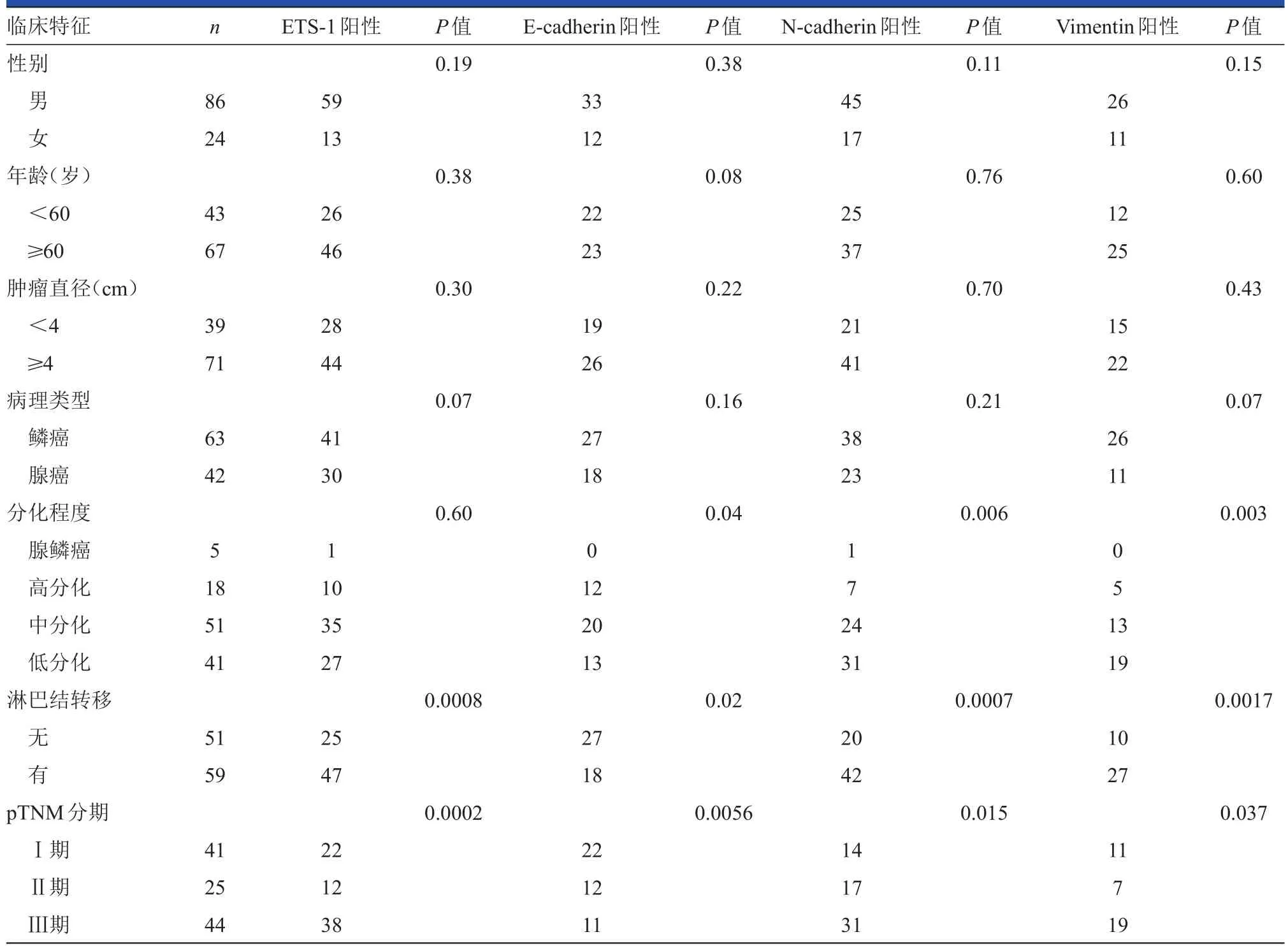
表1 肺癌組織中ETS-1、E-cadherin、N-cadherin與Vimentin的表達與臨床病理特征的關系
E-cadherin屬于鈣依賴性跨膜黏附蛋白家族成員,主要介導同型細胞間的黏附作用,在調節器官、組織的形態發育和維持組織結構的完整性方面具有重要作用。已有相關研究證實E-cadherin基因是一個重要的腫瘤轉移抑制基因,它可能通過促進同型細胞間的黏附而使癌細胞之間保持密切接觸,進而減少癌細胞從原發灶脫離并向遠處轉移[18]。如果E-cadherin表達下降,腫瘤細胞連接松散,也可使腫瘤細胞分裂增殖失控,致腫瘤細胞從原發灶脫落而發生局部或遠處轉移[19]。Lee等[20]的研究表明,E-cadherin在早期、高分化、無浸潤轉移的肺癌細胞中高表達,而在晚期、低分化、有浸潤轉移的肺癌細胞中低表達。提示在腫瘤的發展過程中,E-cadherin的表達下降或消失使腫瘤細胞間的黏附力下降,促進了EMT的發生。本研究結果表明,E-cadherin的表達與腫瘤分化程度、淋巴結轉移和TNM分期呈負相關,與上述文獻報道一致。Shiwu等[21]和Wu等[22]研究證實了E-cadherin表達下調或缺失與肺癌的分化程度、浸潤和轉移密切相關。新近發現E-cadherin基因也是一種抑癌基因,其突變和丟失是腫瘤進展和轉移中一個關鍵性的分子事件[23]。
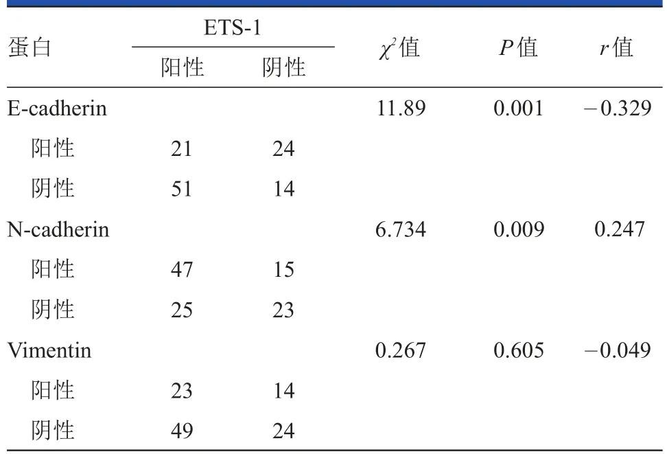
表2 ETS-1與E-cadherin、N-cadherin、Vimentin的相關性分析
N-cadherin由CDH2基因編碼,定位于18q11.2,相對分子量為140 kD。常見于神經外胚層和中胚層組織[24]。N-cadherin對腫瘤上皮細胞、血管平滑肌細胞和外皮細胞之間的相互黏附起重要作用。在正常肺上皮組織中呈陰性。在腫瘤形成的過程中,N-cadherin的表達有助于血管形成及上皮細胞-間質細胞的遷移;還可以通過FGF激活MAPKERK信號轉導途徑,誘導MMP-9基因表達。從而有利于腫瘤血管的形成,使腫瘤易于生長和轉移。近年來有多項研究表明,N-cadherin在惡性黑色素瘤[25]、胰腺癌[26]、乳腺癌[27]、前列腺癌[28]、胃癌[29]及口腔鱗癌[30]等諸多惡性腫瘤中表達上調或重新表達,與腫瘤的浸潤和轉移關系密切。本研究也發現,N-cadherin在肺癌組織中的表達與腫瘤分化程度、淋巴結轉移和TNM分期呈正相關,而與患者的性別、年齡、腫瘤直徑、組織學類型無相關性。
Vimentin是一種在間質表達的Ⅲ型中間絲狀體蛋白,是腫瘤EMT的重要標志物。研究表明,在需要上皮細胞遷移的一系列病理和生理過程中,上皮細胞會表達Vimentin[31]。而Vimentin在上皮細胞遷移及腫瘤侵襲中具有重要的作用[31]。另有文獻報道,Vimentin在膀胱癌中的高表達與腫瘤的浸潤轉移密切相關,Vimentin高表達預示著腫瘤預后不良[32]。已有的研究結果表明,大部分NSCLC患者均有Vimentin表達,而蛋白表達增加是預測腫瘤轉移的一個重要標志[5]。本試驗中,肺癌組織中Vimentin的表達率為34.0%,而在正常癌旁組織中不表達或極低表達,與國內外報道基本一致。
人類ETS(E26 tansformation-specific)基因屬于原癌基因[33]。其編碼產物構成了一個龐大的轉錄調控因子家族,參與調控細胞的增殖分化、遷移凋亡、血管發生和器官形成等。最近的研究顯示,ETS-1在許多惡性腫瘤中普遍高表達[34]。ETS-1還可以調節部分ECM的靶基因包括基質蛋白及其他參與細胞-基質反應的細胞成分,當內源性ETS-1的表達被抑制之后,基質相關蛋白MMP-1及MMP-2在RNA水平表達下調[35]。周梅等[8]采用免疫組化及RT-PCR方法檢測出ETS-1蛋白在NSCLC組織中過表達,ETS-1與肺癌的發生、浸潤、轉移和預后密切相關,并且有可能是肺癌的獨立預后因素。亦有一項研究顯示ETS-1可以作為前列腺癌患者,尤其是高風險患者評估預后的一項重要的分子標志物[36]。孫翠云等[6]利用mRNA FISH法和組織芯片技術,檢測肺癌中ETS-1 mRNA的表達情況,結果有70.4%的患者ETS-1表達陽性,而在正常肺標本中未見表達,差異有統計學意義(P<0.05);該研究表明ETS-1 mRNA表達與肺癌的侵襲轉移有關。本研究發現ETS-1在淋巴結轉移和TNM分期晚的肺癌患者中呈高表達,表明肺癌組織中ETS-1的表達與臨床病理特征具有明顯的相關性。因此,ETS-1可能成為肺癌患者預后判斷及指導臨床治療的重要指標。
本研究結果表明在NSCLC患者中,ETS-1與E-cadherin的表達呈負相關,與N-cadherin的表達呈正相關,與Vimentin的表達無相關性。通過檢測NSCLC中ETS-1與EMT的標志分子E-cadherin、N-cadherin、Vimentin的表達情況,可初步了解肺癌組織中EMT的發生及發展狀況,從而為進一步判斷腫瘤的侵襲轉移能力及制定有效的治療方案提供幫助。
[1]Siegel R,Ma,J,Zou Z,et al.Cancer statistics[J].CA Cancer JClin,2014.64(1):9-29.
[2]張天澤,徐光偉.腫瘤學[M].天津:天津科學技術出版社,1996:1180.
[3]Travis WD.Pathology of lung cancer[J].Clinics in Chest Medicine,2011,32(4):669-692.
[4]de Craene B,Berx G.Regulatory networks defining EMT during cancer initiation and progression[J].Nat Rev Cancer,2013,13(2):97-110.
[5]李巖,李欣,吳愛萍,等.波形蛋白在非小細胞肺癌組織的表達及其意義[J].中華實驗外科雜志,2013, 30(11):2427-2429.
[6]孫翠云,王新允,李艷,等.應用FISH和組織芯片方法研究肺癌組織[J].腫瘤防治研究,2005,32(10): 616-618.
[7]Fernandez-Galilea M,Perez-Matute P,Prieto-Hontoria PL,et al.Effects of lipoic acid on lipolysis in 3T3-L1adipocytes[J].JLipid Res,2012,53(11):2296-2306.
[8]周梅,雷淑慧,胡興勝.ETS-1在非小細胞肺癌中的表達及其臨床意義[J].生命科學研究,2011,15(4): 334-338.
[9]M iao Y,Liu N,Zhang Y,et al.p120ctn isoform 1 expression significantly correlates w ith abnormal expression of E-cadherin and poor survival of lung cancer patients[J].Med Oncol,2010,27(3):880-886.
[10]耿秀東.N-cadherin和E-cadherin在非小細胞肺癌中的表達及相關性研究[D].山東:泰山醫學院,2013.
[11]Hur K,Toiyama Y,Takahashi M,et al.M icroRNA-200c modulates epithelial-to-mesenchymal transition (EMT)in human colorectal cancer metastasis[J].Gut, 2013,62(9):1315-1326.
[12]Shirakihara TM,Saitoh,M iyazono K.Differential regulation of epithelial and mesenchymal markers by delta EF1 proteins in epithelial mesenchymal transition induced by TGF-beta[J].Mol Biol Cell,2007,18(9): 3533-3544.
[13]Okano K,Hibi A,M iyaoka T,et al.Inhibitory effects of the transcription factor Ets-1 on the expression of type I collagen in TGF-beta1-stimulated renal epithelial cells[J].Mol Cell Biochem,2012,369(1-2):247-254.
[14]de Craene B,Berx G.Regulatory networks defining EMT during cancer initiation and progression[J].Nat Rev Cancer,2013,13(2):97-110.
[15]Hou JM,Krebs M,Ward T,et al.Circulating tumor cells as a w indow on metastasis biology in lung cancer [J].Am JPathol,2011,178(3):989-996.
[16]Kokkinos M I,Wafai R,Wong MK,et al.Vimentin and epithelial-mesenchymal transition in human breast cancer-observations in vitro and in vivo[J].Cells Tissues Organs,2007,185(1-3):191-203.
[17]Perl AK,Wilgenbus P,Dahl U,et al.A causal role for E-cadherin in the transition from adenoma to carcinoma [J].Nature,1998,392(6672):190-193.
[18]Danen EH,van Muijen GN,Ruiter DJ.Role of integrins and other cell adhesion molecules in tumor progression and metastasis[J].Lab Invest,1993,68(1):4-17.
[19]Dorudi S,Sheffield JP,Poulsom R,et al.E-cadherin expression in colorectal cancer.An immunocytochem ical and in situ hybridization study[J].Am J Pathol,1993, 142(4):981-986.
[20]Lee YC,Wu CT,Chen CS,et al.The significance of E-cadherin and alpha-,beta-,and gamma-catenin expression in surgically treated non-small cell lung cancers of 3 cm or less in size[J].J Thorac Cardiovasc Surg, 2002,123(3):502-507.
[21]Shiwu Wu,Lan Y,Wenqing S,et al.Expression and clinical significance of CD82/KAI1 and E-cadherin in non-small cell lung cancer[J].Arch Iran Med,2012,15 (11):707-712.
[22]Wu Y,Liu HB,Ding M,etal.The impactof E-cadherin expression on non-small cell lung cancer survival:ameta analysis[J].Mol BiolRep,2012,39(9):9621-9628.
[23]Le Bras GF,Taubenslag KJ,Andl CD.The regulation of cell-cell adhesion during epithelial-mesenchymal transition,motility and tumor progression[J].Cell Adh M igr,2012,6(4):365-373.
[24]Stemm ler MP.Cadherins in development and cancer[J]. Mol Biosyst,2008,4(8):835-850.
[25]Sandig M,Voura EB,Kalnins VI,et al.Role of cadherins in the transendothelialmigration of melanoma cells in culture[J].Cell Moti Cytoskeleton,1997,38(4):351-364.
[26]Nakajima S,DoiR,Toyoda E,etal.N-cadherin expression and epithelial-mesenchymal transition in pancreatic carcinoma[J].Clin CancerRes,2004,10(12Pt1):4125-4133.
[27]翟明翠.N-cadherin與mn23-Hl在乳腺癌中的表達及意義[J].黑龍江醫學,2010,34(8):561-563.
[28]Nalla AK,Estes N,Patel J,et al.N-cadherin mediates angiogenesis by regulating monocyte chemoattractant protein-1 expression via PI3K/Akt signaling in prostate cancer cells[J].Exp Cell Res,2011,317(17):2512-2521.
[29]Kamikihara Y,Ueno S,Natsugoe S.Clinical implications of N-cadherin expression in gastric cancer[J].Pathol Int,2012,62(3):161-166.
[30]Di Domenico M,Pierantoni GM,Feola A,et al.Prognostic significance of N-cadherin expression in oral squamous cell carcinoma[J].Anticancer Research, 2011,31(12):4211-4218.
[31]Chernyatina AA,Nicolet S,Aebi U,et al.Atomic structure of the vimentin central alpha-helical domain and its implications for intermediate filament assembly[J].Proc Natl Acad Sci USA,2012,109(34):13620-13625.
[32]董大海,趙軍,孫立江,等.E鈣黏著素與波形蛋白在膀胱癌中的表達及意義[J].現代泌尿外科雜志, 2010,15(1):22-24.
[33]Oikawa T.ETS transcription factors:possible targets for cancer therapy[J].Cancer Sci,2004,95(8):626-633.
[34]Gambarotta G,Boccaccio C,Giordano S,et al.Ets upregulates MET transcription[J].Oncogene,1996,13(9): 1911-1917.
[35]Dittmer J,Vetter M,Blumenthal SG,et al.Importance of ets1 proto-oncogene for breast cancer progression[J].Zentralbl Gynakol,2004,126(4):269-271.
[36]Li B,Shimizu Y,Kobayashi T,et al.Overexpression of ETS-1 is associated w ithmalignantbiological features of prostate cancer[J].Asian JAndrol,2012,14(6):860-863.
The correlation between expression of ETS-1 and E-cadherin,N-cadherin and Vimentin in non-smallcell lung cancer△
ZHOU Li-li1,4GEXin-guo2CAIMao-huai1#TANG Feng1ZHU Jian-feng3SUN Lin3LIXia3LIU Ju-lin1WANG Cai-lian4#
1Departmentof Oncology,2Departmentof Oncological Surgery,3Departmentof Pathology,the Second People's Hospitalof Yancheng/The Tumour Hospitalof Yancheng,Yancheng 224003,Jiangsu,China4DepartmentofOncology,Zhongda Hospital,MedicalCollegeof SoutheastUniversity,Nanjing 210009,Jiangsu,China
Objective To observe the expression between ETS-1 and neuronal cadherin(E-cadherin,N-cadherin) and Vimentin in non-small cell lung cancer(NSCLC)tissues and to study the clinical correlation of thosemolecules.Method 110 cases of NSCLC patients who had undergone surgical resection were confirmed pathologically and were included in this study,of which the tumor tissues and normal lung tissues thatwere at least 3 cm away from the tumor were selected.The immunohistochem istry SPmethod was used to detect the expression of ETS-1,E-cadherin,N-cadherin and Vimentin,then the correlation was investigated.Result In NSCLC tissues,the positive expression rate of ETS-1,N-cadherin and Vimentin[65%(72/110),56%(62/110),and 34%(37/110),respectively]were significantly higher than those in normal lung tissues[6%(3/50),8%(4/50),2%(1/50),respectively](all P<0.01);The positive expression rate of E-cadherin was 41%(45/110),which was significantly lower than the 84%(42/50)of normal lung tissues.The expression of ETS-1 was positively correlated w ith the lymph node metastasis and TNM staging(P<0.05),butwas not associated w ith gender,age,tumor diameter,histological type and differentiation degree(P>0.05).The expression of E-cadherin was negatively correlated w ith tumor differentiation degree,the lymph node metastasis and TNM staging(P<0.05).The expression of N-cadherin and Vimentin were positively correlated w ith tumor differentiation degree,the lymph node metastasis and TNM staging(P<0.05).The expression of E-cadherin,N-cadherin and Vimentin were not correlated w ith gender,age,tumor diameter and histological type(P>0.05).While the expression of ETS-1 was negatively correlated w ith E-cadherin but positively correlated w ith N-cadherin in NSCLC.Conclusion The expression of ETS-1,E-cadherin,N-cadherin and Vimentin are closely related w ith metastasis of NSCLC,thus the detection on the expression of those molecules w ill be effective in estimating the metastasis and prognosis of NSCLC.
ETS-1;mucins;vimentin;non-small cell lung cancer;neoplasmsmetastasis
R734.2
A
10.11877/j.issn.1672-1535.2015.13.05.11
鹽城市醫學科技發展計劃項目(YK 2014032)
#通信作者(corresponding author),蔡茂懷e-mail:wangcailian65@hotmail.com;王彩蓮e-mail:cmhyc001@163.com
2015-01-12)
猜你喜歡
保健醫苑(2023年2期)2023-03-15 09:03:04
新少年(2022年9期)2022-09-17 07:10:54
中國臨床醫學影像雜志(2022年2期)2022-05-25 13:24:34
體育科技文獻通報(2022年3期)2022-05-23 13:46:54
遼金歷史與考古(2021年0期)2021-07-29 01:06:54
小天使·一年級語數英綜合(2020年6期)2020-12-16 02:56:41
科技傳播(2019年22期)2020-01-14 03:06:54
民用飛機設計與研究(2019年4期)2019-05-21 07:21:24
醫學研究雜志(2015年12期)2015-06-10 06:57:46
北極光(2014年8期)2015-03-30 02:50:51
