磁共振彌散加權(quán)成像對(duì)宮頸癌盆腔淋巴結(jié)轉(zhuǎn)移診斷的應(yīng)用價(jià)值
2017-10-20 10:37:10董樂丹肖琴琴葉瓊許化致楊運(yùn)俊
中國現(xiàn)代醫(yī)生 2017年25期
董樂丹+肖琴琴+葉瓊+許化致+楊運(yùn)俊
[摘要] 目的 探討磁共振彌散加權(quán)成像對(duì)宮頸癌盆腔淋巴結(jié)轉(zhuǎn)移診斷的臨床應(yīng)用價(jià)值。 方法 收集2015年8月~2016年10月我院收治的26例行盆腔淋巴結(jié)清掃術(shù)的宮頸癌患者的術(shù)前MRI檢查資料,術(shù)后均經(jīng)病理檢查證實(shí)。比較分析宮頸癌原發(fā)灶、轉(zhuǎn)移淋巴結(jié)及非轉(zhuǎn)移淋巴結(jié)的ADC值差異;比較轉(zhuǎn)移淋巴結(jié)與非轉(zhuǎn)移淋巴結(jié)最長徑、最短徑及短/長徑之比。 結(jié)果 ①26例宮頸癌患者中轉(zhuǎn)移性淋巴結(jié)56枚,非轉(zhuǎn)移性淋巴結(jié)78枚;②轉(zhuǎn)移性淋巴結(jié)ADC均值為(874.19±158.72)×10-6 mm2/s),較非轉(zhuǎn)移性淋巴結(jié)ADC均值[(921.45±232.98)×10-6 mm2/s]略降低,但兩者間差異無統(tǒng)計(jì)學(xué)意義(P>0.05);宮頸癌原發(fā)灶A(yù)DC均值為(918.78±146.06)×10-6 mm2/s),與轉(zhuǎn)移淋巴結(jié)、非轉(zhuǎn)移淋巴結(jié)ADC均值兩兩比較,差異均無統(tǒng)計(jì)學(xué)意義(P>0.05);③轉(zhuǎn)移性淋巴結(jié)與非轉(zhuǎn)移性淋巴結(jié)最長徑中位數(shù)分別為16.50 mm、7.00 mm,最短徑中位數(shù)分別為8.50 mm、5.00 mm,短長徑比的中位數(shù)為0.55、0.65,兩兩比較差異有統(tǒng)計(jì)學(xué)意義(P<0.05)。 結(jié)論 DWI有利于盆腔淋巴結(jié)的檢出,但是ADC值測量對(duì)轉(zhuǎn)移性淋巴結(jié)的臨床診斷價(jià)值并不大。
[關(guān)鍵詞] 宮頸癌;磁共振彌散加權(quán)成像;轉(zhuǎn)移性淋巴結(jié);ADC值
[中圖分類號(hào)] R737.33 [文獻(xiàn)標(biāo)識(shí)碼] B [文章編號(hào)] 1673-9701(2017)25-0101-04
The application value of magnetic resonance diffusion weighted imaging in the diagnosis of pelvic lymph nodes metastasis of cervical cancer
DONG Ledan XIAO Qinqin YE Qiong XU Huazhi YANG Yunjun
Department of Radiology, the First Affiliated Hospital of Wenzhou Medical University, Wenzhou 325000, China
[Abstract] Objective To explore the clinical application value of MR diffusion weighted imaging in the diagnosis of pelvic lymph node metastasis of cervical cancer. Methods The preoperative MRI examination data of 26 patients with cervical cancer who underwent pelvic lymphadenectomy in our hospital from August 2015 to October 2016 were enrolled in this study. The diagnosis of all cases was confirmed by postoperative MRI findings. The differences of ADC values among primary cervical cancer, metastatic lymph nodes and non-metastatic lymph nodes were analyzed. The longest diameter, the shortest diameter and the ratio of the short/long diameter were compared. Results ①There were 56 metastatic lymph nodes and 78 non-metastatic lymph nodes in 26 cases of cervical cancer. ②The mean value of metastatic lymph node ADC[(874.19±158.72)×10-6 mm2/s] was slightly lower than that of non-metastatic lymph node [(921.45±232.98)×10-6 mm2/s], but there was no significant difference between the two groups(P>0.05). There was no significant difference in the ADC value among the primary tumor[(918.78±146.06)×10-6 mm2/s], metastatic lymph node and non-metastatic lymph node of cervical cancer(P>0.05). ③The median of the longest diameter of the metastatic lymph nodes and the non-metastatic lymph nodes was 16.50 mm and 7.00 mm, respectively. The median of the shortest diameter was 8.50 mm and 5.00 mm, respectively. The median of short diameter/long diameter was 0.55, 0.65 respectively. There was statistically significant difference among the groups(P<0.05). Conclusion DWI is helpful for the detection of pelvic lymph nodes, but the clinical diagnostic value of ADC measurement is not significant.endprint
[Key words] Cervical cancer; Magnetic resonance diffusion weighted imaging; Metastatic lymph node; ADC value
宮頸癌是我國女性生殖系統(tǒng)最常見的惡性腫瘤[1]。按照國際FIGO系統(tǒng),宮頸癌的臨床分期與淋巴結(jié)是否轉(zhuǎn)移并無直接關(guān)聯(lián)[2],但是具體治療方案、療效及預(yù)后情況與淋巴結(jié)是否轉(zhuǎn)移密切相關(guān)[3]。國內(nèi)外大量文獻(xiàn)已經(jīng)證實(shí)淋巴結(jié)轉(zhuǎn)移是宮頸癌患者重要的獨(dú)立危險(xiǎn)因素之一[4]。因此,盆腔淋巴結(jié)轉(zhuǎn)移是判斷預(yù)后及指導(dǎo)治療方案的重要依據(jù)[5,6],目前臨床工作中MRI診斷主要依據(jù)淋巴結(jié)形態(tài)學(xué)改變來判斷是否存在轉(zhuǎn)移。國內(nèi)外許多的研究認(rèn)為DWI對(duì)盆腔各類惡性腫瘤的淋巴結(jié)檢出及是否轉(zhuǎn)移具有較高的臨床價(jià)值[7-9],但在確診盆腔轉(zhuǎn)移性淋巴結(jié)尚存在諸多爭議[10],本研究主要探討MRI檢查對(duì)宮頸癌盆腔淋巴結(jié)是否轉(zhuǎn)移的診斷價(jià)值,并探討ADC值的臨床價(jià)值。
1 資料與方法
1.1一般資料
選擇2015年8月~2016年10月于我院行MRI檢查且經(jīng)病理組織檢查證實(shí)的宮頸癌患者26例作為研究對(duì)象,年齡31~76歲,平均53歲,其中鱗癌24例,腺癌1例,其他(原始神經(jīng)外胚葉腫瘤)1例。納入標(biāo)準(zhǔn):(1)經(jīng)宮頸活檢病理診斷明確,臨床分期為Ⅰa~Ⅱa期;(2)術(shù)前無放化療等輔助治療;(3)術(shù)前常規(guī)行盆腔MRI檢查;(4)均行全子宮切除加淋巴結(jié)清掃術(shù),術(shù)中清楚記錄淋巴結(jié)部位及數(shù)目,術(shù)后均有病理結(jié)果。排除標(biāo)準(zhǔn):(1)術(shù)前行放化療檢查患者;(2)術(shù)前MRI檢查時(shí)間與手術(shù)時(shí)間超過20 d。
1.2 儀器與方法
本組所有患者均使用飛利浦Achieva 3.0T TX超導(dǎo)型磁共振掃描,行盆腔MRI常規(guī)檢查,采用腹部16通道相控陣線圈,依次行DWI、T1WI、軸位T2WI、矢狀位T2WI及冠狀位T2WI。
1.3 圖像處理及評(píng)定標(biāo)準(zhǔn)
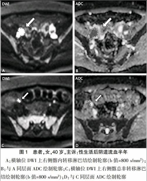
所有數(shù)據(jù)處理均由具備臨床工作經(jīng)驗(yàn)的兩名放射科醫(yī)師進(jìn)行。根據(jù)手術(shù)記錄資料、病理結(jié)果及常規(guī)MR圖像,確定子宮原發(fā)灶、轉(zhuǎn)移淋巴結(jié)及非轉(zhuǎn)移淋巴結(jié)。轉(zhuǎn)移淋巴結(jié)判定:轉(zhuǎn)移淋巴結(jié)指影像上存在1個(gè)或1個(gè)以上轉(zhuǎn)移征象,手術(shù)中確認(rèn)部位及病理結(jié)果確診,對(duì)不能確認(rèn)者予以剔除。首先將軸位T2WI圖像上所有淋巴結(jié)短徑(Short-axis diameter)≥3 mm者納入本組研究,同時(shí)測量淋巴結(jié)長徑(Long-axis diameter)。聯(lián)合常規(guī)T1WI及T2WI圖像,使用Image Analyzer軟件結(jié)合DWI于ADC圖上繪制宮頸癌原發(fā)灶、轉(zhuǎn)移及非轉(zhuǎn)移淋巴結(jié)的每一層面感興趣區(qū)而獲得病灶體積感興趣(VOI),然后通過使用Matelab軟件計(jì)算獲得ADC值,如圖1所示。
1.4 統(tǒng)計(jì)學(xué)方法
應(yīng)用SPSS23.0軟件包進(jìn)行數(shù)據(jù)分析,盆腔淋巴結(jié)長徑、短徑測量值為偏態(tài)分布計(jì)量資料,淋巴結(jié)最長徑、最短徑采用中位數(shù)及四分位數(shù)表示,兩組最長徑、最短徑及短長比采用獨(dú)立樣本秩和檢驗(yàn);兩組原發(fā)病灶及淋巴結(jié)所測ADC均值為計(jì)量資料,宮頸癌原發(fā)病灶、轉(zhuǎn)移淋巴結(jié)及非轉(zhuǎn)移淋巴結(jié)三組之間的比較采用方差分析,檢驗(yàn)水準(zhǔn)α=0.05,P<0.05為差異有統(tǒng)計(jì)學(xué)意義。
2 結(jié)果
2.1 盆腔淋巴結(jié)基本資料分析結(jié)果
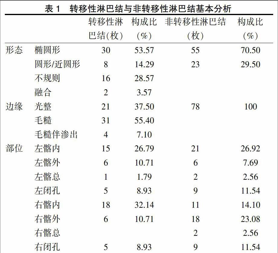
轉(zhuǎn)移性淋巴結(jié)與非轉(zhuǎn)移性淋巴結(jié)基本分析(部位、形態(tài)及邊緣)見表1。共收集盆腔淋巴結(jié)134枚,其中非轉(zhuǎn)移淋巴結(jié)78枚,轉(zhuǎn)移淋巴結(jié)56枚。結(jié)果提示非轉(zhuǎn)移性淋巴結(jié)形態(tài)以橢圓形為主,邊緣基本光整,淋巴結(jié)在盆腔掃描中均可顯示。轉(zhuǎn)移性淋巴結(jié)仍以橢圓形為主,但在影像上不規(guī)則或融合形態(tài)更能提示轉(zhuǎn)移性;邊緣顯示則以毛糙改變?yōu)橹鳎枨徊课徊o特殊性。
2.2 轉(zhuǎn)移性淋巴結(jié)與非轉(zhuǎn)移性淋巴結(jié)最長徑、最短徑及短長徑比
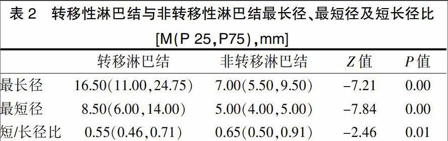
轉(zhuǎn)移性淋巴結(jié)與非轉(zhuǎn)移性淋巴結(jié)最長徑、最短徑及短長徑比分析見表2。結(jié)果表明,盆腔轉(zhuǎn)移性淋巴結(jié)與非轉(zhuǎn)移性淋巴結(jié)最長徑、最短徑及短/長徑比之間的差異有統(tǒng)計(jì)學(xué)意義(P均<0.05)。
2.3 宮頸癌原發(fā)灶及盆腔淋巴結(jié)ADC值分析結(jié)果
宮頸癌原發(fā)灶、盆腔轉(zhuǎn)移性淋巴結(jié)和非轉(zhuǎn)移性淋巴結(jié)之間ADC均值結(jié)果比較,見表3。結(jié)果提示宮頸癌原發(fā)病、轉(zhuǎn)移性淋巴結(jié)與非轉(zhuǎn)移性淋巴結(jié)之間兩兩比較,差異均無統(tǒng)計(jì)學(xué)意義(P>0.05)。
3 討論
宮頸癌是女性生殖系統(tǒng)中最常見的惡性腫瘤之一[1],嚴(yán)重威脅女性的身心健康。宮頸癌的早期診斷、正確分期及淋巴結(jié)是否轉(zhuǎn)移的評(píng)判對(duì)手術(shù)方式的選擇以及療效、預(yù)后都具有重要意義[2,11]。目前臨床上宮頸癌手術(shù)均采用全子宮切除加盆腔淋巴結(jié)清掃,對(duì)患者的創(chuàng)傷較大,會(huì)伴有神經(jīng)損傷、淋巴管囊腫等并發(fā)癥,因此術(shù)前正確評(píng)估對(duì)臨床選擇個(gè)體化治療具有重要意義[12],部分患者可以避免盆腔淋巴結(jié)的清掃。
磁共振擴(kuò)散加權(quán)成像(DWI)是臨床上應(yīng)用較廣泛的功能成像之一,也是目前影像學(xué)檢查中唯一能夠觀察組織水分子活動(dòng)的無創(chuàng)性檢查手段[13,14]。DWI成像原理的主要病理性改變是正常組織局部發(fā)生病變時(shí),細(xì)胞密度、細(xì)胞代謝等發(fā)生變化,致使水分子的擴(kuò)散也發(fā)生不同程度的變化[7,15]。基于這樣的原理磁共振DWI最早是應(yīng)用于中樞神經(jīng)系統(tǒng),尤其是急性腦卒中患者的檢查,這是因?yàn)榧毙阅X卒中會(huì)導(dǎo)致水分子擴(kuò)散受限[16]。目前也證實(shí)DWI對(duì)于組織器官良惡性腫瘤的鑒別是很重要的檢查手段之一[17],因?yàn)閻盒阅[瘤細(xì)胞比正常組織密集以及細(xì)胞體積較大[18],因此DWI圖像上呈明顯高信號(hào),ADC圖上呈明顯低信號(hào)。另外,很多的研究也認(rèn)為淋巴結(jié)在DWI上可以清楚顯示高信號(hào)[19]。因此關(guān)于DWI檢查來鑒別轉(zhuǎn)移性淋巴結(jié)與非轉(zhuǎn)移性淋巴結(jié)的臨床價(jià)值尚有很多爭議[3],部分研究認(rèn)為于DWI上測量ADC值可以從影像上定量性區(qū)分轉(zhuǎn)移及非轉(zhuǎn)移性淋巴結(jié),但目前尚無明確標(biāo)準(zhǔn)值參考范圍[7-8];也有部分研究認(rèn)為DWI聯(lián)合T2WI上描述淋巴結(jié)形態(tài)、大小及邊緣可以大致檢出及評(píng)估轉(zhuǎn)移性淋巴結(jié)與非轉(zhuǎn)移性淋巴結(jié)[18,20]。endprint
本研究結(jié)果顯示評(píng)估盆腔淋巴結(jié)是否轉(zhuǎn)移,T2WI對(duì)淋巴結(jié)形態(tài)、邊緣及大小的評(píng)判可以提供重要的信息,轉(zhuǎn)移性淋巴結(jié)形態(tài)常顯示不規(guī)則或融合狀態(tài),邊緣常常顯示毛糙或伴有滲出的改變,同時(shí)也可以呈橢圓形或圓形以及邊緣光整,因此對(duì)淋巴結(jié)形態(tài)及邊緣的描述可以提供一定的信息,但不足以評(píng)判是否轉(zhuǎn)移。本研究結(jié)果還表明轉(zhuǎn)移性淋巴結(jié)與非轉(zhuǎn)移信淋巴結(jié)兩組間的最長徑、最短徑及短/長徑比之間具有明顯差異,且兩組間的差異有統(tǒng)計(jì)學(xué)意義。這與賀李等[21]研究者認(rèn)為淋巴結(jié)的大小可以作為影像評(píng)判的主要指標(biāo)之一相符,該研究還認(rèn)為淋巴結(jié)短徑>20 mm,常規(guī)MRI基本可以準(zhǔn)確診斷為轉(zhuǎn)移性淋巴結(jié),但同時(shí)也表明尚不能將短徑>10 mm作為評(píng)判標(biāo)準(zhǔn)之一,許多研究者通過嚴(yán)格的“點(diǎn)對(duì)點(diǎn)”影像及病理結(jié)合研究發(fā)現(xiàn)<10 mm的轉(zhuǎn)移性淋巴結(jié)并不少見[13,18],本研究中也發(fā)現(xiàn)此現(xiàn)象。另外,許多的研究[22]認(rèn)為ADC值可以反映病變組織的特性,可以定量評(píng)估轉(zhuǎn)移性淋巴結(jié)[23]。本研究發(fā)現(xiàn)轉(zhuǎn)移性淋巴結(jié)ADC均值低于非轉(zhuǎn)移性淋巴結(jié)及宮頸癌原發(fā)灶,但是組間差異無統(tǒng)計(jì)學(xué)意義。雖然目前有研究發(fā)現(xiàn)盆腔轉(zhuǎn)移與非轉(zhuǎn)移性淋巴結(jié)之間ADC值差異具有顯著性差壓,但是有研究[24]結(jié)果顯示ADC值測量對(duì)診斷淋巴結(jié)是否轉(zhuǎn)移并沒有很大貢獻(xiàn)。但就本研究分析原因可能如下:(1)大部分研究所測的ADC值是淋巴結(jié)或?qū)m頸癌原發(fā)病灶單層圖像上所畫取的興趣區(qū)(region of interest,ROI)ADC值,本研究所測的ADC值為淋巴結(jié)或?qū)m頸癌原發(fā)灶全體積ADC值的均值,因此可能與研究者使用的方法不同存有一定的關(guān)系,而目前尚較少文獻(xiàn)使用該方法對(duì)淋巴結(jié)的是否轉(zhuǎn)移情況進(jìn)行研究,因此該方法還需要更多的研究。(2)可能與本研究樣本不足有一定關(guān)系,因此下一步研究需要擴(kuò)大樣本量,此外還可能與研究工作中研究者與的手工畫取感興趣區(qū)存有一定的誤差有關(guān)。
綜上所述,3.0T磁共振DWI對(duì)宮頸癌盆腔淋巴結(jié)的顯示具有明顯優(yōu)勢,轉(zhuǎn)移性及非轉(zhuǎn)移性淋巴結(jié)的最長徑、最短徑及短/長徑比之間的差異有統(tǒng)計(jì)學(xué)意義,結(jié)合淋巴結(jié)形態(tài)及邊緣情況的綜合評(píng)判可以提高淋巴結(jié)轉(zhuǎn)移性診斷的準(zhǔn)確性。
[參考文獻(xiàn)]
[1] Waggoner SE. Cervical cancer[J]. The Lancet,2003,361(9376):2217-2225.
[2] Pecorelli S. Revised figo staging for carcinoma of the vulva,cervix,and endometrium[J]. International Journal of Gynecology & Obstetrics,2009,105(2):103-104.
[3] Wang YT,Li YC,Yin LL,et al. Can diffusion-weighted magnetic resonance imaging predict survival in patients with cervical cancer? A meta-analysis[J]. European Journal of Radiology, 2016,85(12):2174-2181.
[4] Kim JK,Kim KA,Park BW,et al. Feasibility of diffusion-weighted imaging in the differentiation of metastatic from nonmetastatic lymph nodes:Early experience[J]. Journal of Magnetic Resonance Imaging:JMRI,2008,28(3):714-719.
[5] Liu Y,Liu H,Bai X,et al. Differentiation of metastatic from non-metastatic lymph nodes in patients with uterine cervical cancer using diffusion-weighted imaging[J]. Gynecologic Oncology,2011,122(1):19-24.
[6] Martí-Bonmatí L. Lymph node assessment by diffusion weighted imaging in cervical cancer[J]. European Radiology,2011,21(3):474-477.
[7] Papalia R,Simone G,Grasso R,et al. Diffusion-weighted magnetic resonance imaging in patients selected for radical cystectomy:Detection rate of pelvic lymph node metastases[J]. BJU International,2012,109(7):1031-1036.
[8] Demirbas T,Cimilli T,Bayramoglu S,et al. Contribution of diffusion-weighted imaging to diagnosis and staging of cervical cancer[J]. Balkan Medical Journal,2014,31(2):154-157.
[9] Kuang F,Ren J,Zhong Q,et al. The value of apparent diffusion coefficient in the assessment of cervical cancer [J].European Radiology,2012,23(4):1050-1058.endprint
[10] Chen YB,Liao J,Xie R,et al. Discrimination of metastatic from hyperplastic pelvic lymph nodes in patients with cervical cancer by diffusion-weighted magnetic resonance imaging[J]. Abdominal Imaging,2011,36(1):102-109.
[11] Marzi S,Piludu F,Sanguineti G,et al. The prediction of the treatment response of cervical nodes using intravoxel incoherent motion diffusion-weighted imaging[J]. EuroPean Journal of Radiology,2017,92(6):93-102.
[12] Vandecaveye V,Dresen R,De Keyzer F. Novel imaging techniques in gynaecological cancer[J]. Current Opinion in Oncology,2017,29(5):335-342.
[13] Choi HJ,Roh JW,Seo SS,et al. Comparison of the accuracy of magnetic resonance imaging and positron emission tomography/computed tomography in the presurgical detection of lymph node metastases in patients with uterine cervical carcinoma:A prospective study[J]. Cancer, 2006,106(4):914-922.
[14] Liu B,Gao S,Li S. A comprehensive comparison of ct,mri,positron emission tomography or positron emission tomography/ct,and diffusion weighted imaging-mri for detecting the lymph nodes metastases in patients with cervical cancer:A Meta-analysis based on 67 studies[J]. Gynecologic and Obstetric Investigation,2017,82(3):209-222.
[15] Godeny M,Lengyel Z,Polony G,et al. Impact of 3t multiparametric mri and fdg-pet-ct in the evaluation of occult primary cancer with cervical node metastasis[J]. Cancer Imaging:The Official Publication of the International Cancer Imaging Society,2016,16(1):38.
[16] von Kummer R,Dzialowski I. Imaging of cerebral ischemic edema and neuronal death[J]. Neuroradiology,2017, 59(6):545-553.
[17] Oh E,Yoon YC,Kim JH,et al. Multiparametric approach with diffusion-weighted imaging and dynamic contrast-enhanced mri:A comparison study for differentiating between benign and malignant bone lesions in adults[J]. Clinical radiology,2017,72(7):552-559.
[18] Roy C,Bierry G,Matau A,et al.Value of diffusion-weighted imaging to detect small malignant pelvic lymph nodes at 3 t[J]. European radiology,2010,20(8):1803-1811.
[19] Liang L,Luo X,Lian Z,et al. Lymph node metastasis in head and neck squamous carcinoma:Efficacy of intravoxel incoherent motion magnetic resonance imaging for the differential diagnosis[J]. European Journal of Radiology,2017, 90(5):159-165.
[20] Kitajima K,Yamasaki E,Kaji Y,et al. Comparison of dwi and pet/ct in evaluation of lymph node metastasis in uterine cancer[J]. World Journal of Radiology,2012,4(5):207-214.
[21] 賀李,余深平,莊曉盟,等. 3.0T磁共振DWIBS診斷宮頸癌轉(zhuǎn)移性淋巴結(jié)初探[J].影像診斷與介入放射學(xué),2012,19(5):273-276.
[22] Himoto Y,F(xiàn)ujimoto K,Kido A,et al. Pretreatment mean apparent diffusion coefficient is significantly correlated with event-free survival in patients with international federation of gynecology and obstetrics stage ib to iiib cervical cancer[J]. International Journal of Gynecological cancer:Official Journal of the International Gynecological Cancer Society,2015,25(7):1079-1085.
[23] Ju FJ. Evaluation of the efficacy of chemoradiotherapy in cervical cancer using diffusion-weighted imaging and apparent diffusion coefficient[J]. OncoTargets and Therapy, 2016,9(11):7555-7561.
[24] Nakai G,Mitsuru M,Inada Y,et al. Detection and evaluation of pelvic lymph nodes in patients with gynecologic malignancies using body diffusion-weighted magnetic resonance imaging[J]. J Comput Assist Tomogr,2008,32(5):764-768.endprint
