牛蒡子苷調控血管內皮生長因子抑制糖尿病視網膜病變的機制研究
2020-02-14 03:14:36閆媛媛劉平
中國醫藥生物技術 2020年1期
關鍵詞:糖尿病
閆媛媛,劉平
論著
牛蒡子苷調控血管內皮生長因子抑制糖尿病視網膜病變的機制研究
閆媛媛,劉平
450003 河南,鄭州人民醫院眼科
探討牛蒡子苷(Arc)是否通過調控血管內皮生長因子(VEGF)表達進而抑制糖尿病視網膜病變。
體外培養人視網膜微血管內皮細胞(HRCECs),用不同濃度的 Arc 處理 HRCECs,同時將 si-VEGF 或 pcDNA3.1-VEGF 分別轉染入 HRCECs,MTT 法檢測HRCECs 增殖能力;Western blot 檢測 p21、細胞周期蛋白D1(cyclinD1)、腫瘤壞死因子-α(TNF-α)、人單核細胞趨化蛋白-1(MCP-1)、載脂蛋白 M(ApoM)、VEGF 蛋白表達。
與 LG 組相比,HG 組 HRCECs 的值明顯升高(< 0.05),cyclinD1、TNF-α、MCP-1、ApoM、VEGF 水平均顯著升高(< 0.05),而 p21 表達下調(< 0.05),Arc 作用后可顯著逆轉 HG 對 HRCECs 增殖、cyclinD1、TNF-α、MCP-1、ApoM、VEGF 及 p21 表達的作用,且 Arc 不同劑量組間均存在顯著性差異(< 0.05);抑制 VEGF 表達可明顯減弱高糖作用下的 HRCECs 增殖能力并降低相關炎性因子表達;過表達 VEGF 能逆轉 Arc 對高糖誘導的 HRCECs 增殖及炎癥反應的抑制作用。
牛蒡子苷可能通過降低高糖誘導的 HRCECs 細胞中 VEGF 的高表達進而抑制HRCECs 增殖,并可通過減輕炎癥反應保護細胞免受損傷。
血管內皮生長因子類; 糖尿病視網膜病變; 牛蒡子苷; 人視網膜微血管內皮細胞
糖尿病視網膜病變是糖尿病晚期患者的主要并發癥之一,可降低患者生活質量甚至導致患者失明,其主要病理特點為新血管生成,而血管內皮細胞異常增殖可促進新血管生成[1-2]。糖尿病患者體內持續性高糖環境下可引發機體產生氧化應激、炎癥反應并可促進血管生成因子的釋放進而導致視網膜毛細血管內皮細胞異常增生最終導致糖尿病視網膜病變的發生[3]。如何抑制新血管生成可能成為治療糖尿病視網膜病變的新思路。有研究發現,牛蒡子苷(arctiin,Arc)能夠改善糖尿病大鼠視網膜結構,對大鼠視網膜病變具有一定的治療作用[4]。血管內皮生長因子(vascular endothelial growth factor,VEGF)在糖尿病視網膜病變患者血漿中上調表達,在糖尿病視網膜病變中起著重要的作用[5]。有研究證明,大黃素對高糖條件下人視網膜血管內皮細胞增殖具有抑制作用,其可為糖尿病視網膜病變的預防和治療提供新的方法[6]。相關研究報道指出,抗 VEGF 藥物與二甲雙胍聯合使用可有效治療糖尿病視網膜病變[7]。然而,關于牛蒡子苷對糖尿病視網膜病變過程的作用機制及其是否可通過調控 VEGF 表達進而發揮作用仍未可知。因此,本研究觀察高糖作用下人視網膜微血管內皮細胞(human retinal capillary endothelial cells,HRCECs)中 VEGF 表達變化,探討牛蒡子苷是否可通過調控 VEGF 表達而發揮作用,以期為牛蒡子苷治療糖尿病視網膜病變奠定理論基礎。
1 材料與方法
1.1 材料
HRCECs 細胞購自上海妙順生物科技有限公司;si-VEGF 及其陰性對照均購自上海吉凱基因化學公司;牛蒡子苷購自成都尚科藥業有限責任公司;D-葡萄糖、胎牛血清(FBS)、DMEM 培養基均購自美國 Sigma 公司;蛋白提取試劑盒購自上海貝博生物科技有限公司;蛋白印跡(Western blot)實驗所需相關試劑盒均購自上海碧云天生物技術有限公司;噻唑藍(MTT)購自廣州朗日生物技術有限公司;鼠抗人 TNF-α、MCP-1、ApoM 單克隆抗體均購自武漢友聯特生物技術有限公司;辣根過氧化物酶(HRP)標記的山羊抗鼠 IgG 二抗購自美國 Proteintech 公司;p21、cyclinD1、VEGF 一抗均購自美國 Santa Cruz 公司;Lipofectamine2000 轉染試劑購自美國 Invitrogen 公司。
1.2 方法
1.2.1 HRCECs 培養及實驗分組 常規復蘇 HRCECs,培養于含有 10% FBS 與 5.5 mmol/LD-葡萄糖的 DMEM 培養基,放入 37 ℃、5% CO2培養箱內,穩定傳代后將細胞分為 5 組,LG 組:用 5.5 mmol/L D-葡萄糖的 DMEM 培養基培養;HG 組:用 30 mmol/L 葡萄糖培養細胞[8];HG + Arc 20 mg/L 組:用濃度為 20 mg/L 的牛蒡子苷處理細胞,并用高糖培養基培養;HG + Arc 30 mg/L 組:濃度為 30 mg/L 的牛蒡子苷處理細胞,并用高糖培養基培養;HG + Arc 40 mg/L 組:濃度為40 mg/L 的牛蒡子苷處理細胞,并用高糖培養基培養[9]。各組處理時間均為 48 h。
1.2.2 VEGF 轉染及實驗分組 為驗證 VEGF 對 HRCECs 增殖及炎癥反應的影響,將細胞分為 si-NC 組:用不含 VEGF 序列的陰性對照質粒感染正常培養的細胞,高糖培養基培養;si-VEGF 組:用含有 siRNA-VEGF 序列的質粒感染正常培養的細胞,高糖培養基培養;HG + Arc 30 mg/L + pcDNA3.1 組:不含有VEGF 序列的空載體感染細胞,用含有 30 mg/L 濃度的牛蒡子苷高糖培養基培養;HG + Arc 30 mg/L + pcDNA3.1-VEGF 組:含有 VEGF 序列的載體感染細胞,用含有 30 mg/L濃度的牛蒡子苷高糖培養基培養。培養 48 h 后更換培養基繼續培養,收集對數生長期細胞進行后續實驗。
1.2.3 MTT 法檢測HRCECs 增殖能力 用 0.25% 胰蛋白酶消化各組對數生長期HRCECs,制備單細胞懸液,調整細胞密度,按照每孔 1 × 10⁴個細胞的密度接種于 96 孔板,各組細胞分別于培養 24、48、72 h 時,向每孔內分別加入 15 μl 質量濃度為 10 g/L 的 MTT 溶液,放入恒溫培養箱繼續培養 4 h,棄培養液,向每孔中分別加入 200 μl 二甲基亞砜(DMSO)溶液,低速振蕩 15 min 以溶解結晶,每個時間點每組細胞均設置 5 個復孔,以 490 nm 為檢測波長,應用酶標儀檢測各孔光密度值()。
1.2.4 Western blot 檢測相關蛋白表達 按照蛋白提取試劑盒說明書提取各組 HRCECs 總蛋白,嚴格按照試劑盒說明書進行操作,采用 BCA 法檢測蛋白濃度,煮沸變性后加入 SDS-PAGE 凝膠孔內進行電泳反應,時間為 2 h,采用濕法電轉移法將分離的蛋白轉移至 PVDF 膜,使用 5% 脫脂奶粉封閉 1 h,加入 p21、cyclinD1、TNF-α、MCP-1、ApoM、VEGF 一抗(稀釋比 1:1000),4 ℃孵育 24 h,TBST 洗膜,加入二抗(稀釋比 1:3000),室溫孵育 2 h,TBST 洗膜,滴加 ECL 化學發光試劑,放入凝膠成像系統并應用 Quantity One 軟件檢測各蛋白條帶灰度值。
1.3 統計學處理
2 結果
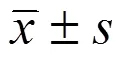
2.1 牛蒡子苷Arc 對高糖作用下的 HRCECs 增殖的影響
與 LG 組相比,HG 組血管內皮細胞 HRCECs 的值明顯升高(< 0.05),p21表達下調(< 0.05),而 cyclinD1 表達上調(< 0.05);與 HG 組相比,HG + Arc 20 mg/L 組、HG + Arc30 mg/L 組、HG + Arc 40 mg/L 組血管內皮細胞 HRCECs 增殖能力顯著降低(< 0.05),p21 的表達水平顯著升高(< 0.05),而 cyclinD1 的表達水平顯著降低(< 0.05),Arc 不同劑量組間均存在顯著性差異(< 0.05),見圖 1。表明牛蒡子苷可明顯抑制高糖作用下的血管內皮細胞 HRCECs 增殖。
2.2 牛蒡子苷 Arc 對高糖作用下的 HRCECs 中相關炎性因子表達的影響
與 LG 組相比,HG 組血管內皮細胞 HRCECs中 TNF-α、MCP-1、ApoM 水平均顯著升高(< 0.05);對比 HG 組,HG + Arc 20 mg/L 組、HG + Arc 30 mg/L 組、HG + Arc 40 mg/L 組血管內皮細胞 HRCECs 中 TNF-α、MCP-1、ApoM 水平均明顯降低(< 0.05),Arc 不同劑量組間均存在顯著性差異(< 0.05),見圖 2。表明牛蒡子苷可明顯抑制高糖作用下 HRCECs 內炎癥反應。
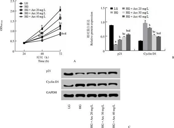
圖 1 Arc 對高糖作用下 HRCECs 增殖的影響(A:不同濃度的 Arc 對高糖作用下 HRCECs 增殖的影響;B:不同濃度的 Arc 對高糖作用下 HRCECs 增殖蛋白表達的影響;C:增殖相關蛋白的表達;與 LG 組比較,aP < 0.05;與 HG 組比較,bP < 0.05;與 HG + Arc 20 mg/L 組比較,cP < 0.05;與 HG + Arc 30 mg/L 組比較,dP < 0.05)
Figure 1 Effect of Arc on the proliferation of HRCECs under high glucose (A:Effect of different concentrations of Arc on the proliferation of HRCECs under high glucose; B:Effect of different concentrations of Arc on the expression of proliferation protein of HRCECs under high glucose; C:Expression of proliferation related protein;a< 0.05, compared with LG group;b< 0.05, compared with HG group;c< 0.05, compared with HG + Arc 20 mg/L group;d< 0.05, compared with HG + Arc 30 mg/L group)

相對蛋白表達Relative protein expression1.5 1.0 0.5 0 TNF-α MCP-1 ApoM GAPDH LG HG HG + Arc 20 mg/L HG + Arc 30 mg/L HG + Arc 40 mg/L TNF-α MCP-1 ApoMA B
Figure 2 Effect of Arc on the expression of related inflammatory factors in HRCECs under high glucose (A:Effect of different concentrations of Arc on the expression of HRCECs related inflammatory factors under high glucose; B:Expression of related proteins;a< 0.05, compared with LG group;b< 0.05, compared with HG group;c< 0.05, compared with HG + Arc 20 mg/L group;d< 0.05, compared with HG + Arc 30 mg/L group)
2.3 牛蒡子苷 Arc 對高糖作用下 HRCECs 中 VEGF 表達的影響
與 LG 組相比,HG 組 HRCECs 中 VEGF 水平顯著升高(< 0.05);相較于 HG 組,HG + Arc 20 mg/L 組、HG + Arc 30 mg/L 組、HG + Arc40 mg/L 組 HRCECs 中 VEGF 水平均明顯降低(< 0.05),Arc 不同劑量組間均存在顯著性差異(< 0.05),見圖 3。表明牛蒡子苷可明顯抑制高糖作用下 HRCECs 中 VEGF 表達。
2.4 抑制 VEGF 表達對高糖作用下 HRCECs 增殖及相關炎性因子表達的影響
Western blot 實驗檢測 si-VEGF 轉染效果,結果顯示,與 si-NC 組相比,si-VEGF 組高糖作用下 HRCECs 中 VEGF 的表達水平顯著降低(< 0.05),提示轉染成功。進一步檢測結果顯示,si-VEGF 組 HRCECs 的值與 si-NC 組比較顯著降低(< 0.05),cyclinD1、TNF-α、MCP-1、ApoM 表達水平均顯著降低(< 0.05),而 p21 的表達水平顯著升高(< 0.05),見圖 4。
相對 VEGF 蛋白表達Relative VEGF protein expression1.0 0.8 0.6 0.4 0.2 0 VEGF GAPDH LG HG HG + Arc 20 mg/L HG + Arc 30 mg/L HG + Arc 40 mg/L LG HG HG + HG + HG + Arc 20 mg/L Arc 30 mg/L Arc 40 mg/LA B
Figure 3 Effect of Arc on the expression of VEGF in HRCECs under high glucose (A:Effect of different concentrations of Arc on the expression of VEGF in HRCECs under high glucose; B:Expression of VEGF protein;a< 0.05, compared with LG group;b< 0.05, compared with HG group;c< 0.05, compared with HG + Arc 20 mg/L group;d< 0.05, compared with HG + Arc 30 mg/L group)

圖 4 抑制 VEGF 表達對高糖作用下 HRCECs 增殖及相關炎性因子表達的影響(A:抑制 VEGF 表達對高糖作用下 HRCECs 增殖的影響;B:抑制 VEGF 表達對高糖作用下 HRCECs 相關炎性因子表達的影響;C:VEGF 增殖及相關炎性因子蛋白的表達;與 si-NC 組比較,aP < 0.05)
Figure 4 Effect of VEGF inhibition on the proliferation of HRCECs and the expression of related inflammatory factors under high glucose (A:Inhibition of VEGF expression has an effect on the proliferation of HRCECs under high glucose; B:Inhibition of VEGF expression has an effect on the expression of HRCECs related inflammatory factors under high glucose; C:Proliferation of VEGF and the expression of related inflammatory factor protein;a< 0.05, compared with si-NC group)
2.5 過表達 VEGF 能逆轉牛蒡子苷Arc 對高糖作用下HRCECs 增殖的抑制作用
為探究牛蒡子苷是否可通過調控 VEGF 表達進而參與糖尿病視網膜病變機制,用濃度為30 mg/L 的牛蒡子苷預處理高糖作用下的 HRCECs,將 VEGF 的過表達載體轉染入 HRCECs,結果顯示,與 HG + Arc 30 mg/L + pcDNA3.1 組比較,HG + Arc 30 mg/L + pcDNA3.1-VEGF 組 HRCECs 的值顯著增加(< 0.05),p21 表達下調(< 0.05),而 cyclinD1 表達上調(< 0.05),見圖 5。表明 VEGF 過表達可明顯逆轉牛蒡子苷對高糖誘導的 HRCECs 增殖的抑制作用。
2.6 過表達 VEGF 能逆轉牛蒡子苷 Arc 對高糖作用下 HRCECs 相關炎性因子表達的影響
如圖 6 所示,與 HG + Arc 30 mg/L + pcDNA3.1組比較,HG + Arc 30 mg/L + pcDNA3.1-VEGF 組 HRCECs 中 TNF-α、MCP-1、ApoM 表達水平均顯著增加(< 0.05)。表明 VEGF 過表達可明顯逆轉牛蒡子苷對高糖誘導的 HRCECs 炎癥反應的抑制作用。
3 討論
糖尿病視網膜病變過程與炎癥、氧化應激、免疫因素等多種因素有關。既往研究表明,糖尿病視網膜病變屬于慢性低度炎癥性疾病,其發病機制中炎癥反應發揮重要作用[10-11]。研究表明中藥可有效保護微血管,并可有效治療糖尿病視網膜病變,但關于其具體作用機制尚未可知[12]。本研究通過選取牛蒡子苷為治療藥物分析其對糖尿病視網膜病變的保護作用及其相關作用機制,為牛蒡子苷應用于糖尿病視網膜病變的防治提供實驗基礎。

OD490 nm2.0 1.5 1.0 0.5 0 相對蛋白表達Relative protein expression 1.5 1.0 0.5 0 24 48 72 VEGF p21 Cyclin D1 時間(h)Time (h)A B
Figure 5 Overexpression of VEGF can reverse the effect of Arc on the proliferation of HRCECs under high glucose (A:Overexpression of VEGF can reverse the inhibition of Arc on the proliferation of HRCECs under high glucose; B:Overexpression of VEGF can reverse the effect of Arc on the expression of proliferation related proteins of HRCECs under high glucose; C:Expression of VEGF and proliferation related proteins;a< 0.05, compared with HG group;b< 0.05, compared with HG + Arc 30 mg/L + pcDNA3.1 group)

相對蛋白表達Relative protein expression1.5 1.0 0.5 0 TNF-α MCP-1 ApoM GAPDH TNF-α MCP-1 ApoM A HG HG + Arc 30 mg/L HG + Arc 30 mg/L+ pcDNA3.1 HG + Arc 30 mg/L+ pcDNA3.1-VEGFB
Figure 6 Overexpression of VEGF can reverse the effect of Arc on the expression of related inflammatory factors in HRCECs under high glucose (A:Overexpression of VEGF can reverse the effect of Arc on the expression of related inflammatory factors in HRCECs under high glucose; B:Expression of related proteins;a< 0.05, compared with HG group;b< 0.05, compared with HG + Arc 30 mg/L + pcDNA3.1 group)
牛蒡子苷是一種從牛蒡子醇提取物內分離的木脂類化合物,可明顯抑制轉錄因子-核因子 NF-κB 活化進而減輕腎臟病變大鼠內單核細胞趨化蛋白-1 表達進而有效改善腎臟損傷[13]。氧化應激可激活 NF-κB 進而上調糖尿病視網膜病變相關炎癥因子表達進而增加血管通透性導致微血管病變[14]。糖尿病微血管病變的主要原因在于持續性高血糖,研究表明牛蒡子苷降低糖尿病小鼠血糖的具體降糖機制尚需進一步研究[15]。本研究結果表明,高糖誘導后 HRCECs 增殖能力明顯增強,不同濃度的牛蒡子苷處理后可顯著抑制 HRCECs 增殖并有效降低 cyclinD1 蛋白表達,而明顯升高 p21 的表達水平,研究表明 cyclinD1 可正向調控細胞周期并可推動細胞增殖,而 p21 可明顯抑制 cyclinD1 表達[16]。提示牛蒡子苷可通過調控細胞增殖相關蛋白表達進而抑制高糖誘導的 HRCECs 增殖。有研究表明,高糖誘導的炎癥反應可破壞視網膜內皮細胞連接進而破壞血-視網膜屏障[17]。高糖誘導條件下 HRCECs 中 TNF-α、MCP-1、ApoM 表達升高,并可加重視網膜血管內皮損傷程度[18-19]。與上述研究結果相似,本研究結果顯示高糖可上調 TNF-α、MCP-1、ApoM 炎性因子表達,牛蒡子苷可顯著降低 TNF-α、MCP-1、ApoM 炎性因子表達,首次證實牛蒡子苷可有效降低高糖誘導的炎癥反應進而對糖尿病視網膜病變發揮保護作用。
VEGF 可通過與內皮細胞表面的受體結合進而促使細胞膜上相關受體發生磷酸化導致下游信號分子激活最終促進細胞增殖[20]。已有研究表明糖尿病視網膜病變患者血清中 VEGF 水平明顯升高,并可能作為評估患者疾病進展的有效指標[21]。糖尿病視網膜病變發生過程中 VEGF 可促進內皮細胞增殖及遷移而增強血管通透性進而加重疾病進展[22-23]。本研究通過抑制 VEGF 在高糖誘導的 HRCECs 中的高表達,使HRCECs增殖活性明顯降低,cyclinD1、TNF-α、MCP-1、ApoM 表達水平均明顯降低,而 p21 的表達水平明顯升高,提示抑制 VEGF 表達可抑制高糖誘導的 HRCECs 增殖并減輕炎癥反應。本研究結果顯示,過表達 VEGF 能逆轉牛蒡子苷對高糖誘導的 HRCECs 增殖的抑制作用,并逆轉牛蒡子苷對高糖誘導的 HRCECs 中相關炎性因子表達的抑制作用,提示牛蒡子苷可能通過抑制 VEGF 表達進而抑制高糖誘導的 HRCECs 增殖并減輕炎癥反應。
綜上所述,牛蒡子苷可抑制高糖誘導的 HRCECs 增殖及炎癥反應,其可能主要通過降低 VEGF 表達而實現目的,此項研究為牛蒡子苷對糖尿病視網膜病變的治療作用相關分子機制提供理論依據。
[1] Yan N, Li ZL. Effects of Ligustrazine on proliferation of human retinal vascular endothelial cells induced by high glucose. Drug Eval Res, 40(10):1414-1417. (in Chinese)
晏妮, 李振龍. 川芎嗪對高糖誘導的人視網膜血管內皮細胞增殖的影響. 藥物評價研究, 2017, 40(10):1414-1417.
[2] Li B, Wang HS, Li GG, et al. The role of endoplasmic reticulum stress in the early stage of diabetic retinopathy. Acta Diabetol, 2011, 48(2):103-111.
[3] Qin XH, Lu JM, Shao MY, et al. Protective effects of Notch1 signaling on human retinal vascular endothelial cells from apoptosis under high glucose. Recent Adv Ophthalmol, 2015, 35(9):806-809, 815. (in Chinese)
秦秀虹, 盧建民, 邵明陽, 等. Notch信號對高糖誘導人視網膜血管內皮細胞凋亡的保護作用. 眼科新進展, 2015, 35(9):806-809,815.
[4] Liu DL, Ma ST. Therapeutic action on retinopathy and optic neuropathy by arctiin in diabetic rats. China J Traditional Chin Med Pharm, 2013, 28(12):3732-3734. (in Chinese)
劉冬戀, 馬松濤. 牛蒡子苷對糖尿病大鼠視網膜和視神經病變的治療作用. 中華中醫藥雜志, 2013, 28(12):3732-3734.
[5] Luo NP, Huang HB, Xu J, et al. Study of the relation ship between diabetic retinopathy and VEGF, IFN-Ras well as NO. Chin J Diabetes, 2001, 9(1):24-27. (in Chinese)
羅南萍, 黃厚斌, 徐軍, 等. VEGF、IFN-γ及NO與糖尿病視網膜病變關系的研究. 中國糖尿病雜志, 2001, 9(1):24-27.
[6] Liu GH. Effect of emodin on VEGF expression and proliferation of human retinal vascular endothelial cells under high glucose conditions. Hengyang:University of South China, 2012. (in Chinese)
劉光輝. 大黃素對高糖條件下VEGF表達及人視網膜血管內皮細胞增殖的影響. 衡陽:南華大學, 2012.
[7] Zhang Z, Liu ZQ, Liu JP, et al. The synergistic effect of metformin and anti-vascular endothelial growth factor in the treatment of diabetic retinopathy. Chin J Ocul Fundus Dis, 2018, 34(5):453-457. (in Chinese)
張哲, 劉竹青, 劉巨平, 等. 二甲雙胍聯合抗血管內皮生長因子藥物治療糖尿病視網膜病變的可能協同作用. 中華眼底病雜志, 2018, 34(5):453-457.
[8] Ma HJ, Li T, Liang XL, et al. The effect of FK506 on the proliferation of HRCECs. Eye Sci, 2006, 22(4):237-243. (in Chinese)
馬紅婕, 李濤, 梁小玲, 等. FK506對人視網膜微血管內皮細胞增殖及凋亡的影響. 眼科學報, 2006, 22(4):237-243.
[9] Fu YY, Zhao Y. Advances in the mechanism of arctioside on diabetic microvascular disease. Chongqing Med, 2014, 43(21):2813-2815. (in Chinese)
付元元, 趙語. 牛蒡子苷對糖尿病微血管病變的作用機制研究進展. 重慶醫學, 2014, 43(21):2813-2815.
[10] Stitt AW, Lois N, Medina RJ, et al. Advances in our understanding of diabetic retinopathy. Clin Sci (Lond), 2013, 125(1):1-17.
[11] Semeraro F, Cancarini A, dell’Omo R, et al. Diabetic retinopathy:vascular and inflammatory disease. J Diabetes Res, 2015, 2015:582060.
[12] Meng XM, Zhang SS, Duan YC. Observation of tongxinluo capsule combined with laser in the treatment of diabetic retinopathy. J Med Forum, 2011, 32(6):155-156. (in Chinese)
孟憲民, 張書申, 段永暢. 通心絡膠囊聯合激光治療糖尿病性視網膜病變的觀察. 醫藥論壇雜志, 2011, 32(6):155-156.
[13] Yang MZ, Zhang XR. Study of fructus arctii on amelioration of renal injury in diabetic rats. Strait Pharm J, 2009, 21(12):49-50. (in Chinese)
楊明正, 張小如. 牛蒡子改善糖尿病大鼠腎臟病變機制的探討. 海峽藥學, 2009, 21(12):49-50.
[14] Pokharel YR, Liu QH, Oh JW, et al. 4-Hydroxykobusin inhibits the induction of nitric oxide synthase by inhibiting NF-kappaB and AP-1 activation. Biol Pharma Bull, 2007, 30(6):1097-1101.
[15] An YG. Effect of arctiin on glucose and lipid metabolism in diabetic mice. Henan Traditional Chin Med, 2013, 33(2):193-195. (in Chinese)
安益國. 牛蒡子苷對糖尿病小鼠糖脂代謝的影響. 河南中醫, 2013, 33(2):193-195.
[16] Yuan Q, Cheng Y, Yang YF, et al. Effect on cell proliferation of mouse dorsal root ganglion cells co-culture with human microvascular endothelial cells. Chin J Exp Surg, 2015, 32(1):135-138. (in Chinese)
原泉, 程揚, 楊亞帆, 等. 小鼠脊髓背根神經節細胞與人微血管內皮細胞共培養對細胞增殖的影響. 中華實驗外科雜志, 2015, 32(1):135-138.
[17] Rangasamy S, McGuire PG, Das A. Diabetic retinopathy and inflammation:novel therapeutic targets. Middle East Afr J Ophthalmol, 2012, 19(1):52-59.
[18] Shi H, Carion TW, Jiang Y, et al. VIP protects human retinal microvascular endothelial cells against high glucose-induced increases in TNF-α and enhances RvD1. Prostaglandins Other Lipid Mediat, 2016, 123:28-32.
[19] Tang H, Luo GH, Yao S, et al. Inhibitory effects of apolipoprotein M on inflammatory factors induced by high glucose in human retinal vascular endothelial cells. Chin J Exp Ophthalmol, 2018, 36(3):194- 198. (in Chinese)
唐睆, 羅光華, 姚霜, 等. 載脂蛋白M對高糖誘導的人視網膜血管內皮細胞中相關炎性因子表達的抑制作用. 中華實驗眼科雜志, 2018, 36(3):194-198.
[20] Vingolo EM, Fragiotta S, Mafrici M, et al. Vitreous and plasma changes of endothelin-1, adrenomedullin and vascular endothelium growth factor in patients with proliferative diabetic retinopathy. Eur Rev Med Pharmacol Sci, 2017, 21(4):662-668.
[21] Wang CY. Changes and clinical significance of serum VEGF and NO in patients with diabetic retinopathy. Chin J Lab Diagn, 2015, 19(1):94-95. (in Chinese)
王春雁. 糖尿病視網膜病變患者血清VEGF、NO的變化及臨床意義. 中國實驗診斷學, 2015, 19(1):94-95.
[22] Le YZ. VEGF production and signaling in Müller glia are critical to modulating vascular function and neuronal integrity in diabetic retinopathy and hypoxic retinal vascular diseases. Vision Res, 2017, 139:108-114.
[23] Zhang ZZ, Qin XH, Zhang J. MicroRNA-183 inhibition exerts suppressive effects on diabetic retinopathy by inactivating BTG1-mediated PI3K/Akt/VEGF signaling pathway. Am J Physiol Endocrinol Metab, 2019, 316(6):E1050-E1060.
Arctiin inhibits diabetic retinopathy through regulating vascular endothelial growth factor (VEGF)
YAN Yuan-yuan, LIU Ping
We aim to investigate whether arctiin (Arc) inhibits diabetic retinopathy by regulating the expression of vascular endothelial growth factor (VEGF).
Human retinal microvascular endothelial cells (HRCECs) were cultured, and HRCECs were treated with different concentrations of Arc, and si-VEGF or pcDNA3.1-VEGF were transfected into HRCECs. The proliferation of HRCECs was detected by MTT assay. The expressions of p21, cyclinD1, TNF-α, MCP-1, ApoM and VEGF protein were detected by Western blot.
Compared with LG group, thevalue of HRCECs in HG group was significantly increased (< 0.05), and cyclinD1, TNF-α, MCP-1, ApoM, VEGF levels were significantly increased (< 0.05), while p21 expression was down-regulated (< 0.05). After Arc treatment, the effect of HG on the proliferation of HRCECs, cyclinD1, TNF-α, MCP-1, ApoM, VEGF and p21 was significantly reversed, and there were significant differences between different doses of Arc (< 0.05). Inhibition of VEGF expression significantly attenuated the proliferation of HRCECs under high glucose and decreased the expression of related inflammatory factors. Overexpression of VEGF reversed the inhibitory effect of Arc on high glucose-induced proliferation and the inflammatory response of HRCECs.
Arc inhibits the proliferation of HRCECs by reducing the high expression of VEGF in HRCECs induced by high glucose, and may protect cells from damage by reducing inflammation.
Vascular endothelial growth factors; Diabetic retinopathy; Arctiin; Human retinal capillary endothelial cells
YAN Yuan-yuan, Email:Yshengvip222@163.com
10.3969/j.issn.1673-713X.2020.01.008
Author Affiliation:Department of Ophthalmology, Zhengzhou People's Hospital, Henan 450003, China
河南省科學技術基金(182102311208)
閆媛媛,Email:Yshengvip222@163.com
2019-07-01
猜你喜歡
中老年保健(2022年5期)2022-08-24 02:35:42
中老年保健(2022年1期)2022-08-17 06:14:56
中老年保健(2021年5期)2021-08-24 07:07:20
中老年保健(2021年9期)2021-08-24 03:51:04
中老年保健(2021年7期)2021-08-22 07:42:16
中老年保健(2021年3期)2021-08-22 06:49:56
中老年保健(2021年11期)2021-08-22 03:15:16
中國生殖健康(2020年2期)2021-01-18 02:51:44
中國生殖健康(2018年2期)2018-11-06 07:11:04
基層中醫藥(2018年2期)2018-05-31 08:45:04
