二維剪切波彈性成像在頸部不同病理類型淋巴結中的鑒別診斷價值
2020-11-10 04:36:33梁奎王曉榮宋濤
中國醫(yī)藥導報 2020年25期
梁奎 王曉榮 宋濤
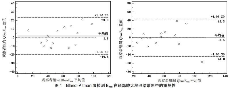
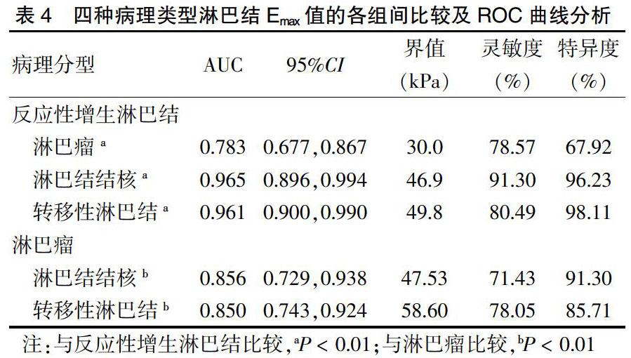
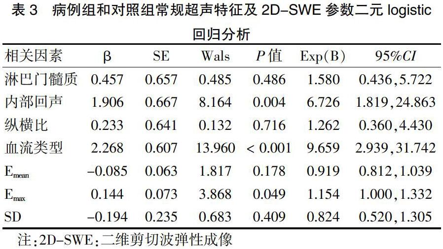
[摘要] 目的 探討二維剪切波彈性成像(2D-SWE)在頸部不同病理類型淋巴結鑒別診斷中的價值。 方法 選取2017年10月—2019年5月新疆醫(yī)科大學第一附屬醫(yī)院101例共145個頸部腫大淋巴結的患者,對其行常規(guī)超聲及2D-SWE檢查,將其按淋巴結病理或隨訪6個月后的結果進行分組,將淋巴結結核、淋巴瘤、轉移性淋巴結作為病例組(92個),反應性增生淋巴結作為對照組(53個)。通過二元logistic回歸篩選出對病例組和對照組鑒別價值較高的彈性參數(shù),并通過該參數(shù)在各組間的比較及ROC曲線確定診斷界值。 結果 二元logistic回歸顯示淋巴結血流類型、內部回聲及2D-SWE參數(shù)Emax在病例組和對照組間鑒別診斷價值較高。各組間比較顯示淋巴瘤、淋巴結結核、轉移性淋巴結的Emax值均高于反應性增生淋巴結(均P < 0.01),且逐級增高。鑒別淋巴瘤與反應性增生淋巴結Emax界值為30.0 kPa(P < 0.01),AUC、靈敏度、特異度分別為0.783、78.57%、67.92%,鑒別淋巴結結核與淋巴瘤Emax界值為47.53 kPa(P < 0.01),AUC、靈敏度、特異度分別為0.856、71.43%、91.30%,轉移性淋巴結Emax值與淋巴結結核比較,差異無統(tǒng)計學意義(P > 0.05)。 結論 頸部不同病理類型淋巴結的硬度值存在差異,2D-SWE參數(shù)Emax是反映這一硬度差異的首選彈性參數(shù),可作為頸部淋巴結疾病超聲鑒別診斷的重要補充指標。
[關鍵詞] 頸部淋巴結;病理類型;鑒別診斷;二維剪切波彈性成像
[中圖分類號] R445.1? ? ? ? ? [文獻標識碼] A? ? ? ? ? [文章編號] 1673-7210(2020)09(a)-0029-05
[Abstract] Objective To investigate the differential diagnosis value of two dimensional shear wave elastography (2D-SWE) in different pathological types of cervical lymph nodes. Methods A total of 101 patients with 145 enlarged cervical lymph nodes from October 2017 to May 2019 in the First Affiliated Hospital of Xinjiang Medical University were selected, and they underwent routine ultrasound and 2D-SWE examination. They were grouped according to lymph node pathology or results after six months of follow-up. Nodal tuberculosis, lymphoma and metastatic lymph nodes were selected as the case group (92 cases) and reactive hyperplasia lymph nodes as the control group (53 cases). Binary logistic regression was used to screen the elastic parameter with high differential value between the case group and the control group, and the diagnostic boundary value was determined by the comparison of this parameter between the groups and the ROC curve. Results Binary logistic regression showed that lymph node blood flow type, internal echo and 2D-SWE parameter Emax had higher differential diagnostic value between the case group and the control group. The Emax values of lymphomas, nodal tuberculosis and metastatic lymph nodes were higher than those of reactive hyperplasia lymph nodes (all P < 0.01) and increased step by step. The Emax boundary value of lymphomas and reactive hyperplasia lymph nodes was 30.0 kPa (P < 0.01), and the AUC, sensitivity and specificity were 0.783, 78.57% and 67.92%, respectively. The Emax boundary value of nodal tuberculosis and lymphoma was 47.53 kPa (P < 0.01), and the AUC, sensitivity and specificity were 0.856, 71.43% and 91.30%, respectively. There was no significant difference in Emax value between reactive hyperplasia lymph nodes and nodal tuberculosis (P > 0.05). Conclusion The 2D-SWE parameter Emax is the preferred elastic parameter to reflect the difference in hardness of cervical lymph nodes of different pathological types, which can be used as an important supplementary indicator for the ultrasonic differential diagnosis of cervical lymph node diseases.
轉移性淋巴結在四種病理類型中硬度最高,與癌細胞侵蝕,淋巴結結構被破壞,纖維組織增生及壞死物沉積有關[17-21]。轉移性淋巴結Emax值與淋巴結結核比較,差異無統(tǒng)計學意義(P > 0.05),對二者的鑒別仍需要結合臨床及超聲特征綜合判斷[22-24]。
有研究[25-27]采用Q-Trace描記出淋巴結最大截面進行測量,描記法對發(fā)現(xiàn)淋巴結局灶性轉移可能更具優(yōu)勢,但會將淋巴結內鈣化灶和液化壞死區(qū)包含在內,由于剪切波不能在液體中傳播,彈性圖上液化壞死區(qū)內出現(xiàn)大片“空洞”區(qū),影響圖像質量及測量,而鈣化灶會使測量結果高于淋巴結的真實硬度。因此選擇避開鈣化灶和液化壞死區(qū)的感興趣區(qū),使用Q-Box法測量,結果可信度更高。
本研究局限性有:①樣本量較小,未按淋巴結結核分型和轉移淋巴結腫瘤組織學類型進行亞組分析[28];②頸部固有曲率導致彈性圖容易出現(xiàn)偽影,頸動脈搏動可能會帶動周圍組織振動產生橫波,這些因素都可能影響測值準確性[29]。
綜上所述,頸部不同病理類型淋巴結硬度存在差異,2D-SWE參數(shù)Emax是反映這一硬度差異的首選彈性參數(shù),可作為頸部淋巴結疾病超聲鑒別診斷的重要補充指標。
[參考文獻]
[1]? 宋爽,張麒,韓紅,等.基于特權信息學習的淋巴結病變計算機輔助診斷[J].自動化儀表,2019,40(12):61-65.
[2]? 葛喜鳳,李磊,崔立剛,等.超聲彈性對比指數(shù)在淋巴結良惡性鑒別診斷中的應用價值[J].中國醫(yī)學科學院學報,2018,40(5):680-684.
[3]? 李強.超聲剪切波彈性成像的技術進展[J].中國醫(yī)療設備,2017,32(7):101-105,123.
[4]? Desmots F,F(xiàn)akhry N,Mancini J,et al. Shear Wave Elastography in Head and Neck Lymph Node Assessment:Image Quality and Diagnostic Impact Compared with B-Mode and Doppler Ultrasonography [J]. Ultrasound Med Biol,2016,42(2):387-398.
[5]? Choi YJ,Lee JH,Lim HK,et al. Quantitative shear wave elastography in the evaluation of metastatic cervical lymph nodes [J]. Ultrasound Med Biol,2013,39(6):935-940.
[6]? Dudea SM,Lenghel M,Botar-Jid C,et al. Ultrasonography of superficial lymph nodes:be-nign vs. Malignant [J]. Med Ultrason,2012,14(4):294-306.
[7]? 冉飛武,李建彬,梁超前,等.頭頸部腫瘤頸部淋巴結分區(qū)及其靶區(qū)的勾畫[J].中華放射腫瘤學雜志,2005,14(6):528-534.
[8]? 中華醫(yī)學會超聲醫(yī)學分會介入超聲學組彈性成像評估肝纖維化專家組.二維剪切波彈性成像評估慢性乙型肝炎肝纖維化臨床應用指南[J].臨床肝膽病雜志,2018,34(2):255-261.
[9]? 阮鏡良,梁銘,吳嘉怡,等.淋巴結大小對剪切波彈性定量參數(shù)診斷價值的影響[J].中國超聲醫(yī)學雜志,2017,33(6):492-494.
[10]? Bhatia KS,Cho CC,Tong CS,et al. Shear Wave Elasticity Imaging of Cervical Lymph Nodes [J]. Ultrasound Med Biol,2012,38(2):195-201.
[11]? 王曉榮,劉霞,姚蘭輝,等.頸部淋巴結淋巴瘤皮質回聲及其病理基礎的初步探討[J].中國超聲醫(yī)學雜志,2013, 29(8):676-680.
[12]? Fukuhara T,Matsuda E,Endo Y,et al. Impact of Fibrotic Tissue on Shear Wave Velocity in Thyroid:An Ex Vivo Study with Fresh Thyroid Specimens [J]. Biomed Res Int,2015,2015:569367.
[13]? 軒維鋒,張建興,崔立剛,等.淺表組織超聲與病理診斷[M].北京:人民軍醫(yī)出版,2015:141-180.
[14]? 趙奕文,金正吉,鄭穎,等.頸淋巴結結核的超聲表現(xiàn)與分型[J].上海醫(yī)學影像,2008,17(3):218-219,封三.
[15]? 張更臣,李俊來,曹兵生,等.頸部淋巴結結核超聲表現(xiàn)與病理對照研究[J].中國超聲醫(yī)學雜志,2013,29(10):879-882.
[16]? 白智群,王學梅,黃崑,等.剪切波彈性成像在頸部淋巴結結核診斷及鑒別診斷中的價值[J].中國超聲醫(yī)學雜志,2018,34(10):874-877.
[17]? 陳順軍,宋樂樂,賀軍領,等.剪切波彈性成像診斷甲狀腺癌術后經(jīng)131I治療后頸部淋巴結良惡性的價值[J].中國醫(yī)學影像學雜志,2019,27(2):128-130.
[18]? 江建偉,陳曼.超聲彈性成像聯(lián)合常規(guī)超聲對淋巴瘤性淺表淋巴結腫大的應用價值[J].中國醫(yī)學計算機成像雜志,2019,25(2):176-180.
[19]? 趙大衛(wèi),王明正,劉佳坤,等.功能性頸淋巴結清掃術治療頸部淋巴結核的療效觀察[J].疑難病雜志,2019,18(1):52-56.
[20]? 趙躍,劉春蓉,張桐碩,等.基于危險因素構建的Logistic回歸模型對甲狀腺乳頭狀癌頸部淋巴結轉移的預測價值研究[J].中國醫(yī)藥,2019,14(2):282-286.
[21]? 劉旭,陳曉艷.剪切波彈性成像預測甲狀腺乳頭狀癌頸部淋巴結轉移的價值[J].廣東醫(yī)學,2019,40(5):729-731,735.
[22]? 王思思,王波,謝超,等.甲狀腺乳頭癌淋巴結轉移與甲狀腺球蛋白的關系[J].中國醫(yī)藥導報,2018,15(35):114-117.
[23]? 張雪云,聶芳,呂文豪,等.超聲造影對結核性淋巴結與轉移性淋巴結的鑒別診斷價值[J].中國超聲醫(yī)學雜志,2018,34(5):403-406.
[24]? 黃琨,李玉丹,包明穩(wěn),等.高頻超聲檢查在頸部異常淋巴結中的診斷價值[J].中國醫(yī)藥科學,2019,9(23):231-233.
[25]? Tan S,Miao LY,Cui LG,et al. Value of Shear Wave Elastography Versus Contrast-Enhanced Sonography for Differentiating Benign and Malignant Superficial Lymphadenopathy Unexplained by Conventional Sonography [J]. J Ultrasound Med,2017,36(1):189-199.
[26]? 馬玉峰,張昕,張微,等.Q-box手動描繪模式剪切波彈性成像對乳腺癌新輔助化療后腋窩淋巴結轉移的早期預測[J].中國醫(yī)學創(chuàng)新,2018,15(26):129-133.
[27]? 杜宗艷,孫詠梅,寧春平,等.不同ROI和彈性模量值對剪切波彈性成像診斷頸部良惡性淋巴結效能的影響[J].中國醫(yī)學影像技術,2019,35(1):50-54.
[28]? 趙穎燕,奚佳穎,蔣海波,等.VTIQ技術對不同病理類型的鼻咽癌頸部轉移性淋巴結的診斷價值回顧性研究[J].中國超聲醫(yī)學雜志,2018,34(9):772-774.
[29]? 喬曉慧,刑晉放.剪切波超聲彈性成像的原理及臨床應用現(xiàn)狀[J].中國介入影像與治療學,2015,12(8):512-515.
(收稿日期:2020-05-13)
