
近紅外線透照齲齒探測儀對面齲的診斷效果
2017-05-11 17:18:16張麗麗牛赟武峰李憲起
中國醫學創新 2017年12期
張麗麗+牛赟+武峰+李憲起
【摘要】 目的:探討應用近紅外線透照齲齒探測儀(DIAGNOcam)檢查面齲損,對比探視診、體視顯微鏡的檢查結果,評估DIAGNOcam的診斷效果。方法:收集20顆離體牙分別通過探視診檢查、DIAGNOcam儀器檢查,不同操作者分別記錄結果。齲齒染色液染色后顯微鏡下檢查齲損,并作為金標準,評估探視診與儀器檢查齲損的一致性、特異性、靈敏性。結果:兩種檢查方法在D0水平一致性較差,D1-2水平一致性一般,D3-4水平有高度的一致性。探視診檢查在D1-2水平靈敏性最低,D3-4水平特異性最高;DIAGNOcam檢查在D1-2水平靈敏性明顯高于探視診檢查,在D3-4水平特異性最高。結論:近紅外線透照齲齒探測儀相對傳統方法,更有利于面早期齲的診斷,但仍需要進一步臨床研究分析其效果。
【關鍵詞】 近紅外線透照齲齒探測儀; 面齲; 探視診
Diagnostic Effect of DIAGNOcam in Early Occlusal Caries/ZHANG Li-li,NIU Yun,WU Feng,et al.//
Medical Innovation of China,2017,14(12):008-011
【Abstract】 Objective:To explore the effect of DIAGNOcam in early occlusal caries, by comparing the results of visual examination and stereomicroscope,to evaluate the diagnostic effect of DIAGNOcam. Method:Twenty extracted teeth were examined by visual examination and DIAGNOcam examination,different examiners recorded the results separately.Staining the teeth,then observing under the stereomicroscope,the results recognized as gold standard to evaluate the sensitivity,specificity and coherence between the two groups.Result:The consistency of the two methods in the D0 level was poor,the D1-2 level was generally consistent,and the D3-4 level was highly consistent.The sensitivity of visual examination at D1-2 was the lowest,and the specificity of D3-4 was the highest;the sensitivity of DIAGNOcam at D1-2 was significantly higher than that of visual examination, and the specificity at D3-4 was the highest.Conclusion:The DIAGNOcam is helpful to diagnose the early occlusal caries compared to traditional methods,further clinical investigations are needed to analyze its evaluation.
【Key words】 DIAGNOcam; Occlusal caries; Visual examination
First-authors address:Shanxi Medical University Stomatological Hospital,Taiyuan 030001,China
doi:10.3969/j.issn.1674-4985.2017.12.003
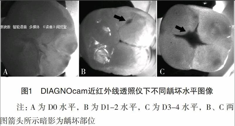
在臨床口腔檢查與保健工作中,早期齲的發現和治療對于口腔科醫生來說是一個挑戰,容易發生漏診而延誤早期治療[1]。因早期齲時,牙齒色形質改變不明顯,無明顯齲洞,因此多數情況下,需要醫生具備較為豐富的臨床經驗才敢于做出診斷[2]。近年來,為了改善這一狀況,學者們提出了很多新的診斷新思路,但這些新的診斷方法也并不完全可靠[3-6]。近年來,出現一種利用近紅外光透照診斷齲齒的儀器,即DIAGNOcam(Kavo公司,德國)[7-8],通過透照暗影反應齲損的程度。目前國內尚無此儀器檢測齲損效果的研究,國外也較少,因此本試驗在離體牙上通過探視診觀察頜面齲壞程度,以顯微鏡下觀察齲損結果作為金標準,評估該設備診斷面齲的準確性及臨床應用價值,現報道如下。
1 材料與方法
1.1 儀器與材料 近紅外線透照齲齒探測儀(DIAGNOcam,Kavo Germany)、口腔顯微鏡(ZEISS Germany)、生理鹽水、雙氧水、齲齒染色液(Sable Seek caries indicator, Ultradent USA)。
1.2 方法
1.2.1 離體牙的收集 收集20顆離體前磨牙與磨牙,牙體完整,肉眼未見明顯齲洞或充填體,沖洗清理,編號,雙氧水浸泡24 h后生理鹽水保存。
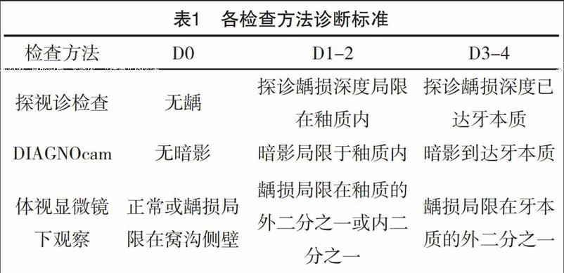
1.2.2 探視診檢查 取出檢測牙,清潔頜面溝裂,干凈后吹干,一名有臨床經驗醫生檢查,按照齲病診斷標準分為D0、D1-2、D3-4,視診結合探診初步判定是否有齲及齲損程度,記錄結果,見表1。
