改良胰管支架用于內鏡逆行胰膽管造影取凈膽總管結石后膽道引流的安全性及有效性分析
2023-04-29 20:25:39李佳林任笠坤冉勛夏紹萱周昱彤黃鑫韓民
臨床肝膽病雜志 2023年8期
關鍵詞:支架
李佳林 任笠坤 冉勛 夏紹萱 周昱彤 黃鑫 韓民
摘要:目的探討改良7Fr胰管塑料支架用于膽道內引流的臨床應用價值。方法收集2021年4月—2022年6月在貴州醫科大學附屬醫院肝膽外科行內鏡逆行胰膽管造影(ERCP)取石后行膽道引流的121例膽總管結石患者的臨床資料,根據術后膽道引流方式分為改良支架組(n=59)和鼻膽引流組(n=60),其中改良支架組采用改良7Fr胰管塑料支架引流,鼻膽引流組采用鼻膽管引流,回顧性分析兩組患者臨床資料,觀察改良支架組中支架自行脫落情況,比較兩組臨床療效、患者術后舒適度及術后并發癥發生率。符合正態分布的計量資料兩組間比較采用成組t檢驗;非正態分布的計量資料兩組間比較采用Mann-Whitney U檢驗;計數資料兩組間比較采用χ2檢驗。結果兩組結石清除率為100%。兩組患者術后住院天數、總住院天數比較,差異均有統計學意義(t值分別為-3.997、2.317,P值均<0.05)。兩組患者術后血清TBil、DBil、ALP、AST及GGT均較術前明顯下降(P值均<0.05)。兩組患者TBil、DBil、ALP、ALT、AST、GGT術前及術后比較,差異均無統計學意義(P值均>0.05)。改良支架組與鼻膽引流組術后-術前生化指標差值比較,差異均無統計學意義(P值均>0.05)。兩組術后24 h內舒適度評分、術后首次進食、進飲時間比較,差異均有統計學意義(t值分別為2.001、3.579、4.604,P值均<0.05)。兩組并發癥比較,差異均無統計學意義(P值均>0.05)。改良支架組支架自行脫落率為83.05%,未發生支架阻塞、移位、斷裂、穿孔及感染并發癥。結論ERCP取凈膽總管結石后,應用改良7Fr胰管塑料支架行膽道內引流與鼻膽管引流效果相當,可縮短患者術后住院時長、提升患者術后舒適度,加速患者康復,且支架自行脫落率高,臨床應用安全、有效。
關鍵詞:胰膽管造影術,? 內窺鏡逆行;? 引流術;? 支架
Safety and efficacy of modified pancreatic duct stent in biliary drainage after complete bile duct stone removal by endoscopic retrograde cholangiopancreatography
LI Jialin REN Likun RAN Xun XIA Shaoxuan ZHOU Yutong HUANG Xin HAN Min(1. College of Clinical Medicine, Guizhou Medical University, Guiyang 550004, China; 2. Department of Hepatobiliary Surgery, The Affiliated Hospital of Guizhou Medical University, Guiyang 550004, China)
Corresponding author:HAN Min, 409582096@qq.com? (ORCID: 0000-0001-7218-5276)
Abstract:ObjectiveTo investigate the clinical application value of modified 7Fr pancreatic duct plastic stent in biliary drainage. MethodsClinical data were collected from 121 patients with choledocholithiasis who underwent endoscopic retrograde cholangiopancreatography (ERCP) lithotomy and biliary drainage in Department of Hepatobiliary Surgery, The Affiliated Hospital of Guizhou Medical University, from April 2021 to June 2022, and according to the method for postoperative biliary drainage, they were divided into modified stent group with 59 patients and nasobiliary drainage group with 60 patients. The patients in the modified stent group received drainage with the modified 7Fr pancreatic duct plastic stent, and those in the nasobiliary drainage group received nasobiliary drainage. A retrospective analysis was performed for their clinical data, and stent dislodgement was observed for the modified stent group. The two groups were compared in terms of clinical outcome, postoperative comfort, and postoperative complications. The independent-samples t test was used for comparison of normally distributed continuous data between groups, and the Mann-Whitney U test was used for comparison of non-normally distributed continuous data between groups; the chi-square test was used for comparison of categorical data between groups. ResultsBoth groups achieved a stone clearance rate of 100%. There were significant differences between the two groups in the length of postoperative hospital stay and the total length of hospital stay (t=-3.997 and 2.317, both P<0.05). After treatment, both groups had significant reductions in total bilirubin (TBil), direct bilirubin (DBil), indirect bilirubin (IBil), alkaline phosphatase (ALP), aspartate aminotransferase (AST), and gamma-glutamyl transpeptidase (all P<0.05), and there were no significant differences between the two groups in the changes in TBil, DBil, IBil, ALP, alanine aminotransferase, and AST after treatment (all P>0.05). Also, no significant differences were observed between the two groups in the changes in biochemical parameters after treatment (all P>0.05). There were significant differences between the two groups in comfort score within 24 hours after surgery and the time to first eating and drinking after surgery (t=2.001, 3.579, and 4.604, all P<0.05). There were no significant differences in complications between the two groups (all P>0.05). In the modified stent group, the rate of spontaneous stent dislodgement was 83.05%, and there were no complications such as stent occlusion, displacement, rupture, perforation, and infection. ConclusionAfter complete bile duct stone removal by ERCP, biliary drainage using the modified 7Fr pancreatic duct plastic stent has a similar effect to nasobiliary drainage and can shorten the length of postoperative hospital stay, improve postoperative comfort, and accelerate postoperative recovery, with a relatively high spontaneous dislodgement rate. Therefore, it is safe and effective in clinical practice.
Key words:Cholangiopancreatography,? Endoscopic Retrograde;? Drainage;? Stents
內鏡逆行胰膽管造影(ERCP)已成為治療膽總管結石的主要方法,經ERCP取凈結石后,為預防相關并發癥,常規膽道引流方式有經內鏡鼻膽管引流術(endoscopic nasobiliary drainage,ENBD)及內鏡下膽道支架引流術(endoscopic rerograde biliary drainage,ERBD)[1-3]。用于膽道引流的普通支架較少有自發脫落特性,指南[4]建議3 ~ 6個月后取出,但二次取出增加治療過程費用及心理負擔,甚至增加并發癥發生風險。成婧等[5]研究報道,放置胰管塑料支架行膽道內引流,引流效果顯著。He等[6]研究報道,應用改良胰管支架用于預防ERCP術后胰腺炎(post-ERCP pancreatitis,PEP)發生,14天自行脫落率為84.21%。本院膽總管結石患者經ERCP取凈結石后,應用去除側翼錨定結構的改良7Fr胰管塑料支架行膽道內引流,療效滿意,現分析報道如下。
1資料與方法
1.1研究對象回顧性分析2021年7月—2022年6月于貴州醫科大學附屬醫院肝膽外科接受ERCP取石治療的121例膽總管結石患者的臨床資料。
1.2納入標準(1)經腹部B超,上腹部CT、MRI或MRCP證實膽總管原發或繼發結石;(2)接受ERCP且行膽總管取石術;(3)術后采用改良胰管塑料支架或鼻膽引流管引流;(4)年齡>18歲;(5)術后經腹部B超,上腹部CT、MRI或MRCP復查未見結石征象。
1.3排除標準(1)合并急、慢性胰腺炎或既往胰腺炎病史;(2)合并膽胰腫瘤;(3)上消化道狹窄;(4)既往上消化道重建病史;(5)對造影劑過敏;(6)合并嚴重心肺腎功能不全;(7)失訪臨床資料不全。
1.4方法
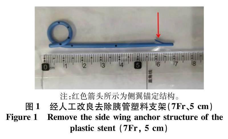
1.4.1操作器械日本Olympus公司TJF260V電子十二指腸鏡,南京微創公司0.035英寸(0.889 mm)黃斑馬導絲、和諧夾,美國BostonScientific公司乳頭括約肌切開刀、鼻膽引流管,胰管塑料支架(7Fr、5 cm,COOK公司),球囊擴張器(BostonScientific公司),取石網籃(COOK公司)。
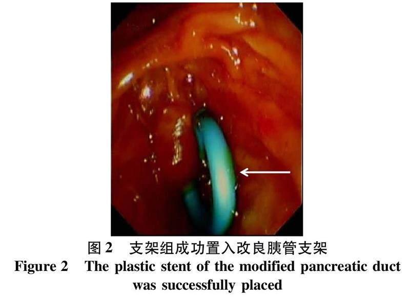
1.4.2操作步驟常規靜脈麻醉,經口腔插入十二指腸鏡,進鏡巡至十二指腸降段尋到乳頭,導絲引導下經乳頭插管,注入20%碘佛醇造影劑,C臂透視,觀察膽總管結石情況,術中根據結石位置、大小及膽管直徑情況,行內鏡下乳頭括約肌切開術(endoscopic sphincterotomy,EST)、內鏡下球囊擴張術(endoscopic papillary balloon dilation,EPBD),采用球囊或網籃取石清理膽道后,再次造影證實無結石殘留后循導絲膽管內置入改良胰管塑料支架(7Fr、5 cm,COOK公司),鼻膽引流組經導絲引導下鼻膽管插入膽總管,鼻膽管尖端置入左右肝管開口處,見膽汁流出并再次C臂透視證實支架或鼻膽引流管固定在位,檢查乳頭及視野所見無活動性出血,退鏡結束手術。術后予抗感染、抑酸、抑酶及補液等支持治療(圖1、2)。
1.5觀察指標與隨訪兩組患者術后24 h內均完成視覺模擬疼痛評分(VAS評分,1~10分)以評估患者術后舒適度[7],評分越低者術后舒適化程度越高。記錄兩組患者結石清除情況、術后惡心嘔吐情況、首次進食進飲時間、并發癥發生情況,同時監測術后24 h TBil、DBil、ALT、AST、ALP及GGT變化水平。采用腹部X線、超聲或CT隨訪觀察改良支架組患者術后1、2、4、8、12周支架脫落情況。若術后大于3個月支架仍未脫落者,經十二指腸鏡取出。
1.6相關定義(1)PEP:修訂版亞特蘭大國際共識標準[8]PEP定義為下列3項中≥2項:胰腺炎引起的持續上腹部疼痛;術后24 h淀粉酶或脂肪酶較正常值上限升高3倍以上;CT影像學檢查急性胰腺炎特征性表現,如胰腺腫大、壞死、胰周積液。重癥PEP:以患者經腹部CT掃描[9]提示存在胰腺周圍滲液以及胸腹腔積液,需進行經皮穿刺引流術或手術治療;(2)高淀粉酶血癥:術24 h血淀粉酶水平升高至3倍正常值上限,且未出現發熱、腹痛等癥狀。(3)出血:術后出現黑便、嘔血癥狀,或鼻膽引流管中可見新鮮血液流出,并出現血壓下降,需進行二次內鏡下止血;(4)膽管炎:術后72 h出現體溫升高(>38 ℃),白細胞計數升高,伴有腹痛癥狀,并排除其他部位感染[10]。
1.7統計學方法采用 SPSS 26.0軟件進行數據統計分析。符合正態分布的計量資料采用x±s表示,兩組間比較采用成組t檢驗;非正態分布的計量資料用M(P25~P75)表示,兩組間比較采用Mann-Whitney U檢驗;計數資料兩組間比較采用χ2檢驗。P<0.05為差異有統計學意義。
2結果
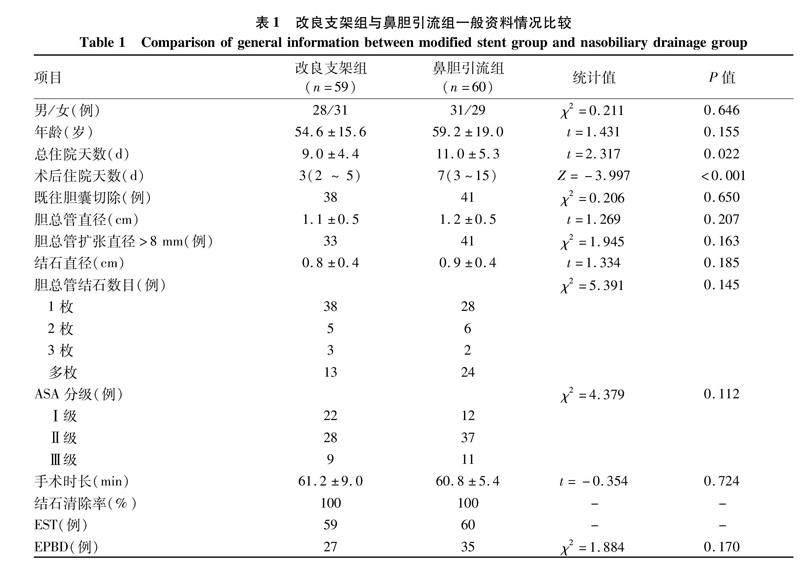
2.1一般資料121例患者中2例失訪,最終119例患者納入本研究,根據術后膽道引流方式分為改良支架組(n=59)和鼻膽引流組(n=60)。兩組患者術后住院天數、總住院天數比較,差異均有統計學意義(P值均<0.05)(表1)。
2.2術前及術后24 h生化指標組內比較情況兩組患者術后血清TBil、DBil、ALP、ALT、AST及GGT均較術前明顯下降(P值均<0.05)(表2)。
2.3兩組間術前及術后24 h生化指標組間比較兩組患者TBil、DBil、ALP、ALT、AST、GGT術前及術后比較,差異均無統計學意義(P值均>0.05)(表2)。
2.4兩組間術后-術前生化指標差值比較改良支架組與鼻膽引流組術后-術前生化指標差值比較,差異均無統計學意義(P值均>0.05)(表3)。
2.5兩組間術后情況比較兩組術后24 h內舒適度評分(VAS評分)、術后首次進食、進飲時間比較,差異均有統計學意義(P值均<0.05)(表4)。
2.6兩組患者術后并發癥比較兩組患者發生PEP、高淀粉酶血癥、出血的情況比較,差異均無統計學意義(P值均>0.05)(表5)。兩組均無膽管炎、穿孔、死亡和30 d再入院的患者。
2.7改良支架組患者支架脫落情況改良支架組59患者中50例完成復查,未發生阻塞、移位、斷裂、穿孔及感染等支架相關并發癥,其中某患者脫落前后復查腹部立位平片對比如圖3所示。第1周脫落3例,第2周脫落14例,第4周脫落23例,第8周脫落6例,第12周脫落3例。1例患者術后第12周門診復查,腹部立位平片提示支架未自行脫落,再次經內鏡取出,無不良事件發生。9例患者未復查腹部X片、B超或CT,電話隨訪患者本人或家屬無特殊不適。支架脫落率為83.05%(49/59)。
3討論
ERCP取石后常規膽道引流方式有ENBD及ERBD,兩種引流方式效果相當[11-13]。ENBD為外引流,會導致患者術后鼻咽部不適、膽汁及水電解質體外丟失,拔管時可引起惡心、嗆咳刺激不適,降低患者術后舒適度,外接引流袋影響美觀。對于不耐受者,有自行拔管、脫管等可能[14]。ERBD為內引流,無膽汁及水電解質流失,更符合人體生理學特點。普通膽道塑料支架較少有自發脫落特點,長期留置會因細菌被膜覆蓋或膽泥形成引發膽道梗阻[15],甚至移位致腸穿孔[16]、膽管穿孔可能,且長期留置可引起膽管炎發生。因此,歐洲婦科內鏡學會[17]建議3~6個月需再次取出。應用胰管支架能有效預防PEP發生[18-19],但胰管支架用于膽道內引流預防ERCP相關并發癥的臨床研究報道較少。成婧等[5]研究報道應用5Fr胰管支架用于ERCP取凈膽總管結石后膽道引流,支架自行脫落率為86.00%,多數患者無需二次行ERCP將支架取出,降低了患者的手術風險、心理及經濟負擔。He等[6]多中心隨機對照研究比較應用改良5Fr胰管支架與普通胰管支架用于PEP預防,改良胰管支架2周自行脫落率為84.21%,且未增加PEP或其他并發癥發生風險。
目前沒有指南明確規定ERCP取石后應放置何種引流方式。Lee等[7]研究認為經ERCP取石治療后無需放置鼻膽引流管。但為預防相關并發癥,多數術者選擇留置鼻膽管引流。本研究中改良支架組結石清除率為100%,結石取凈后應用改良7Fr胰管塑料支架行膽道內引流,術后膽紅素明顯下降,與鼻膽引流組比較,療效同樣顯著,與Kawashima等[20]報道一致。改良支架組療效同樣顯著的原因:(1)膽管結石完全清除,解除梗阻,膽道通暢性恢復;(2)改良胰管支架與鼻膽管直徑大小一致,可有效引流,同時也避免支架過早脫落而導致引流失敗的風險。
在并發癥方面,改良支架組發生PEP 1例,經積極保守治療后好轉。兩組患者均有高淀粉酶血癥發生,其發生率與相關研究[21-22]報道一致。高淀粉酶血癥與PEP的主要發生機制相似,與手術器械對乳頭及胰管機械性損傷、外界細菌移位或逆行感染產生炎性損傷、注入造影劑對胰管及其內容物造成流體及化學性動力損傷、各種術源性刺激激活胰酶原、水解胰腺組織產生酶學損傷、術中熱力性損傷有關[23]。也有研究[24]報道,與膽道支架及鼻膽管置入有關,而Cotton等[25]多因素分析認為,膽道支架及鼻膽管置入與高淀粉酶血癥發生并無直接關系。本研究中兩組患者行ERCP取石前聯合行EST,部分聯合行EPBD,可能系術中插管、乳頭擴張持續性機械性刺激及熱力損傷導致乳頭水腫引起胰管內壓力增高、胰腺實質損傷有關。兩組高淀粉酶血癥患者經積極保守治療后痊愈出院,均未進一步發展為PEP。出血是ERCP術后最嚴重的并發癥之一,有研究[26]報道其發生率為0.3%~2.0%,其主要原因為EST。對術后出血者,可再次內鏡下止血或介入、手術方法止血。鼻膽引流組有3例發生出血,術中出血者均予噴灑腎上腺素及止血夾夾閉處理,術后出血者再次內鏡檢查后乳頭周圍未見活動性出血,積極保守治療后痊愈。
Bajbouj等[27]指出長期留置支架會引起支架移位、堵塞、膽管炎及穿孔等并發癥可能。研究[28-29]表明,因支架堵塞發生支架相關膽管炎發病率為3.5%~40%。Pisello等[30]研究指出長期膽道支架引流引起支架相關膽管炎病死率可達6.7%。改良支架組1例患者術后第12周支架未自行脫落,為避免長期留置支架相關并發癥發生,再次經十二指腸鏡取出。Sofuni等[31]多中心隨機對照研究認為,應用可自行脫落胰管支架能有效預防PEP,相關并發癥發生風險低。但Nishiwaki等[32]報道,為預防PEP而留置自行脫落胰管支架可引起腹膜后穿孔。在本研究中改良支架組未發生支架移位、堵塞、斷裂、感染、穿孔等并發癥,可能是因改良7Fr胰管支架腸腔端的單豬尾結構能有效防止支架近端移位、減少腸腔黏膜壓迫,避免膽道及十二指腸穿孔發生。經人工改良去除膽管端側翼結構能增加自發脫落可能[33]、降低人為干預率、足時有效引流同時避免長期留置支架引發膽管堵塞、損傷及膽管炎。對隨訪時間>3個月未自行脫落者,及時二次內鏡取出可避免支架相關并發癥發生。
本研究中改良支架組術后舒適度高于鼻膽引流組,可能是因為采用改良胰管支架行膽道內引流后,減少帶管、拔管帶來的鼻咽部刺激。《肝膽胰外科術后加速康復專家共識(2015版)》[34]認為減少應激是加速康復外科理念的核心原則,是加速患者術后康復的基礎。改良支架組術后首次進飲、進食時間早于對照組,術后早期經口進食、進飲符合加速康復外科理念[35],可促進胃腸功能恢復、減輕腸道應激、減少液體負荷。加速患者康復不是單純追求縮短住院時長,本研究中兩組患者經評估達出院標準,改良支架組術后住院時間短于鼻膽管引流組。
改良支架組中膽道引流支架多數自行脫落,自行脫落率為83.05%,但有9例患者未復查,自行脫落率可能高于文獻[5-6]報道水平。改良支架自行脫落分析可能與下列因素有關:(1)EST破壞乳頭括約肌功能;(2)胰管支架側翼錨定結構在置入膽道前經人工改良去除;(3)單豬尾結構置于十二指腸腔內增大與食物、流體運動摩擦面積;(4)改良胰管支架具有多發側孔,膽汁隨側孔引出降低支架與膽管內壁的摩擦系數;(5)十二指腸自身蠕動因素;(6)多數患者合并膽總管擴張。
綜上所述,膽管結石患者經ERCP取盡結石后,應用去除側翼錨定結構改良的7Fr胰管塑料支架行膽道內引流與鼻膽管引流療效相當,且能提升患者術后舒適度、加速患者術后康復,且自行脫落率高,臨床應用具有安全性及有效性。需指出的是,此研究為單中心回顧性研究,樣本量較少,存在選擇偏倚,仍有一定局限性,還需多中心、大樣本前瞻性隨機對照研究驗證。
倫理學聲明:本研究方案經由貴州醫科大學附屬醫院倫理委員會審批,批號:2023倫審第344號。ERCP前均獲得每位患者及家屬的書面知情同意。利益沖突聲明:本文不存在任何利益沖突。作者貢獻聲明:李佳林負責課題設計,資料分析,撰寫論文;任笠坤、冉勛、夏紹萱、周昱彤、黃鑫參與核查數據,修改論文;韓民負責擬定寫作思路,指導撰寫文章并最后定稿。
參考文獻:
[1]MUKAI S, ITOI T, BARON TH, et al. Indications and techniques of biliary drainage for acute cholangitis in updated Tokyo Guidelines 2018[J]. J Hepatobiliary Pancreat Sci, 2017, 24(10): 537-549. doi: 10.1002/jhbp.496.
[2]LIN Y, LIN XY, CHEN R, et al. Application of ERCP in treatment of common bile duct stones after cholecystectomy[J/OL]. Chin J Hepat Surg(Electronic Edition), 2021, 10(5): 502-505. DOI: 10.3877/cma.j.issn.2095-3232.2021.05.015.林穎, 林顯藝, 陳榮, 等. ERCP在膽囊切除術后膽總管結石治療中的應用[J/OL]. 中華肝臟外科手術學電子雜志, 2021, 10(5): 502-505. DOI: 10.3877/cma.j.issn.2095-3232.2021.05.015.
[3]DONG WF, PANG EJ, DAI ZL. Clinical efficacy of ERCP combined with LC in treatment of patients with cholecystolithiasis combined with choledocholithiasis and influencing factors for recurrence of choledocholithiasis after surgery[J]. Clin Misdiagn Misther, 2021, 34(5): 85-90. DOI: 10.3969/j.issn.1002-3429.2021.05.017.董維峰, 龐爾君, 代鎮嶺. ERCP聯合LC治療膽囊結石合并膽總管結石臨床效果及術后膽總管結石復發影響因素分析[J]. 臨床誤診誤治, 2021, 34(5): 85-90. DOI: 10.3969/j.issn.1002-3429.2021.05.017.
[4]DUMONCEAU JM, TRINGALI A, BLERO D, et al. Endoscopic biliary stenting: indications, choice of stents, and results: European Society of Gastrointestinal Endoscopy (ESGE) clinical guideline-updated october 2017[J]. Endoscopy, 2018, 50(9): 910-930. DOI: 10.1055/a-0659-9864.
[5]CHENG J, LI QL, XU MD, et al. Clinical value of using pancreatic duct stent for bile duct drainage after endoscopic removal of common bile duct stones[J]. Chin J Dig Endosc, 2019, 36(9): 686-688. DOI: 10.3760/cma.j.issn.1007-5232.2019.09.015.成婧, 李全林, 徐美東, 等. 內鏡取凈膽總管結石后應用胰管支架行膽管引流的臨床價值[J]. 中華消化內鏡雜志, 2019, 36(9): 686-688. DOI: 10.3760/cma.j.issn.1007-5232.2019.09.015.
[6]HE QB, WANG L, PENG CY, et al. Modified prophylactic 5-fr pancreatic duct stent enhances the rate of spontaneous dislodgement: A multicenter randomized controlled trial[J]. United European Gastroenterol J, 2018, 6(10): 1519-1526. DOI: 10.1177/2050640618804729.
[7]LEE JK, LEE SH, KANG BK, et al. Is it necessary to insert a nasobiliary drainage tube routinely after endoscopic clearance of the common bile duct in patients with choledocholithiasis-induced cholangitis? A prospective, randomized trial[J]. Gastrointest Endosc, 2010, 71(1): 105-110. DOI: 10.1016/j.gie.2009.08.009.
[8]BANKS PA, BOLLEN TL, DERVENIS C, et al. Acute Pancreatitis Classification Working Group. Classification of acute pancreatitis-2012: revision of the Atlanta classification and definitions by international consensus[J]. Gut, 2013, 62(1): 102-111. DOI: 10.1136/gutjnl-2012-302779.
[9]BALTHAZAR EJ, ROBINSON DL, MEGIBOW AJ, et al. Acute pancreatitis: value of CT in establishing prognosis[J]. Radiology, 1990, 174(2): 331-336. DOI: 10.1148/radiology.174.2.2296641.
[10]COTTON PB, EISEN GM, AABAKKEN L, et al. A lexicon for endoscopic adverse events: report of an ASGE workshop[J]. Gastrointest Endosc, 2010, 71(3): 446-454. DOI: 10.1016/j.gie.2009.10.027.
[11]PARK SY, PARK CH, CHO SB, et al. The safety and effectiveness of endoscopic biliary decompression by plastic stent placement in acute suppurative cholangitis compared with nasobiliary drainage[J]. Gastrointest Endosc, 2008, 68(6): 1076-1080. DOI: 10.1016/j.gie.2008.04.025.
[12]LEE DW, CHAN AC, LAM YH, et al. Biliary decompression by nasobiliary catheter or biliary stent in acute suppurative cholangitis: a prospective randomized trial[J]. Gastrointest Endosc, 2002, 56(3): 361-365. DOI: 10.1016/s0016-5107(02)70039-4.
[13]OTANI K, UEKI T, MATSUMURA K, et al. Comparison between endoscopic biliary stenting and nasobiliary drainage in patients with? acute cholangitis due to choledocholithiasis: is endoscopic biliary stenting useful?[J]. Hepatogastroenterology, 2015, 62(139): 558-563.
[14]SHARMA BC, KUMAR R, AGARWAL N, et al. Endoscopic biliary drainage by nasobiliary drain or by stent placement in patients with acute cholangitis[J]. Endoscopy, 2005, 37(5): 439-443. DOI: 10.1055/s-2005-861054.
[15]PERRI V, FAMILIARI P, TRINGALI A, et al. Plastic biliary stents for benign biliary diseases[J]. Gastrointest Endosc Clin N Am, 2011, 21(3): 405-433, viii. DOI: 10.1016/j.giec.2011.04.012.
[16]GROMSKI MA, BICK BL, VEGA D, et al. A rare complication of ERCP: duodenal perforation due to biliary stent migration[J]. Endosc Int Open, 2020, 8(11): E1530-E1536. DOI: 10.1055/a-1231-4758.
[17]MANES G, PASPATIS G, AABAKKEN L, et al. Endoscopic management of common bile duct stones: European Society of Gastrointestinal Endoscopy (ESGE) guideline[J]. Endoscopy, 2019, 51(5): 472-491. DOI: 10.1055/a-0862-0346.
[18]RASHDAN A, FOGEL EL, MCHENRYL Jr, et al. Improved stent characteristics for prophylaxis of post-ERCP pancreatitis[J]. Clin Gastroenterol Hepatol, 2004, 2(4): 322-329. DOI: 10.1016/s1542-3565(04)00062-x.
[19]TSUCHAYA T, ITOI T, SOFULI A, et al.Temporary pancreatic stent to prevent post endoscopic retrograde cholangiopancreatography pancreatitis: a preliminary, single-center, randomized controlled trial[J]. J Hepatobiliary Pancreat Surg, 2007, 14(3): 302-307. DOI: 10.1007/s00534-006-1147-8.
[20]KAWASHIMA H, ITOH A, OHNO E, et al. Is nasobiliary drainage unnecessary for drainage of acute suppurative cholangitis? Our experience[J]. Dig Endosc, 2010, 22(Suppl 1): S118-S122. DOI: 10.1111/j.1443-1661.2010.00959.x.
[21]CHRISTOFORIDIS E, GOULIMARIS I, KANELLOS I, et al. Post-ERCP pancreatitis and hyperamylasemia: patient-related and operative risk factors[J]. Endoscopy, 2002, 34(4): 286-292. DOI: 10.1055/s-2002-23630.
[22]ANDRIULLI A, CLEMENTE R, SOLMI L, et al. Gabexate or somatostatin administration before ERCP in patients at high risk for post-ERCP pancreatitis: a multicenter, placebo-controlled, randomized clinical trial[J]. Gastrointest Endosc, 2002, 56(4): 488-495. DOI: 10.1067/mge.2002.128130.
[23]FREEMAN ML, GUDA NM. Prevention of post-ERCP pancreatitis: a comprehensive review[J]. Gastrointest Endosc, 2004, 59(7): 845-864. DOI: 10.1016/s0016-5107(04)00353-0.
[24]HE QB, XU T, WANG J, et al. Risk factors for post-ERCP pancreatitis and hyperamylasemia: A retrospective single-center study[J]. J Dig Dis, 2015, 16(8): 471-478. DOI: 10.1111/1751-2980.12258.
[25]COTTON PB, GARROW DA, GALLAGHER J, et al. Risk factors for complications after ERCP: a multivariate analysis of 11,497 procedures over 12 years[J]. Gastrointest Endosc, 2009, 70(1): 80-88. DOI: 10.1016/j.gie.2008.10.039.
[26]BAE SS, LEE DW, HAN J, et al. Risk factor of bleeding after endoscopic sphincterotomy in average risk patients[J]. Surg Endosc, 2019, 33(10): 3334-3340. DOI: 10.1007/s00464-018-06623-8.
[27]BAJBOUJ M, TREIBER M, LUDWIG L, et al. Forgotten biliary endoprosthesis.“Follow up” after 10 years[J]. Endoscopy, 2008, 40 Suppl 2: E221. DOI: 10.1055/s-2008-1077431.
[28]LAWRNCE C, ROMAGNUOLO J, PAYNE KM, et al. Low symptomatic premature stent occlusion of multiple plastic stents for benign biliary strictures: comparing standard and prolonged stent change intervals[J]. Gastrointest Endosc, 2010, 72(3): 558-563. DOI: 10.1016/j.gie.2010.05.029.
[29]LUBBERT C, WENDT K, FEISTHAMMEL J, et al. Epidemiology and resistance patterns of bacterial and fungal colonization of biliary plastic stents: a prospective cohort study[J]. PLoS One, 2016, 11(5): e0155479. DOI: 10.1371/journal.pone.0155479.
[30]PISELLO F, GERACI G, LI VOLSI F, et al. Permanent stenting in "unextractable" common bile duct stones in high risk patients. A prospective randomized study comparing two different stents[J]. Langenbecks Arch Surg, 2008, 393(6): 857-863. DOI: 10.1007/s00423-008-0388-1.
[31]SOFUNI A, MAGUCHI H, ITOI T, et al. Prophylaxis of post-endoscopic retrograde cholangiopancreatography pancreatitis by an endoscopic pancreatic spontaneous dislodgement stent[J]. Clin Gastroenterol Hepatol, 2007, 5(11): 1339-1346. DOI: 10.1016/j.cgh.2007.07.008.
[32]NISHIWAKI M, MIZUNO C, YANO K, et al. Retroperitoneal perforation caused by migration of a pancreatic spontaneous dislodgement stent into periampullary diverticula[J]. Intern Med, 2018, 57(3): 351-355. DOI: 10.2169/internalmedicine.9054-17.
[33]KEHLET H, SLIM K. The future of fast-track surgery[J]. Br J Surg, 2012, 99(8): 1025-1026. DOI: 10.1002/bjs.8832.
[34]Chinese Research Hospital Association, Society for Hepatopancreatobiliary Surgery. Expert consensus on enhanced recovery after hepatobiliary & pancreatic surgery(2015 edition)[J]. J Clin Hepatol, 2016, 32(6): 1040-1045. DOI: 10.3969/j.issn.1001-5256.2016.06.004.中國研究型醫院學會肝膽胰外科專業委員會. 肝膽胰外科術后加速康復專家共識(2015版)[J]. 臨床肝膽病雜志, 2016, 32(6): 1040-1045. DOI: 10.3969/j.issn.1001-5256.2016.06.004.
[35]VARADHAN KK, NEAL KR, DEJONG CH, et al. The enhanced recovery after surgery (ERAS) pathway for patients undergoing major elective open colorectal surgery: a meta-analysis of randomized controlled trials[J]. Clin Nutr, 2010, 29(4): 434-440. DOI: 10.1016/j.clnu.2010.01.004.
收稿日期:2022-12-12;錄用日期:2023-02-07
本文編輯:林姣
引證本文:LI JL, REN LK, RAN X, et al. Safety and efficacy of modified pancreatic duct stent in biliary drainage after complete bile duct stone removal by endoscopic retrograde cholangiopancreatography[J]. J Clin Hepatol, 2023, 39(8): 1911-1918.
猜你喜歡
保健醫苑(2022年5期)2022-06-10 07:46:12
小哥白尼(趣味科學)(2021年8期)2021-11-20 06:08:04
海洋信息技術與應用(2020年3期)2020-08-24 07:25:10
中國臨床醫學影像雜志(2019年5期)2019-08-27 02:48:00
中國生物醫學工程學報(2019年4期)2019-07-16 08:04:16
模具制造(2019年3期)2019-06-06 02:10:54
制造業自動化(2017年2期)2017-03-20 14:26:14
中國繼續醫學教育(2015年6期)2016-01-07 07:38:49
沈陽醫學院學報(2014年4期)2014-12-27 13:44:20
航天器工程(2014年5期)2014-03-11 16:35:55
